


Review
History
• Past medical history: subarachnoid hemorrhage, no trauma.
• PE: (-)
•既往史:蛛网膜下出血,否认外伤史。
• 神经查体:-

图 1. 当地医院头颅CT平扫示桥前池、鞍上池蛛网膜下腔出血,脑组织轻度水肿。

图 2. 6天后复查头颅CT平扫,蛛网膜下腔出血吸收。
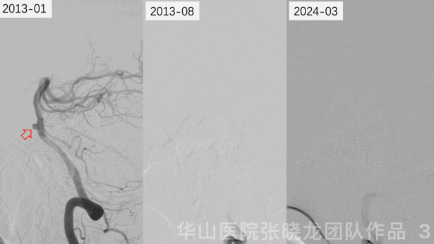
Figure 3 GIF. A basilar dissecting aneurysm was confirmed by DSA in January in 2013. The aneurysm was not detected by 7 month and 11 year angiograms.
图 3 GIF. 2013年1月造影证实基底动脉夹层动脉瘤。然而7个月和11年后的造影均未见动脉瘤显示。
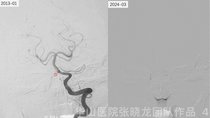
图 4 GIF. 2013年1月造影证实基底动脉下段夹层动脉瘤,11年后再次复查造影未见动脉瘤显影。
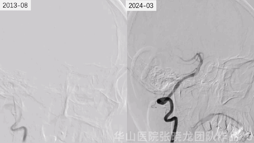
图 5 GIF. 左侧椎动脉造影示颅内血管完好,未见动脉瘤显示。
Video 1. The wall of basilar artery did not present with thickness nor enhancement from HR-MR in March 2024.
视频 1. 2024年3月高分辨率磁共振未见基底动脉管壁增厚或强化。
Summary
•The ruptured inferior segment basilar artery dissecting aneurysm led to the subarachnoid hemorrhage.
•Intracranial dissecting aneurysms more commonly occur in the posterior circulation, mainly at the level of vertebrate artery, and cause subarachnoid haemorrhage and/or cerebellar infarctions.
•Vertebrobasilar system dissecting aneurysms, especially ruptured, harboured high re-bleeding risks, which were suggested treatment rapidly and early.
•Isolated ruptured dissecting aneurysms involving basilar arteries are extremely rare and sparsely reported in the literature. Meyers, P. M. found 3 out of 1587 pediatric patients presented with spontaneous disappearance of basilar artery aneurysms. By 2 to 7-month imaging follow up, thrombosis formed and aneurysms shrunk. In adult groups, complete spontaneous resolution for posterior circulation intracranial dissecting aneurysms reported involved PICA and PCA.
•The cause of intracranial arterial dissections and dissecting aneurysms remains undetectable in majority of the cases. Connective tissue disorders and a number of other conditions, such as systemic infections, hypertension, hyperhomocysteinemia, smoking, oral contraceptive use, and hypercoagulable states, have been associated with intracranial dissections.
•The exact mechanism causing disappearance of dissecting aneurysm is still unclear. Most researchers think mural hematoma has (double) effect to both lumen and wall. It is not only the effect to cease further dissection in the wall (intima-media) of the aneurysms and promote healing, but also have an effect to the lumen of the dissecting aneurysm which results in reduction of inflow blood, create stasis of blood flow, and finally shrinkage (remodeling) of aneurysm.
•If this kind of dissecting aneurysms remained existence, flow diverters can be selected. While the efficacy still needs more cases.
•该病例考虑基底动脉下段夹层动脉瘤破裂致蛛网膜下腔出血。
•颅内段夹层动脉瘤常见于后循环,主要是椎动脉,可引起蛛网膜下腔出血和/或小脑梗死。
•后循环夹层动脉瘤,尤其是已经破裂出血的,再出血风险高,建议尽早治疗。
•单纯的破裂夹层基底动脉瘤自愈率很罕见,其相关文献报道也极其少。Meyers, P. M. 在1587名颅内动脉瘤儿童患者中发现了3例有良好转归的基底动脉夹层动脉瘤。3例患者经过2-7个月的影像学随访,出现了动脉瘤内血栓形成、动脉瘤变小的良好转归。在成年人中后循环颅内段夹层动脉瘤完全自愈的相关文献中,有关于小脑后下动脉和大脑后动脉夹层动脉瘤自愈的病例报道。
•颅内动脉夹层和夹层动脉瘤形成原因,在大部分病例中尚不清楚。结缔组织病和其他一些原因,如全身感染、高血压、高同型半胱氨酸血症,吸烟,口服避孕药、高凝状态等,可能与颅内夹层相关。
•夹层动脉瘤自愈的机制尚不清晰。目前认为壁间血肿的能一方面能缓解血管壁内膜进一步撕裂,促进管壁修复,同时会使腔内血流减慢、血流淤滞、动脉瘤回缩。
•若夹层动脉瘤未自愈,可尝试血流导向支架,但其疗效仍需更多病例证实。
声明:脑医汇旗下神外资讯、神介资讯、脑医咨询、Ai Brain 所发表内容之知识产权为脑医汇及主办方、原作者等相关权利人所有。
投稿邮箱:NAOYIHUI@163.com
未经许可,禁止进行转载、摘编、复制、裁切、录制等。经许可授权使用,亦须注明来源。欢迎转发、分享。
投稿/会议发布,请联系400-888-2526转3。

