Review
History
• Past medical history: DM for one year; smoking and drinking for 25 years, quitted for one month.
• PE: (-)
• 46岁,男性。
• 2天内发作性意识不清伴肢体抽搐2次。
• 既往史:糖尿病1年;吸烟、饮酒25年,已戒1月。
• 查体:-。

Figure 1. Enhanced-MRI revealed a cerebral AVM on the right parietal lobe with ectatic vessels.
图 1. 增强磁共振提示右侧顶叶脑动静脉畸形伴周围血管扩张。


Figure 2. No edema and micro-bleedings were observed.
图 2. 磁共振未见脑水肿及微出血灶。
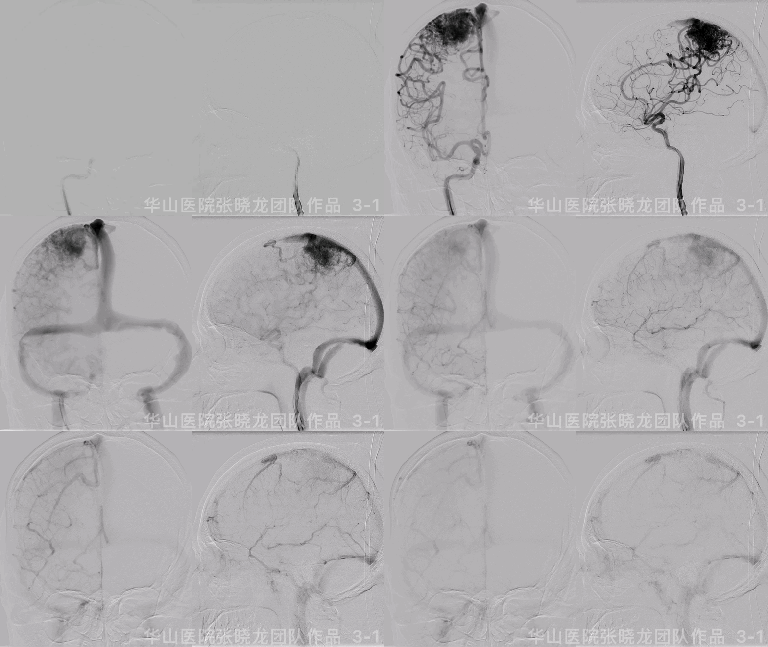
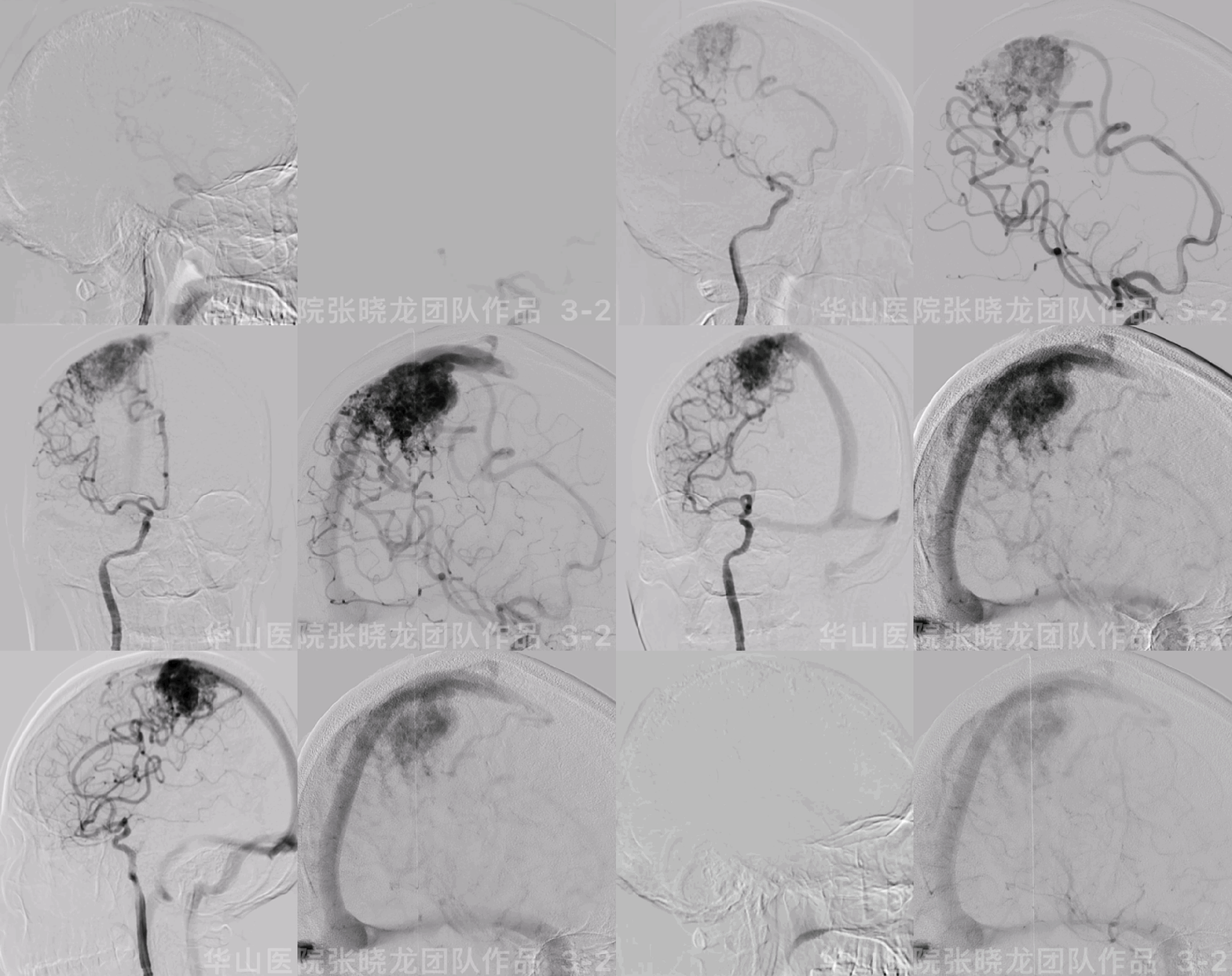
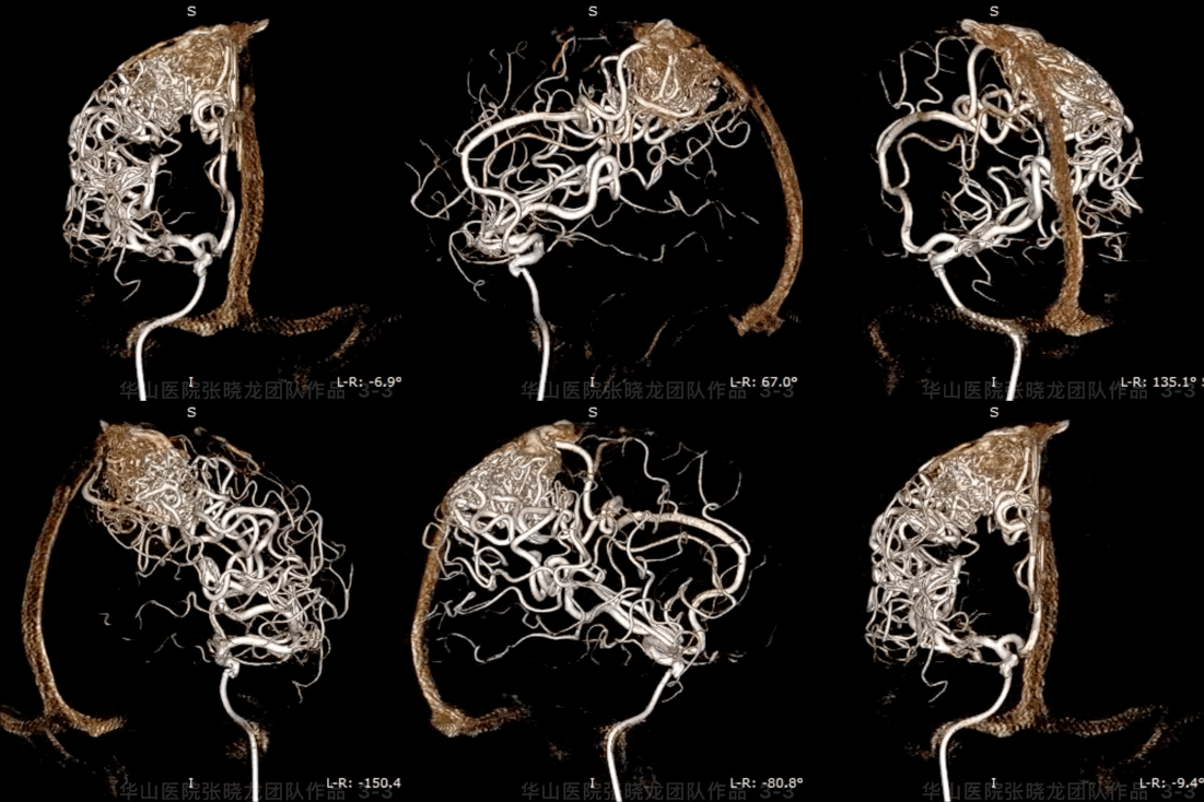
Figure 3 GIF. Right ACA and and right MCA branches fed the nidus and drained to superior sagittal sinus.
图 3 GIF. 造影示右侧大脑前动脉及大脑中动脉供血右侧顶叶畸形团,向上矢状窦引流。
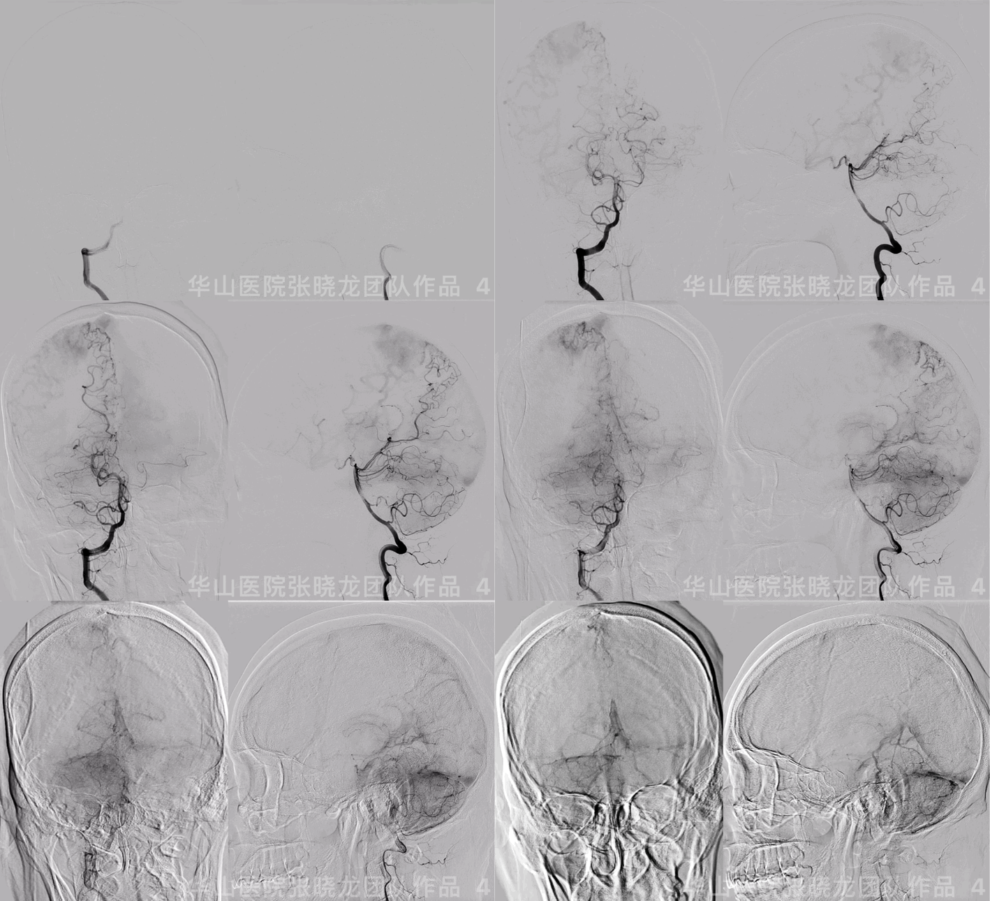
图 4 GIF. 右侧椎动脉造影示右侧大脑后动脉分支也参与畸形团供血。

图 5. 左侧颈内动脉及椎动脉、双侧颈外动脉无供血。右侧大脑前动脉及大脑后动脉直径增粗。
1
Strategy
•Right Juvenile parietal lobe AVM:
1.Feeding arteries: right MCA, right ACA, and right PCA branches.
2.Drainage into SSS.
3.Spetzler scale 3: maximal diameter 3-6cm, 2; function area, right parietal lobe, 1; no deep vein drainage, 0.
•Diluted Glubran is preferred for the juvenile high-flow AVM for staged embolization to decrease the flow and bleeding risk.
1.供血动脉:右侧大脑中动脉、大脑前动脉及右侧大脑后动脉分支。
2.引流静脉:经上矢状窦引流。
3.Spetzler scale 3:最大直径3-6cm,评分2;功能区,右侧顶叶,评分1;无深静脉引流,评分0。
•高流量幼稚型脑动静脉畸形计划分期靶向栓塞,一期栓塞计划采用稀释的Glubran减流,从而缓解临床症状和降低出血风险。
•术中及术后严格控制收缩压,降低出血风险。
2
1st Stage Operation


图 6. 行全身麻醉。将6F Navein导引导管置入右侧颈内动脉海绵窦段,动脉灌注尼莫地平1ml。术中收缩压维持在90-100mmHg。在Mirage .008微导丝支撑下将Marathon微导管经右侧大脑中动脉超选入畸形团供血动脉内,手推造影证明微导管到位。经微导管注入20% Glubran。


Figure 7 GIF. Another Marathon was advanced into the nidus through another right MCA branch. Injected 20% Glubran into the nidus.
图 7 GIF. 将另一根Marathon微导管经另一支大脑中动脉分支供血动脉超选至畸形团内。注入20% Glubran。


Figure 8 GIF. Angiograms showed the nidus was a bit of embolization while the intra-aneurysms still existed. Withdrew the microcatheter slightly, then injected 15% Glubran. Glubran casted well into the nidus.
图 8 GIF. 复查造影示畸形团少许栓塞,畸形团内仍可见瘤样扩张。将微导管头端少许撤回,然后注入15% Glubran,液体胶在畸形团内铸型良好。

图 9. 继续注入15% Glubran。当液体胶向供血动脉返流时停止打胶,撤回微导管。

图 10 GIF. 复查造影畸形团血流明显减低。


Figure 11. Marathon microcatheter was navigated into right ACA branch. Then injected 15% Glubran, casting well in the nidus.
图 11. 将Marathon微导管超选至右侧大脑前动脉分支,证实微导管在位。注入15% Glubran,在畸形团内铸型良好。

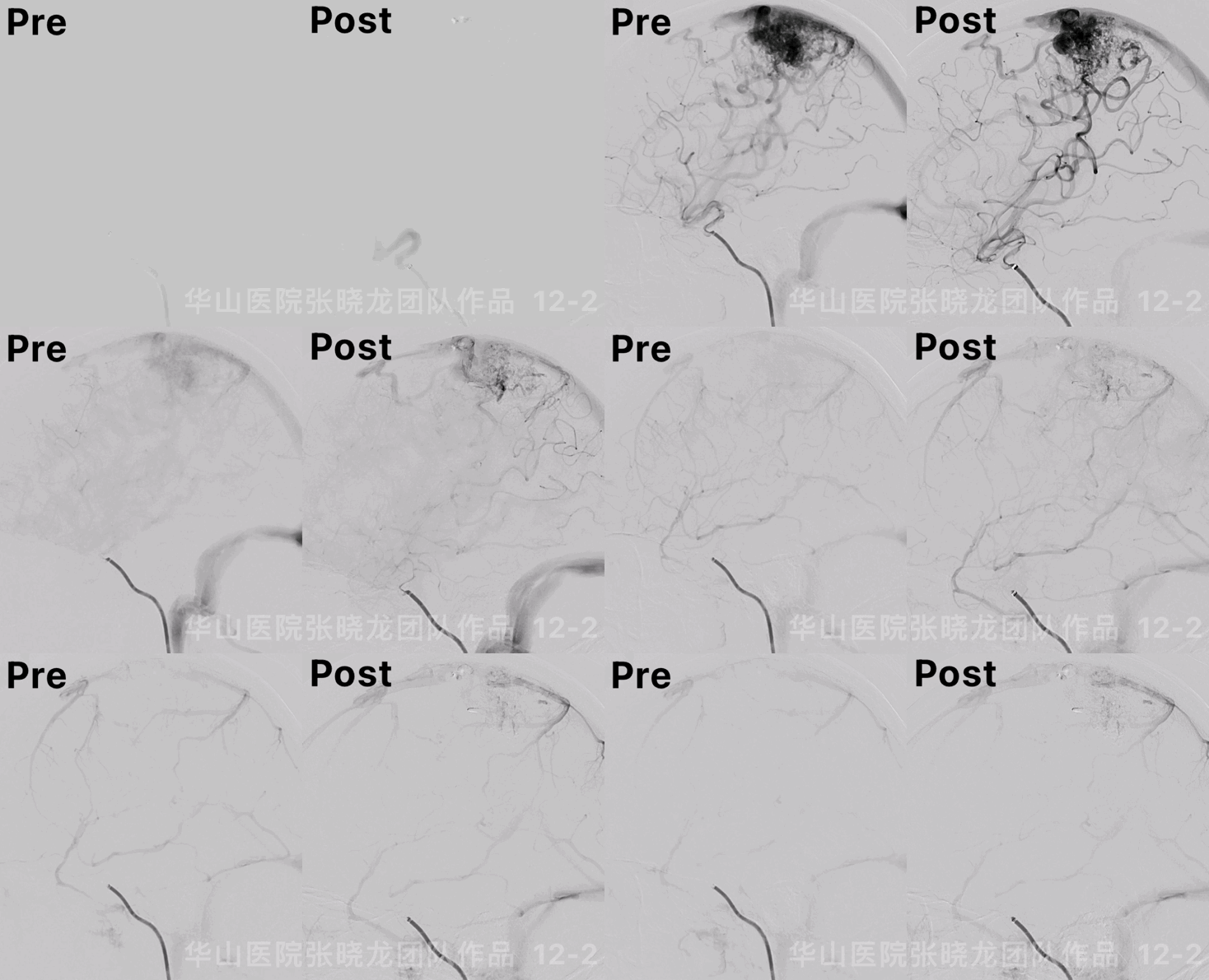
Figure 12 GIF. The nidus was partially embolized and flow decreased comparing with pre-operation.
图 12 GIF. 术后复查造影,畸形团部分栓塞,流量明显降低。
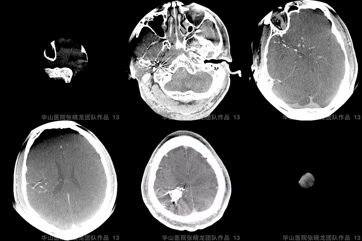
图 13 GIF. 术后即刻Dyna-CT未见出血。
3
Post-Operation
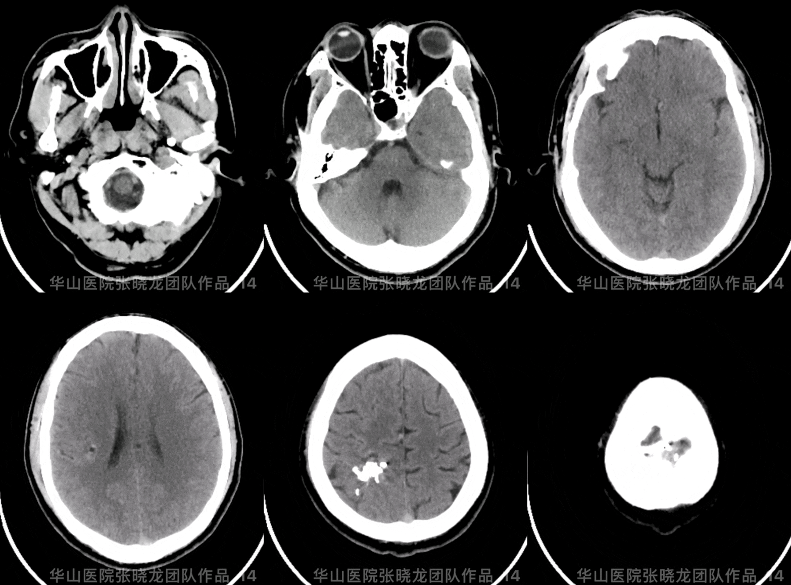
Figure 14 GIF. Post-operative day 1 cranial CT did not demonstrate any hemorrhage. Continue monitoring systolic BP between 90-110mmHg.
图 14 GIF. 术后第一天复查头颅CT平扫未见出血。继续严格控制收缩压,收缩压范围控制在90-110mmhg。
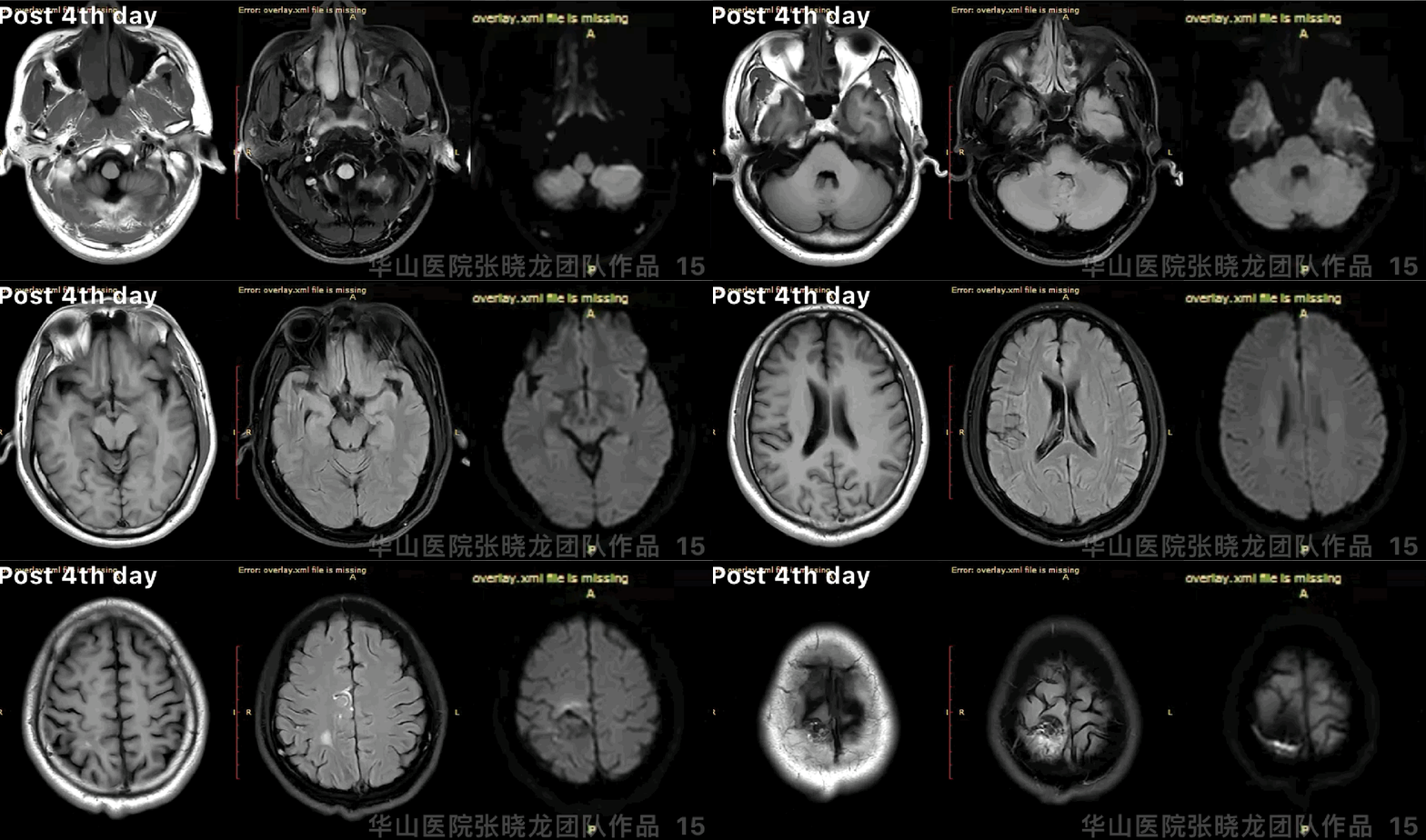
图 15 GIF. 术后第4天复查头颅MRI示畸形团周围少许水肿。患者无明显神经功能缺损症状。
4
Summary
•Right Juvenile parietal lobe AVM:
1.Feeding arteries: right MCA, right ACA, and right PCA branches.
2.Drainage into SSS.
3.Spetzler scale 3: maximal diameter 3-6cm, 2; function area, right parietal lobe, 1; no deep vein drainage, 0.
•Diluted Glubran is preferred for the juvenile high-flow AVM for staged embolization to decrease the flow and bleeding risk.
1.供血动脉:右侧大脑中动脉、大脑前动脉及右侧大脑后动脉分支。
2.引流静脉:经上矢状窦引流。
3.Spetzler scale 3:最大直径3-6cm,评分2;功能区,右侧顶叶,评分1;无深静脉引流,评分0。
•高流量幼稚型脑动静脉畸形计划分期靶向栓塞,一期栓塞计划采用稀释的Glubran减流,缓解临床症状和降低出血风险。
•术中及术后严格控制收缩压(90-100mmHg) ,降低出血风险。
•一期栓塞后血流量显著减少,血管构筑变得更加明晰。
•计划三个月后随访及二期栓塞治疗。
张晓龙
复旦大学附属华山医院
复旦大学附属华山医院放射科主任医师,博士、教授、博士生导师;
斯坦福大学医学院客座临床教授;
主持国家自然科学基金3项,第一作者或通讯作者发表国内外权威期刊文章50余篇;
中华医学会、放射学会、卫生部医政司等组织中担任副主任委员、组长等职务.《中国名医百强榜》神经介入专业中国十强(2012年度、2013年度、2014年度、2015-16年度、2017-18年度);
擅长复杂和疑难脑血管疾病的介入治疗,如复杂脑动脉瘤的栓塞,硬脑膜动静脉瘘栓塞,脑动静脉畸形栓塞,脑梗死的支架,脊髓血管畸形治疗;
自1995年开始从事脑血管疾病介入诊治工作和研究,师从黄祥龙教授、沈天真教授和凌锋教授,是我国最早从事神经介入的专家之一。2010年9月至今连续介入治疗颅内动脉瘤1500余例,无操作致死。
声明:脑医汇旗下神外资讯、神介资讯、脑医咨询、Ai Brain 所发表内容之知识产权为脑医汇及主办方、原作者等相关权利人所有。
投稿邮箱:NAOYIHUI@163.com
未经许可,禁止进行转载、摘编、复制、裁切、录制等。经许可授权使用,亦须注明来源。欢迎转发、分享。
投稿/会议发布,请联系400-888-2526转3。


