Review
History
• 52 y/o female.
• Medication: Aspirin, Clopidogrel, Atorvastatin, and Rabeprazole.
• 52岁,女性。
• 头痛40天,右侧肢体无力伴发作性表达困难1月。
• 当地医院CTA提示左侧颈内动脉闭塞,左侧椎动脉V4段夹层动脉瘤。MRI提示左侧大脑半球急性脑梗死。
• 神经查体:右侧肢体肌力V-,右侧脸部及大拇指麻木。


图 1. 当地医院CTA提示左侧颈内动脉闭塞。CT平扫可见左侧顶叶低密度影,DWI可见顶叶急性梗死灶。

Figure 2. Local hospital CTP depicted the left parietotemporal low perfusion.
No microhemorrhage was detected from SWI.
图 2. 当地医院CTP示左侧顶颞叶低灌注。SWI未见明显微出血灶。


图 3. 1月后复查高分辨率磁共振及头颅MRA示原闭塞左侧颈内动脉再通,左侧椎动脉V4段及左侧颈内动脉可见壁间血肿,管壁增厚及血栓血管形成,左侧ICA海绵窦段局部重度狭窄。

图 4 GIF. DSA证实左侧海绵窦段夹层伴局部重度狭窄,双侧颈内动脉颈段发育不良。
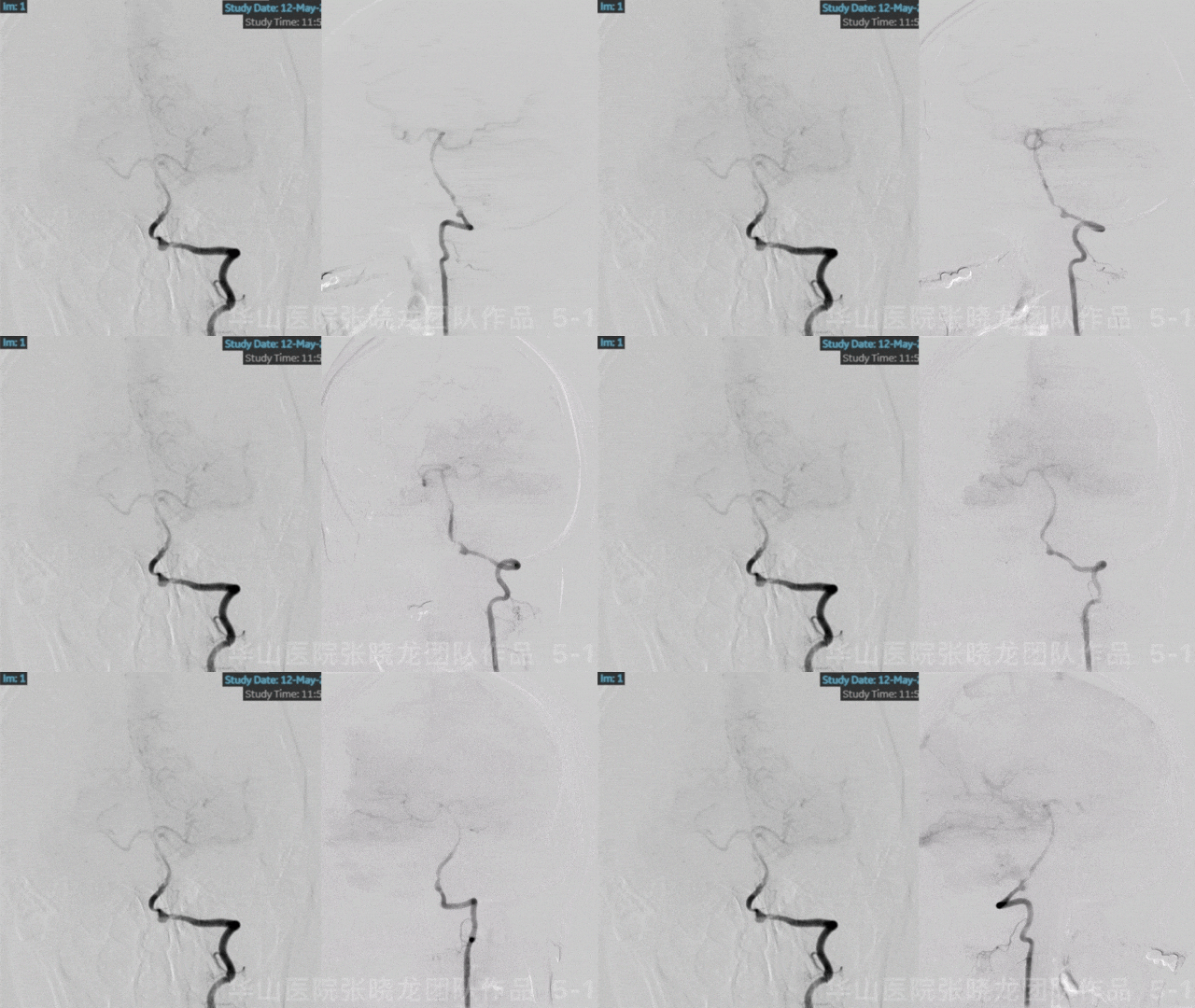

图 5 GIF. 后循环造影证实左侧椎动脉V4段夹层动脉瘤伴载瘤动脉远端狭窄,左侧椎动脉V2段小夹层。后循环经后交通动脉代偿左侧前循环。
1
Strategy
1.The left V4 segment dissecting aneurysm with distal segment stenosis presented growth or rupture risks, which was suggested to treat.
2.Due to the simple procedure and without a perforator arising from the segment, a flow diverter can be employed. The post-stenting large coil technique is an alternative.
3.For better apposition and remodeling effect, the pre-dilation with insufficient pressure should be performed.
4.This patent was considered segmental artery dysplasia due to multiple dissections. The left V2 segment dissection can be stented to lower occlusion risks.
5.The left ICA spontaneously recanalized in one month after antiplatelet administration. Left cavernous segment dissection with severe stenosis and low perfusion should be stented to improve left hemisphere perfusion and decrease re-occlusion risk.
6.SWI: no micro-hemorrhages and CTA: no significant calcifications.
6.风险因素评估:SWI未见明显微出血灶,同时CTA提示未见明显钙化灶。
2
Step 1. Left V4 segment dissection
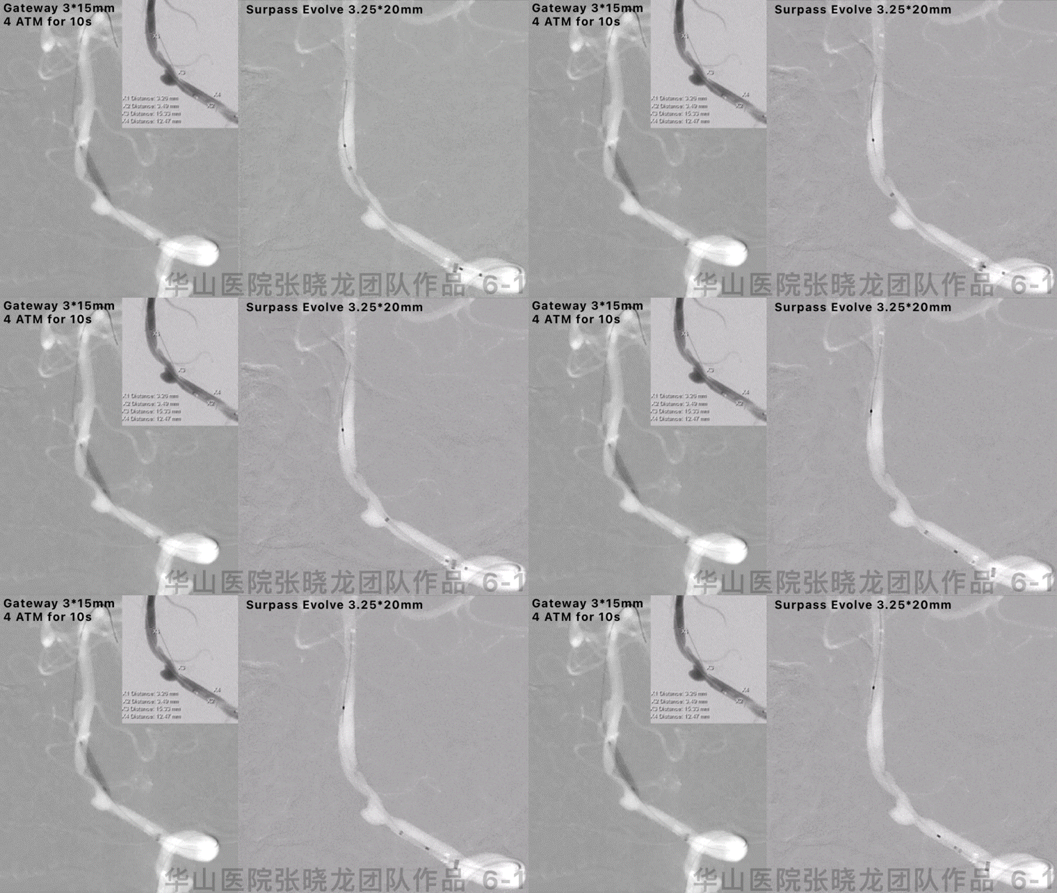
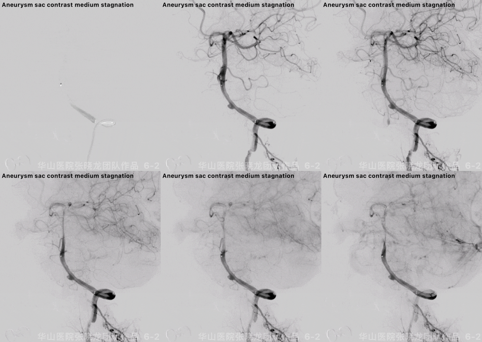
图 6 GIF. 全身麻醉, 全身肝素化。将6F 105cm Navien导引导管在125cm MP和0.035导丝导引下置于V4夹层狭窄段近端提供足够支撑。选用Gateway 3*15mm球囊在Floopy-300导丝支撑下置于夹层狭窄段,予4ATM扩张10s后泄除球囊。XT-27微导管置于基底动脉,Surpass Evolve 3.25*20mm支架于夹层段释放,支架释放满意。复查造影夹层动脉瘤内造影剂滞留,载瘤动脉通畅及支架打开并贴壁良好。
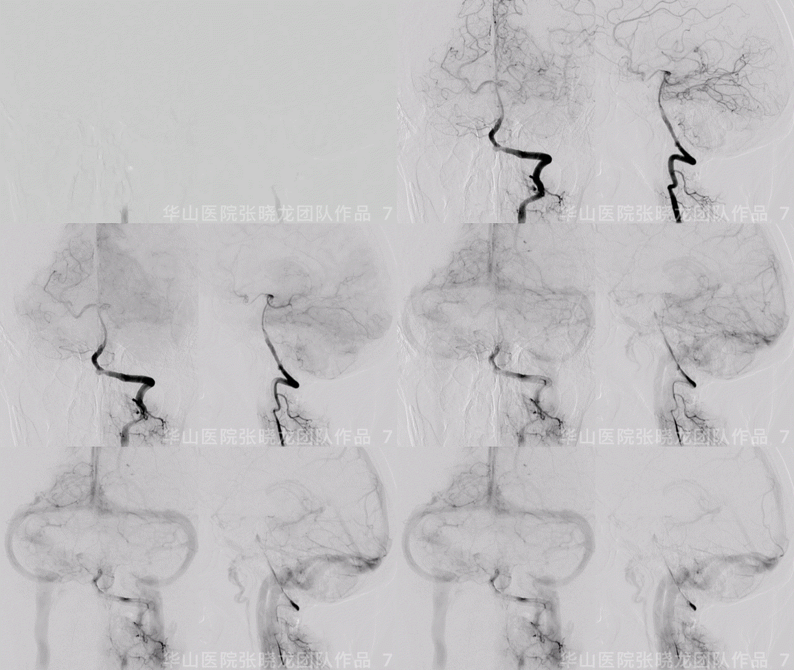
图 7 GIF. 正侧位造影均示颅内血管显影良好。经导引导管予替罗非班5ml和尼莫地平0.5ml。
3
Step 2. Left V2 segment dissection

图 8. 选用Wingspan 3.5*15mm支架于左侧V2段夹层处释放,复查造影夹层段修复满意。
4
Step 3. Left ICA cavernous segment stenosis

图 9. 将6F 105cm Navien置于左侧颈内动脉。经动脉灌注尼莫地平0.5ml预防血管痉挛。Gateway 3*15mm球囊于海绵窦段狭窄处4ATM扩张10s,球囊扩张满意,造影证实未见出血。选用Wingspan 3.5*15mm(剪去头端,避免刺破血管)于狭窄段释放。
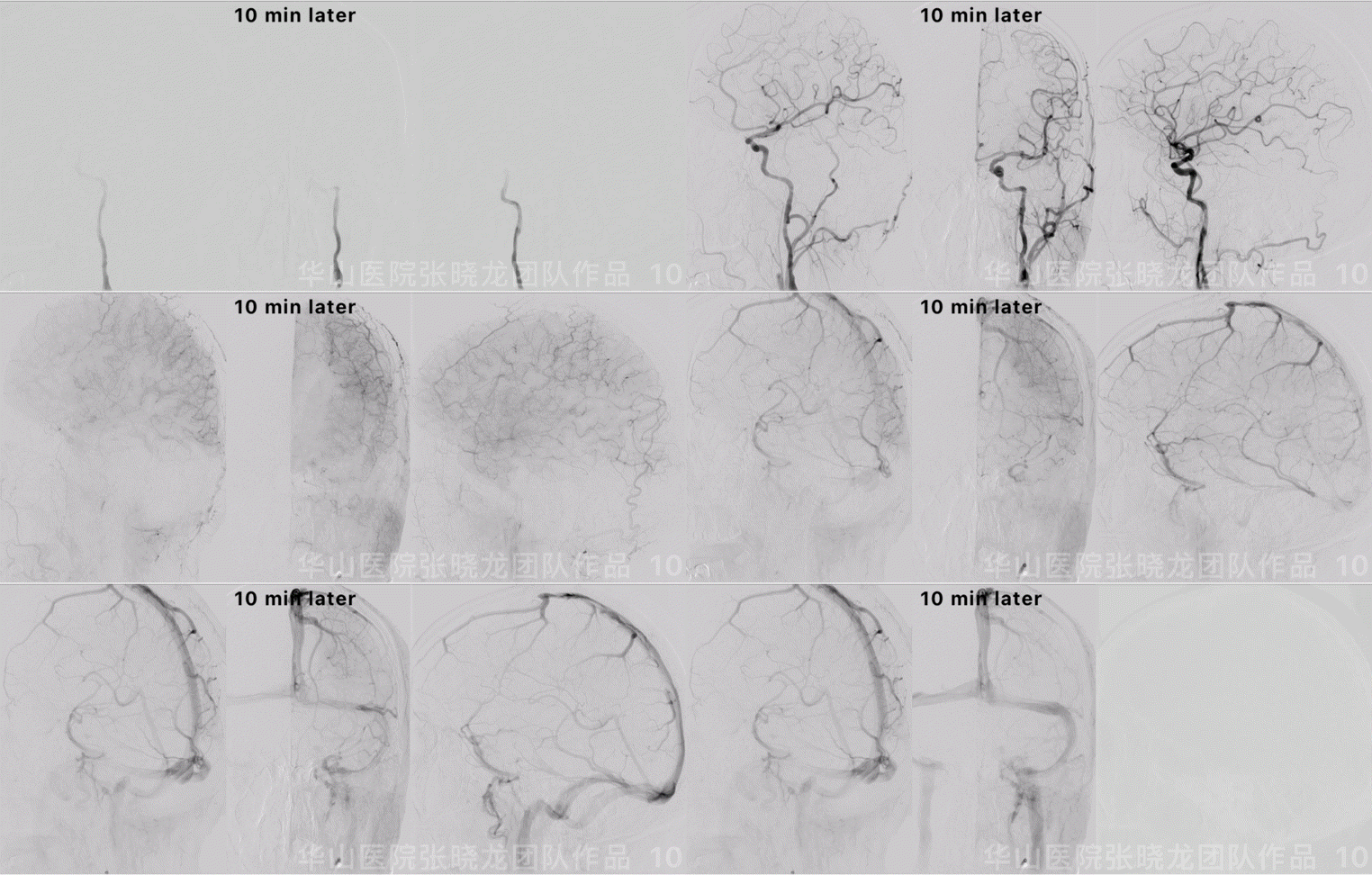
Figure 10 GIF. Angiograms showed the vessels vasospasm. Tirofiban 8ml and Nimodipine in total 2ml were administered. Waiting for 10 minutes. Vasospasm alleviated and left anterior blood flow improved significantly.
图 10 GIF. 复查造影颈段血管痉挛明显。经导引导管给予替罗非班8ml,反复多次共给予尼莫地平2ml。10min后复查造影血管痉挛缓解,左侧前循环血流明显改善。
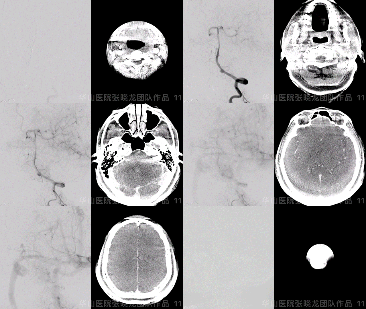
Figure 11 GIF. Re-checking left vertebral artery angiograms visualized the dissecting aneurysm sac contrast stagnation and dissecting segment recovered well. Tirofiban 3ml was administered. DynaCT showed no bleeding.
图 11 GIF. 复查左椎造影:夹层动脉瘤内造影剂滞留明显,夹层段修复良好。经导引导管给予替罗非班3ml。Dyna-CT未见出血。
5
Post-Operation
●NE: GCS 15, bilateral eye movement normal, bilateral muscle strength normal, no swallowing difficulty nor facial paralysis.
●Medication: Stopped Clopidogrel and changed to Ticagrelor 45mg bid. Continue Aspirin 100mg qd. AA 87.5%, ADP 50.3%, CYP2C19 IM.
●神经查体:患者GCS 15,双侧眼球运动正常,四肢肌力正常,无饮水呛口及面瘫。
●药物:停氯吡格雷,改成替格瑞洛45mg bid,继续口服阿司匹林100mg qd。血栓弹力图:阿司匹林抑制率87.5%,氯吡格雷抑制率50.3,氯吡格雷基因代谢中等代谢,酶活性减低。
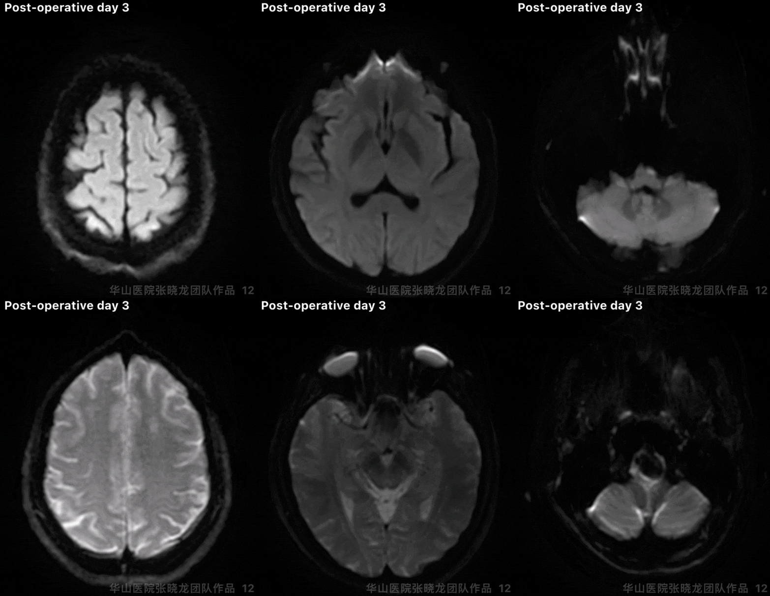
Figure 12 GIF. No acute infarctions were detected from DWI.
图 12 GIF. 术后第3天复查头颅DWI未见急性脑梗死。
6
Follow Up
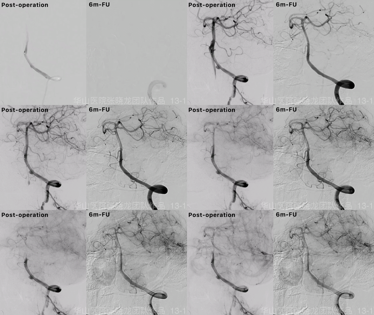

图 13 GIF. 6个月随访复查造影左侧椎动脉夹层修复完好,夹层动脉瘤不显影。

Figure 14. A left V2 segment small dissecting recovered completely by 6 month follow up.
图 14. 6个月随访左侧椎动脉V2段小夹层完全修复。
视频 1. 6个月复查左侧颈内动脉海绵窦段支架内无再狭窄,血流通畅,颅内血管完好。

Figure 15. A left anterior circulation blood flow increased without posterior circulation compensation. Left ICA cervical segment dissection recovered.
图 15. 6个月复查左侧前循环血流增加,不需要后循环代偿。左侧颈内动脉修复。
7
Summary
1.Intracranial dissecting aneurysms harboured growth and rupture risks, which was suggested treatment. Both post stenting large coil technique and flow diverters can be selected. A flow diverter was adopted for the left V4 segment dissecting aneurysm with distal segment stenosis. By 6 month follow up, the dissecting aneurysm was invisible and parent artery recovered well. While more cases were needed to verify the safety and efficacy.
2.Extracranial dissections with severe stenoses can be treated by balloon angioplasty and stenting in order to improve left hemisphere blood flow and decrease re-occlusion risk. Left cavernous segment dissection and severe stenosis was stented. By 6 month follow up, no re-stenosis occurred and left anterior circulation improved significantly.
3.Cervical segment dissections without stenoses can be treated by stenting or medicine alone. The left V2 segment dissection was stented by a Wingspan. The vessel wall recovered completely by 6 month follow up. Bilateral ICA cervical segment dissections were treated by medicine alone.
4.This patent was considered segmental artery dysplasia due to multiple dissections. Aspirin was suggested for long-term. Next DSA follow up was scheduled in 2-3 years.
1.颅内段夹层动脉瘤,存在增大及破裂出血风险,建议治疗,可选择大圈辅助支架后释放或血流导向装置。本病例中左侧椎动脉v4段夹层动脉瘤伴载瘤动脉远端狭窄,采用了血流导向支架,术后6月随访夹层动脉瘤及血管壁修复良好,但血流导向装置的安全性及有效性仍需更多病例来证实。
2.颅外段夹层合并重度狭窄选择球囊辅助支架技术,改善血供同时降低狭窄加重或闭塞风险。本病例中左侧颈内动脉海绵窦段夹层伴局部重度狭窄及低灌注,行球囊扩张+支架成形术。术后6月管壁修复良好,支架内无再狭窄,左侧前循环灌注增加。左侧椎动脉V2段小夹层采用Wingspan支架治疗,6个月随访管壁完全修复。
3.颈内动脉颈段或椎动颈段夹层不合并狭窄可选择支架或药物治疗。本病例中椎动脉V2段小夹层采用Wingspan支架治疗,6个月随访管壁完全修复。双侧颈内动脉颈段小夹层药物治疗。
4.该患者考虑节段性动脉发育不良导致多发夹层病变。建议长期口服阿司匹林。2-3年后再复查脑血管造影。
张晓龙
复旦大学附属华山医院
复旦大学附属华山医院放射科主任医师,博士、教授、博士生导师;
斯坦福大学医学院客座临床教授;
主持国家自然科学基金3项,第一作者或通讯作者发表国内外权威期刊文章50余篇;
中华医学会、放射学会、卫生部医政司等组织中担任副主任委员、组长等职务.《中国名医百强榜》神经介入专业中国十强(2012年度、2013年度、2014年度、2015-16年度、2017-18年度);
擅长复杂和疑难脑血管疾病的介入治疗,如复杂脑动脉瘤的栓塞,硬脑膜动静脉瘘栓塞,脑动静脉畸形栓塞,脑梗死的支架,脊髓血管畸形治疗;
自1995年开始从事脑血管疾病介入诊治工作和研究,师从黄祥龙教授、沈天真教授和凌锋教授,是我国最早从事神经介入的专家之一。2010年9月至今连续介入治疗颅内动脉瘤1500余例,无操作致死。
声明:脑医汇旗下神外资讯、神介资讯、脑医咨询、Ai Brain 所发表内容之知识产权为脑医汇及主办方、原作者等相关权利人所有。
投稿邮箱:NAOYIHUI@163.com
未经许可,禁止进行转载、摘编、复制、裁切、录制等。经许可授权使用,亦须注明来源。欢迎转发、分享。
投稿/会议发布,请联系400-888-2526转3。


