Review
History
• 59 y/o male.
• Suffered from right lower limb weakness and mild urination disturbance for 4 years, aggravated for one year.
Right lower limb V-. Right lower limb numbness.
• 59岁,男性。
• 右下肢无力伴轻度小便困难4年,加重1年。
右下肢肌力V-,右下肢麻木。
Aminoff评分G1M1F0。

图 1. 胸段 MRI提示异常血管流空影及脊髓水肿。
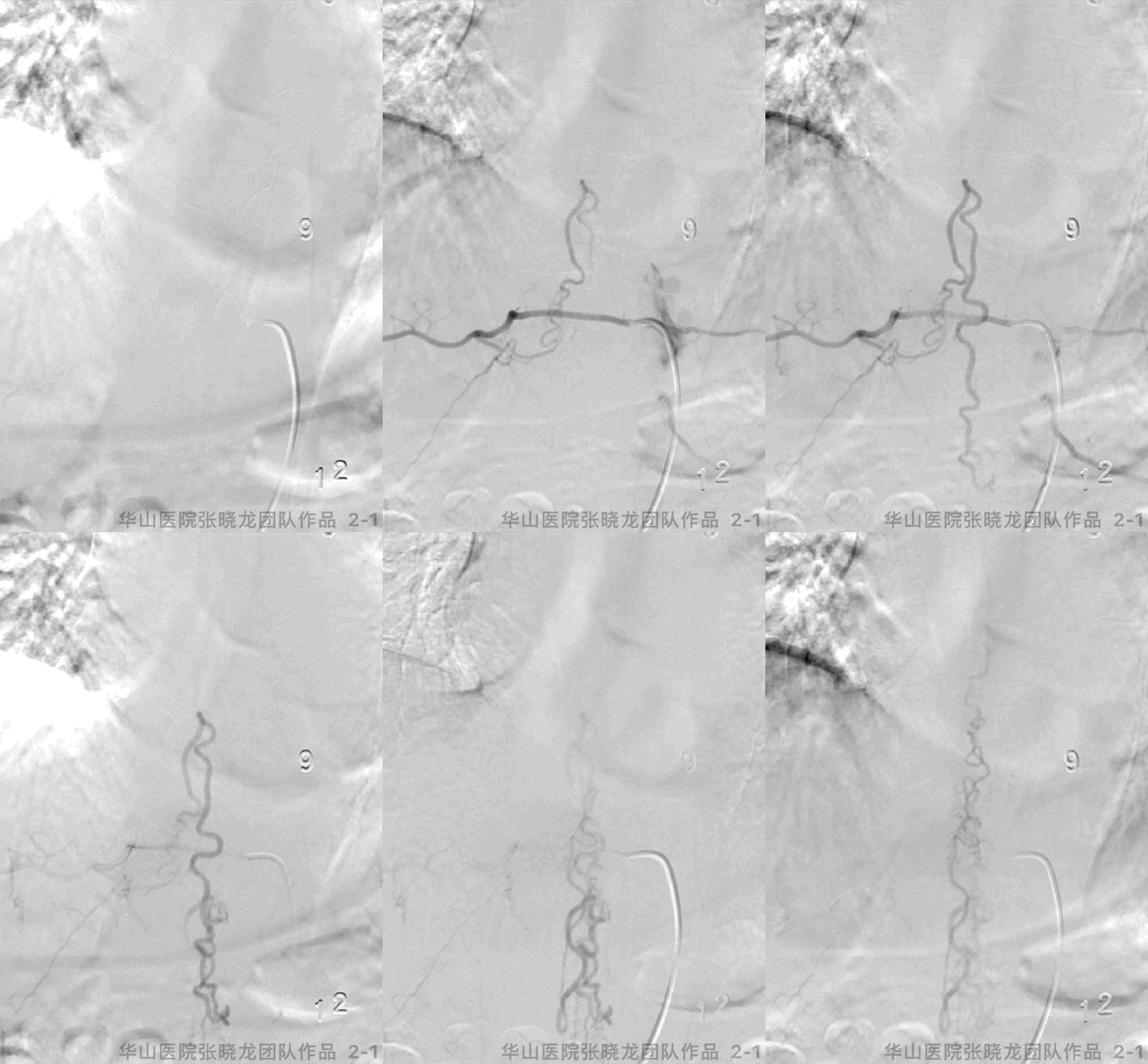
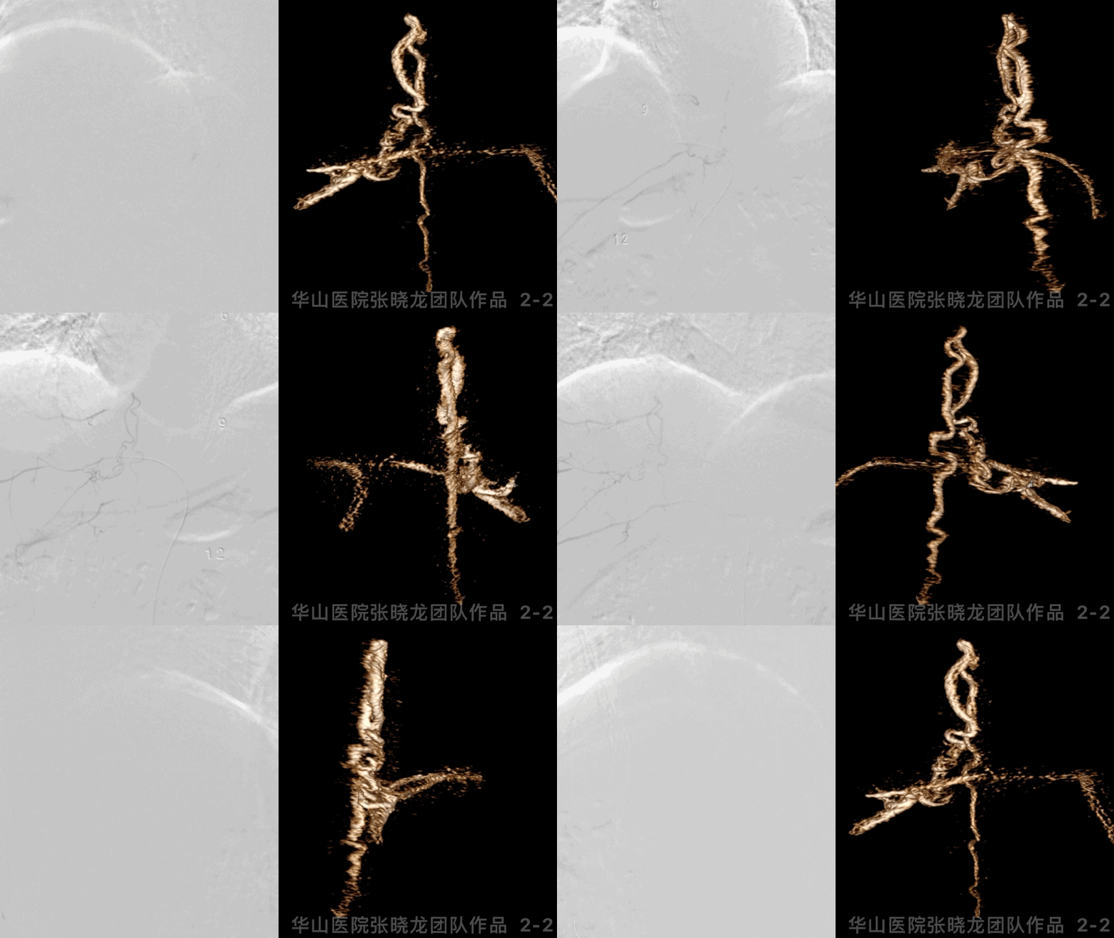
Figure 2 GIF. T10 segment spinal dural arteriovenous fistula was confirmed, which was fed by right radicular arteries and drained to spinal venous. No other feeding arteries were found from spinal angiography.
图 2 GIF. 脊髓血管造影证实T10硬脊膜动静脉瘘,由右侧T10肋间动脉发出的根固有动脉供血向脊髓静脉引流。全脊髓血管造影证实无其他动脉供血。
1
Strategy
The fistula located at right T10 intervertebral foramen. The SDAVF was fed by right radicular arteries and drained to spinal venous.
Microcatheter angiography should be performed to test the reflux and meanwhile rule out functional spinal artery to avoid excessive embolization.
Microcatheter should be navigated to the fistula as far as possible.
Onyx-18 was selected if microcatheter was navigated to the distal part of feeding artery. While Glubran was an alternative if microcatheter cannot be in place.
诊断:硬脊膜动静脉瘘,瘘口位于右侧T10水平椎间孔,由右侧第10肋间动脉发出的根固有动脉供血,向脊髓静脉引流。 栓塞前需微导管造影测试返流情况,同时有助于发现功能型脊髓动脉,避免过度栓塞。 微导管需尽可能向远端超选。
若微导管超选至供血动脉远端,选择Onyx-18打胶;若微导管无法超选到很远,可以选择Glubran。
2
Operation

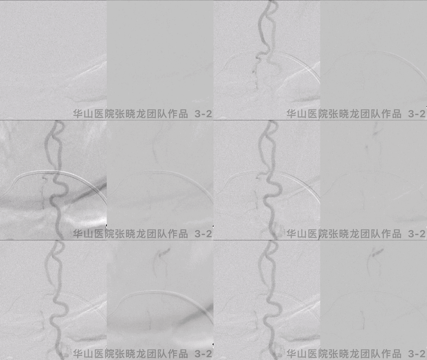

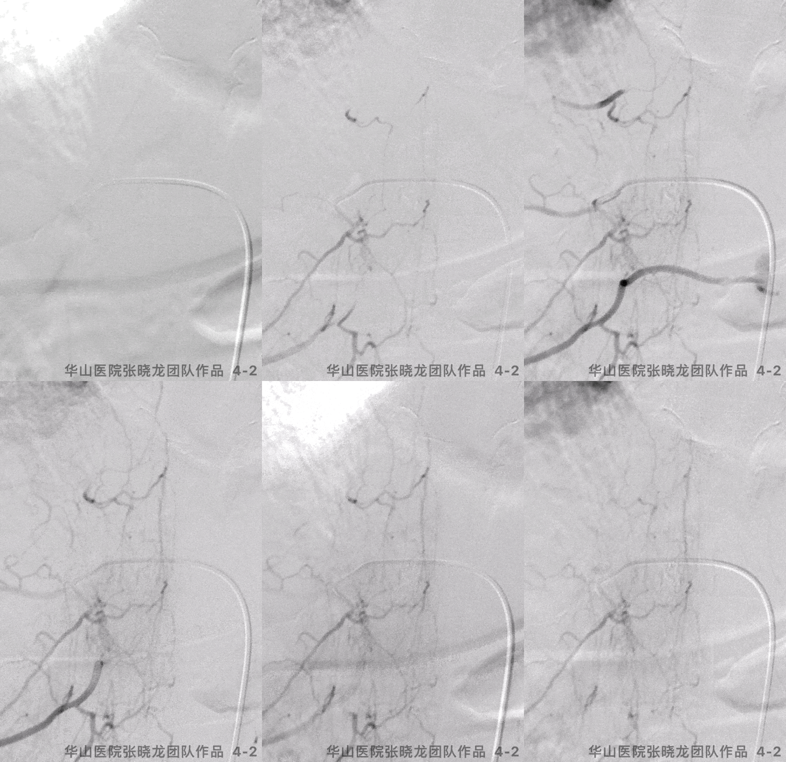
图 4 GIF. 注入Onyx-18 0.3ml,胶在脊髓静脉、动静脉瘘铸型良好。撤出微导管复查造影,瘘口不显影,局部可见造影剂外渗。肝素自然中和。
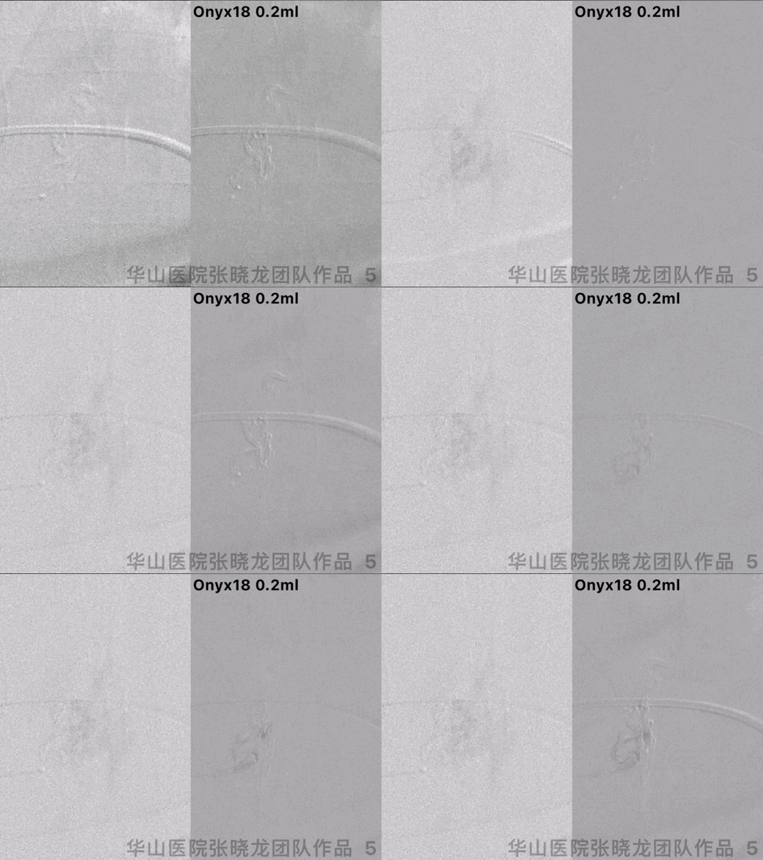
图 5 GIF. 选用Echelon-10 45°微导管超选至供血动脉分支近端,证实非有正常功能的脊髓动脉,注入Onyx-18 0.2ml将供血动脉主干闭塞。
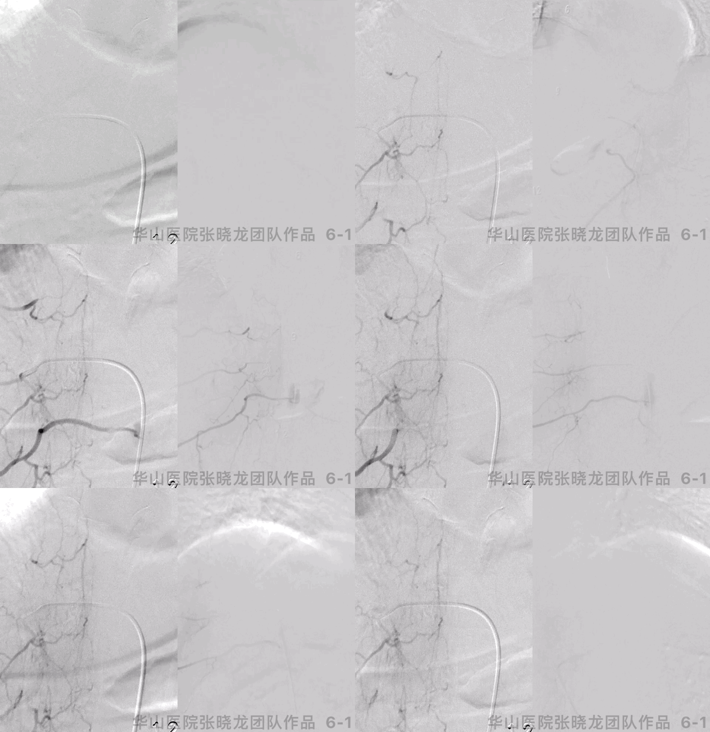

图 6 GIF. 复查造影无活动性出血,瘘口不显影。复查紧邻节段肋间动脉及腰动脉造影未见残余瘘口显影,脊髓前、后动脉显影良好。
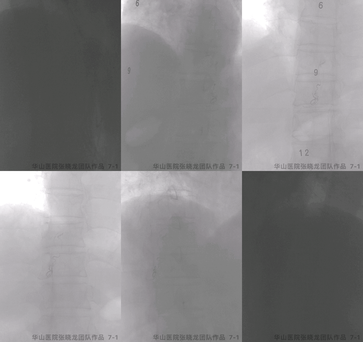


图 8. 术后复查MR提示脊髓水肿较前略好转。
3
Post-Operation
PE: B bilateral limbs V, right lower limb numbness relieved. Aminoff-Logue score: G0M0. Medication: Continue Rivaroxaban 20mg qd (Low molecular heparin was prescribed for 3 days and bridging with Rivaroxaban before operation). Mannitol.
查体:四肢肌力V,右下肢麻木较前好转。Aminoff-Logue score: G0M0.
药物:继续利伐沙班20mg qd(术前3天皮下注射低分子肝素并桥接利伐沙班)。并予甘露醇适当脱水。
4
Summary
The fistula located at right T10 intervertebral foramen. The SDAVF was fed by right radicular arteries and drained to spinal venous.
Microcatheter angiography should be performed to test the reflux and meanwhile rule out functional spinal artery to avoid excessive embolization.
Microcatheter should be navigated to the fistula as far as possible.
Onyx-18 was selected if microcatheter was navigated to the distal part of feeding artery. While Glubran was an alternative if microcatheter cannot be in place.
The fistula and primary venous were casted densely, while the distal spinal veins should be avoided packed.
Adjacent intercostal/lumbar artery angiography (Contralateral and adjacent levels) were performed to rule out other feeding arteries.
Post-operative confirmed onyx casted at the right 10th intervertebral foramen.
Low molecular heparin was prescribed perioperative period for 3 days and bridging with Rivaroxaban. Rivaroxaban was prescribed for the long term.
诊断:硬脊膜动静脉瘘,瘘口位于右侧T10水平椎间孔,由右侧第10肋间动脉发出的根固有动脉供血,向脊髓静脉引流。
栓塞前需微导管造影测试返流情况,同时有助于发现脊髓功能动脉,避免过度栓塞。
微导管需尽可能向远端超选。
若微导管超选至供血动脉远端,选择Onyx-18打胶;若微导管无法超选到很远,则可以选择Glubran。
瘘口及初级静脉端须致密栓塞,避免栓塞脊髓静脉远端。
术后需要复查紧邻节段肋间动脉、腰动脉,排除瘘口残余供血。
术后胸椎CT证实液体胶在右侧第10椎间孔铸型。
术前3天予低分子肝素皮下注射并桥接利伐沙班,术后建议继续长期口服利伐沙班。
* 注:本文作者:张晓龙教授团队,内容来源:上海神经介入论坛,脑医汇获授权转载发布

点击或扫描上方二维码
查看更多“介入”内容
声明:脑医汇旗下神外资讯、神介资讯、脑医咨询、Ai Brain 所发表内容之知识产权为脑医汇及主办方、原作者等相关权利人所有。
投稿邮箱:NAOYIHUI@163.com
未经许可,禁止进行转载、摘编、复制、裁切、录制等。经许可授权使用,亦须注明来源。欢迎转发、分享。
投稿/会议发布,请联系400-888-2526转3。


