

Review
History
• 48 y/o female.
• An ACOM aneurysm was occasionally detected by local hospital CTA.
• Past medical history: No chronic or acute headache; no HTN nor DM; alcohol consumption for 20 years, stopped 1 year.
• Medication: /.
• NE: -.
• 48岁,女性。
• 药物:无。
• 神经查体:-。

图 1. CTA提示前交通动脉瘤,DWI未见明显急性脑梗死。

Figure 2. No sac wall enhancement was visualized from HR-MRI.
图 2. 高分辨率磁共振提示前交通动脉瘤,瘤壁未见明显强化。


图 3 GIF. DSA造影证实前交通动脉瘤,双侧A1段发育良好。右侧压颈试验显示右侧ACA和MCA可经前交通动脉显影。左侧后交通动脉圆锥。

图 4. 3D重建示前交通动脉瘤形态不规则。
1
Strategy

In this case, the AcomA-AN neck completely envelops the anterior communicating artery, and the sac involves bilateral A1~A2 segments, so the treatment of double-stent assisted embolization was adopted.
“H” or “X” stent assisted coiling can be tried for this complex wide-necked aneurysm.
Bilateral A1 should be developed well for the “H” and “X” techniques.
Due to the obvious angulation between right A1 and A2, the release of “H” shaped stent is difficult. "H" shaped stent is filled with the coil and occluded the Acom artery.
"X" shaped stent can form a cross at the anterior communicating artery and increase the stent coverage of the sac.
前交通动脉瘤形态不规则,破裂风险高,建议治疗。
该病例中前交通动脉瘤瘤颈完全累及前交通动脉,瘤腔累及双侧A1-2段,所以采用双支架辅助栓塞策略。
这种复杂的宽颈动脉瘤,可以采用 “H” 或 “X” 支架辅助栓塞。
“H”和 “X” “支架的优劣势:
双侧A1段发育良好时可以采用“H” 或 “X”支架辅助栓塞。
由于右侧A1和A2段成角明显, 微导管进入远端载瘤动脉困难,导致“H” 支架释放困难,但是支架置入后可矫正A1到A2段成角畸形,从而降低动脉瘤的复发。
“X” 型支架在前交通动脉相互交叉重叠,增加了瘤腔处支架的覆盖率,但是第二枚支架需要两次穿孔第一枚支架,可能导致支架局部打开不良,引起血栓形成;同时该方案无法矫正血管的成角畸形,导致复发率增加。
“H” stent technique was selected.
Stenting microcatheter steam shape is important for cute angle between A1 and A2.
Segmentally shaped the microwire is necessary for crossing A1 and A2 initial angle.
Different working projections (3 working projections) were selected for sac embolization and double-stent deployment.
After the angle straightened via Solitaire stent deployment, nimodipine should be routinely used to relieve vasospasm.
For small irregular aneurysms, 3D coil is preferred to form basket to prevent 2D coil from puncturing the aneurysms, and dense packing might not be required due to relative low recurrence rate.
本病例计划采用“H”型支架技术。
由于A1和A2段成角锐利,支架微导管头端的塑形十分重要。
微导丝节段性塑形有助于A1-2段的超选。
选择多个(3个)不同的工作角度分别用于动脉瘤的栓塞、2枚支架的释放。
Solitaire支架释放后载瘤动脉拉直,需常规使用尼莫地平缓解血管痉挛。
2
Operation

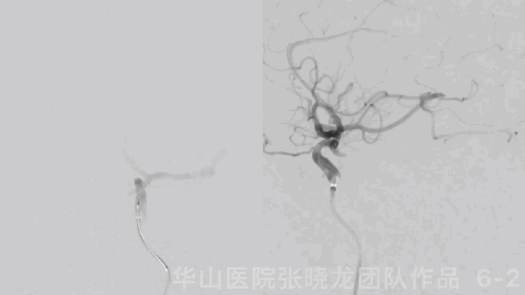
图 6 GIF. 全身肝素化。将导引导管置于左侧颈内动脉海绵窦段,Prowler plus微导管在Synchro-II standard微导丝导引下置于左侧大脑前动脉A2段。Solitaire 4*20mm于左侧A2至A1段释放。透视下证实支架近端位于A1段。经导引导管于尼莫地平0.5ml。复查造影颅内血管显影良好。

Figure 7. Parent artery was straightened significantly.
图 7. 载瘤动脉明显拉直。
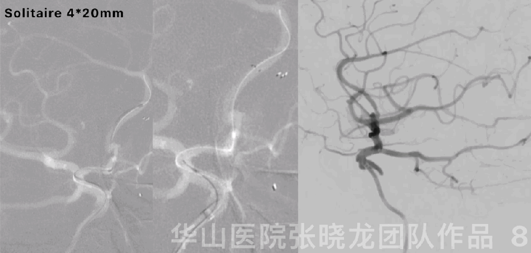
图 8 GIF. 导引导管置于右侧颈内动脉海绵窦段。Prowler plus微导管在Synchro-II standard微导丝导引下置于右侧大脑前动脉A2段。Solitaire 4*20mm于右侧A2至A1段释放。复查造影未见颅内血管出血或血栓形成,前交通动脉瘤仍然显影。


Figure 9. Right ACA straightened obviously.
图 9. 支架后右侧大脑前动脉明显拉直。


图 10. 3D重建显示前交通动脉瘤形态不规则,伴多发子瘤。

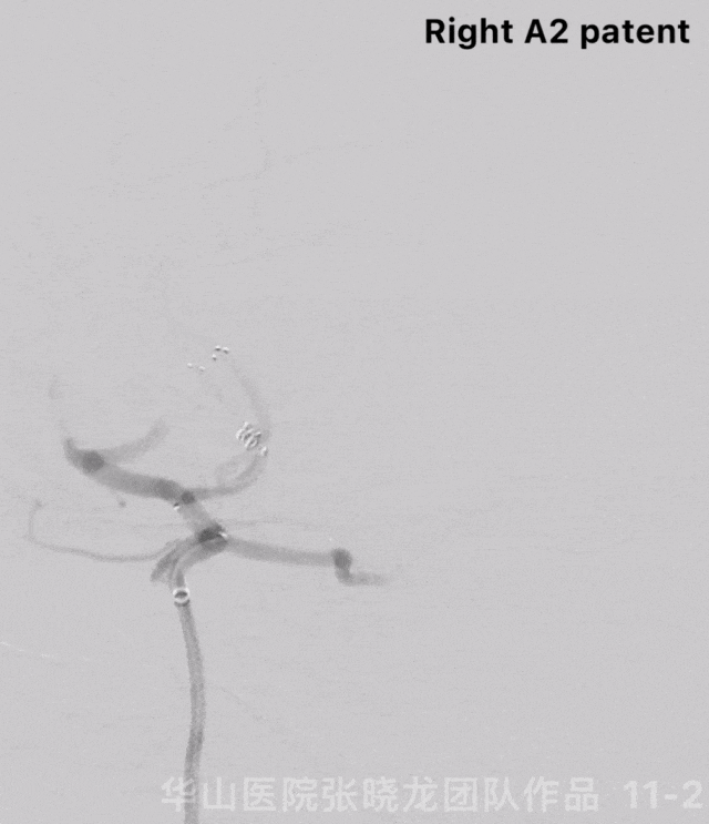
Figure 11 GIF. Angiograms showed no intracranial vessels bleeding and the small aneurysm still visualized. The aneurysm size 2.71*2.04mm, neck 1.51mm. Inserted a Prime 3D 2mm*4cm coil. DSA depicted the parent artery patent and the aneurysm was packed satisfactorily.
图 11 GIF. 复查造影颅内血管完好,动脉瘤仍显影。测量动脉瘤大小2.71*2.04mm,瘤颈1.51mm。经Echelon-10直头微导管填入一枚Prime 3D 2mm*4cm弹簧圈。复查造影动脉瘤不显影,载瘤动脉通畅。
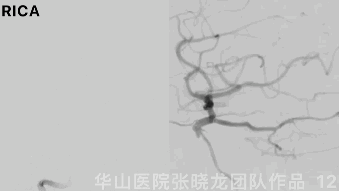
图 12 GIF. 复查右侧颈内动脉造影,颅内血管完好,动脉瘤瘤体少许显影(Raymond IIIa)。
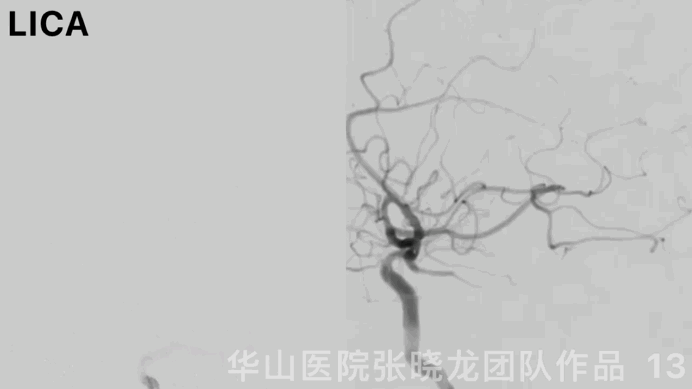
Figure 13 GIF. Left ICA angiograms demonstrated intracranial vessels intact and the aneurysm sac with slight residue.
图 13 GIF. 复查左侧颈内动脉造影,颅内血管完好,动脉瘤体少许残留( Raymond IIIa)。
图 14 GIF. 术后即刻Dyna-CT前纵裂未见明显造影剂滞留。术后1天复查头颅CT平扫,原高密度造影剂吸收,颅内未见出血。
3
Post-operation
NE:
GCS 15, bilateral pupils light reflux normal, normal muscle strength, no neurological deficiency.
Medication:
Tirofiban 6ml/h maintained for 48 hours.
Aspirin and Clopidogrel were prescribed.
TEG ADP 98.5 AA 95.4.
神经查体:
GCS 15,双侧瞳孔等大等圆、对光反射灵敏,四肢肌力正常,未见神经功能缺损。
药物:
替罗非班6ml/h维持48h。
口服阿司匹林及氯吡格雷。
血栓弹力图:氯吡格雷抑制率98.5%,阿司匹林抑制率95.4%。
氯吡格雷基因代谢正常。
Video 1. Right ACA was patent and the aneurysm was not relapsed by 6 month follow up.
视频 1. 6个月复查右侧大脑前动脉通畅,支架内未见狭窄,动脉瘤未见残余。
Video 2. Left ACA was patent and the aneurysm wan no recurrence by 6 month follow up.
视频 2. 6个月复查左侧大脑前动脉通畅,支架内未见狭窄,动脉瘤未见复发。


Figure 15. Bilateral anterior cerebral arteries straightened.
图 15. 双侧大脑前动脉拉直。


Figure 16. No infarctions were detected from MRI.
图 16. 头颅MRI未见脑梗死。
4
Summary
Anterior communicating artery aneurysm with irregular shape was revealed. Due to high rupture risk, this aneurysm should be treated.
In this case, the AcomA-AN neck completely envelops the anterior communicating artery, and the sac involves bilateral A1~A2 segments, so the treatment of double-stent assisted embolization was adopted.
“H” or “X” stent assisted coiling can be tried for this complex wide-necked aneurysm.
“H” and “X” stent pros and cons:
Bilateral A1 should be developed well for the “H” and “X” techniques.
Due to the obvious angulation between right A1 and A2, the release of “H” shaped stent is difficult. While "H" shaped stent can straighten the angle between A1 and A2, which can lower the aneurysm recurrence.
"X" shaped stent can form a cross at the anterior communicating artery and increase the stent coverage of the sac. However, crossing “X” stent assisted coiling needs to pass through the mesh twice, which harboured a relative high risk of intra-stent thrombosis because stents did not fully deployed. Meanwhile, "X" shaped stent can not straighten the parent artery, which increased the recurrence.
前交通动脉瘤形态不规则,破裂风险高,建议治疗。
该病例中前交通动脉瘤瘤颈完全累及前交通动脉,瘤腔累及双侧A1-2段,所以采用双支架辅助栓塞策略。
这种复杂的宽颈动脉瘤,可以采用 “H” 或 “X” 支架辅助栓塞。
“H”和 “X” 支架的优劣势:
双侧A1段发育良好时可以采用“H” 或 “X”支架辅助栓塞。
由于右侧A1和A2段成角明显, 微导管进入远端载瘤动脉困难,导致“H” 支架释放困难,但是支架置入后可矫正A1到A2段成角畸形,从而降低动脉瘤的复发。
“X” 型支架在前交通动脉相互交叉重叠,增加了瘤腔处支架的覆盖率,但是第二枚支架需要两次穿孔第一枚支架,可能导致支架局部打开不良,引起血栓形成;同时该方案无法矫正血管的成角畸形,导致复发率增加。
Summary of operation:
Stenting microcatheter steam shape is important for cute angle between A1 and A2.
Segmentally shaped the microwire is necessary for crossing A1 and A2 initial angle.
Different working projections (3 working projections) were selected for sac embolization and double-stent deployment.
After the angle straightened via Solitaire stent deployment, nimodipine should be routinely used to relieve vasospasm.
The anterior Raymond IIIa sac can be followed up.
Continue Aspirin for 6 months. Next follow up was scheduled in 2-3 years.
由于A1和A2段成角锐利,支架微导管头端的塑形十分重要。
微导丝节段性塑形有助于A1-2段的超选。
选择多个(3个)不同的工作角度分别用于动脉瘤的栓塞、2枚支架的释放。
Solitaire支架释放后载瘤动脉拉直,需常规使用尼莫地平缓解血管痉挛。
小的不规则动脉瘤,首选3D圈成篮,同时由于复发风险相对低,不要求致密栓塞。
术后前交通动脉瘤瘤体少许显影( Raymond IIIa)可以随访,6个月随访动脉瘤完全不显影。
随访后建议继续口服阿司匹林6月后停药。2-3年后再次复查脑血管造影。

张晓龙
复旦大学附属华山医院
复旦大学附属华山医院放射科主任医师,博士、教授、博士生导师;
斯坦福大学医学院客座临床教授;
主持国家自然科学基金3项,第一作者或通讯作者发表国内外权威期刊文章50余篇;
中华医学会、放射学会、卫生部医政司等组织中担任副主任委员、组长等职务.《中国名医百强榜》神经介入专业中国十强(2012年度、2013年度、2014年度、2015-16年度、2017-18年度);
擅长复杂和疑难脑血管疾病的介入治疗,如复杂脑动脉瘤的栓塞,硬脑膜动静脉瘘栓塞,脑动静脉畸形栓塞,脑梗死的支架,脊髓血管畸形治疗;
自1995年开始从事脑血管疾病介入诊治工作和研究,师从黄祥龙教授、沈天真教授和凌锋教授,是我国最早从事神经介入的专家之一。2010年9月至今连续介入治疗颅内动脉瘤1500余例,无操作致死。

点击上方二维码
查看更多“介入”内容
声明:脑医汇旗下神外资讯、神介资讯、脑医咨询、Ai Brain 所发表内容之知识产权为脑医汇及主办方、原作者等相关权利人所有。
投稿邮箱:NAOYIHUI@163.com
未经许可,禁止进行转载、摘编、复制、裁切、录制等。经许可授权使用,亦须注明来源。欢迎转发、分享。
投稿/会议发布,请联系400-888-2526转3。

