Review
History
• Medication: Aspirin, Clopidogrel, Metoprolol, Rosuvastatin, Estazolam (艾司唑仑).
• NE:(-).
• 15天内发作性头晕2次,当地医院CTA提示左侧大脑中动脉下干夹层动脉瘤。
• 药物:阿司匹林,氯吡格雷,美托洛尔,瑞舒伐他汀,艾司唑仑。
• 神经查体:-。

图 1. CTA提示左侧大脑中动脉分叉部夹层动脉瘤。

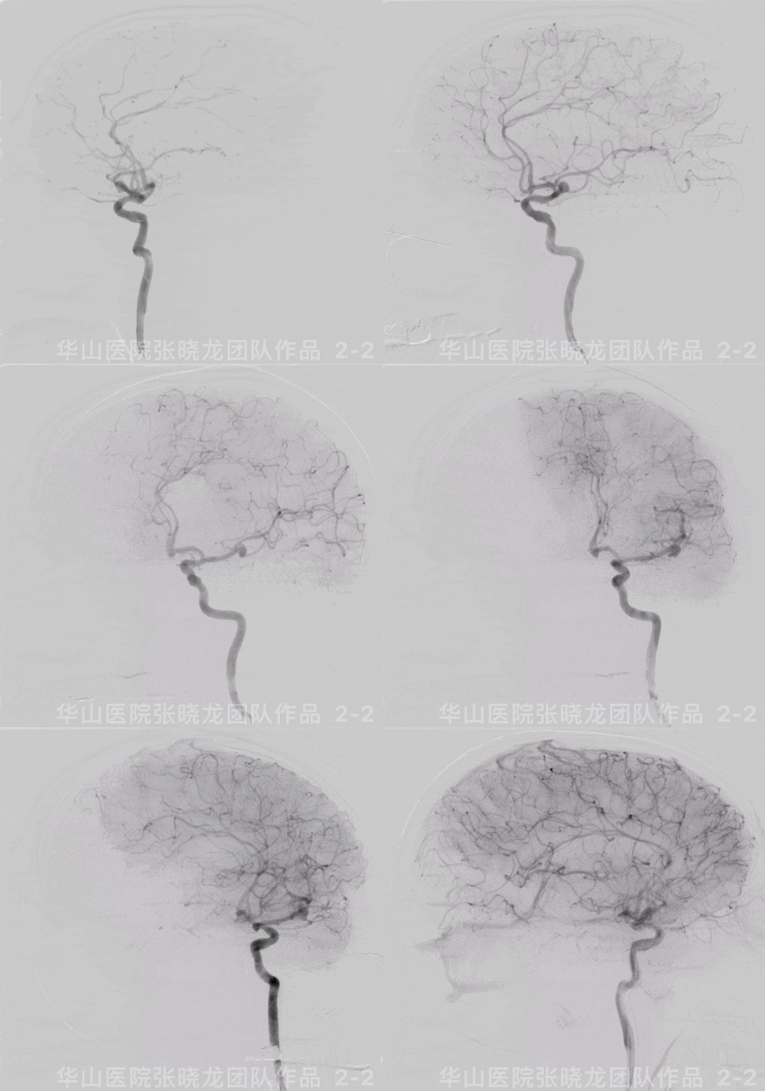
Figure 2 GIF. DSA confirmed a left middle cerebral inferior trunk initial segment dissecting aneurysm with irregular shape and daughter sac.
图 2 GIF. DSA证实左侧大脑中动脉下干起始部夹层动脉瘤,形态不规则,伴子瘤形成。

Figure 3. A large left MCA inferior trunk initial segment dissecting aneurysm with daughter sac was observed. The inferior trunk initial segment presented with an acute curve.
图 3. 3D重建提示左侧大脑中动脉下干起始部夹层动脉瘤伴子瘤,大脑中动脉下干起始部有急弯。
1
Strategy
1.A large left MCA inferior trunk initial segment dissecting aneurysm with daughter sacs indicated a high rupture risk which should be treated.
2.Post large coils stenting technique will be adopted to protect the inferior trunk.
3.Due to an acute curve of inferior trunk initial segment, Atlas stent was preferred. Solitaire stent harboured a relative high ischemic or occlusion risk.
1.左侧大脑中动脉下干起始部大夹层动脉瘤伴子瘤,破裂风险高,建议治疗。
2.计划采用大圈支架后释放技术保护大脑中动脉下干。
4.选用Solitaire支架置于上干,拉直成角,改变血流冲击方向,降低复发风险。
5.若随访时动脉瘤复发,可上干置入pipeline支架。
2
Operation

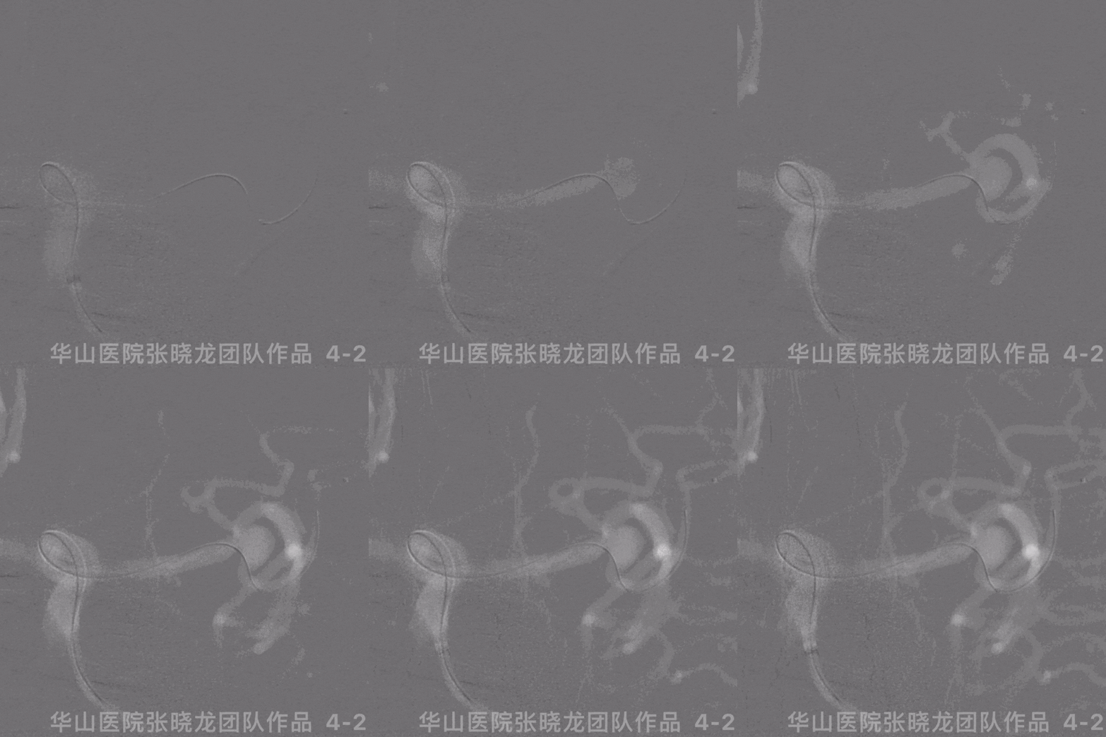
Figure 4 GIF. Working projection selection: An size 5.8*4.69mm, neck 1.92mm, proximal parent artery diameter 2.22mm, distal parent artery diameter 1.73mm. 6F Envoy DA guiding catheter was placed into left cavernous segment and Nimodipine 1ml was administered. Synchro-II microwire and spiral-curved SL-10 microcatheter were navigated into inferior trunk via skating technique (Noting: Kept the microcatheter tension and persistently advanced the microwire). Re-roadmap confirmed no hemorrhage. General heparization and Nimodipine 1ml were administered.

图 5. 将头端塑“C”弯的Prowler plus微导管置于大脑中动脉上干, Solitaire 4*20mm支架释放保护上干及瘤颈。支架释放后上干拉直。

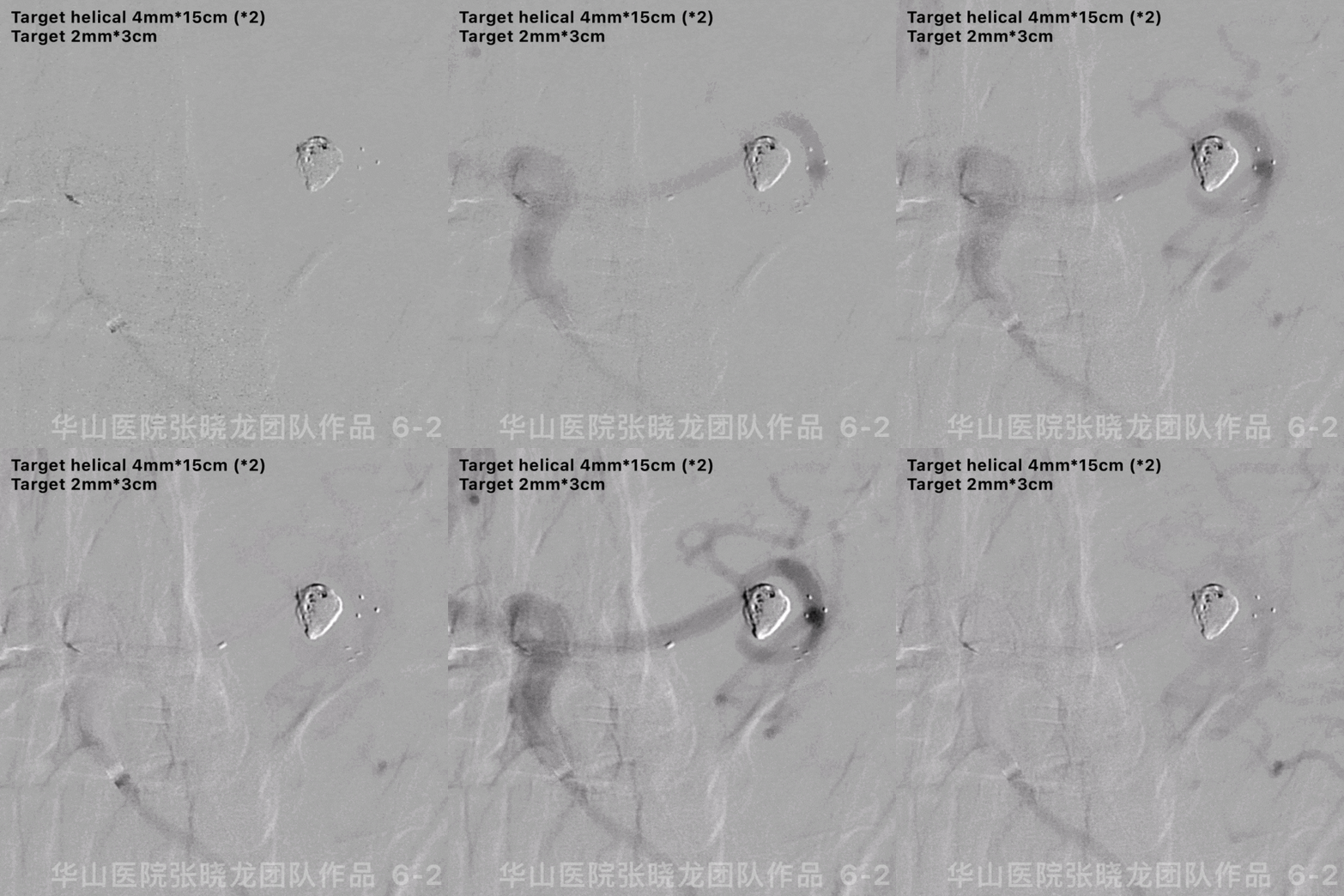

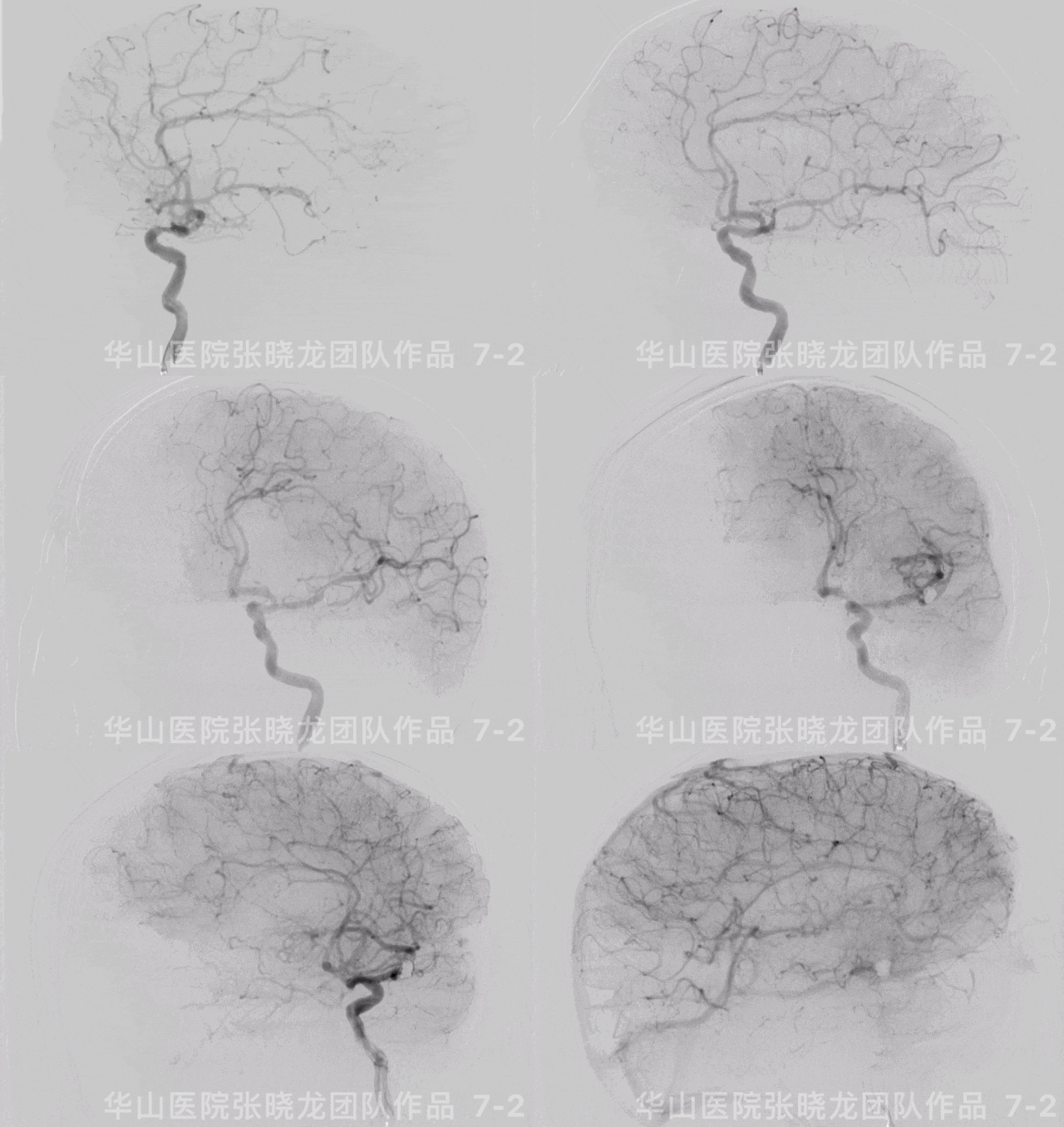
Figure 7 GIF. Angiograms showed the aneurysm was packed satisfactorily. Tirofiban 7ml was administered and waited.
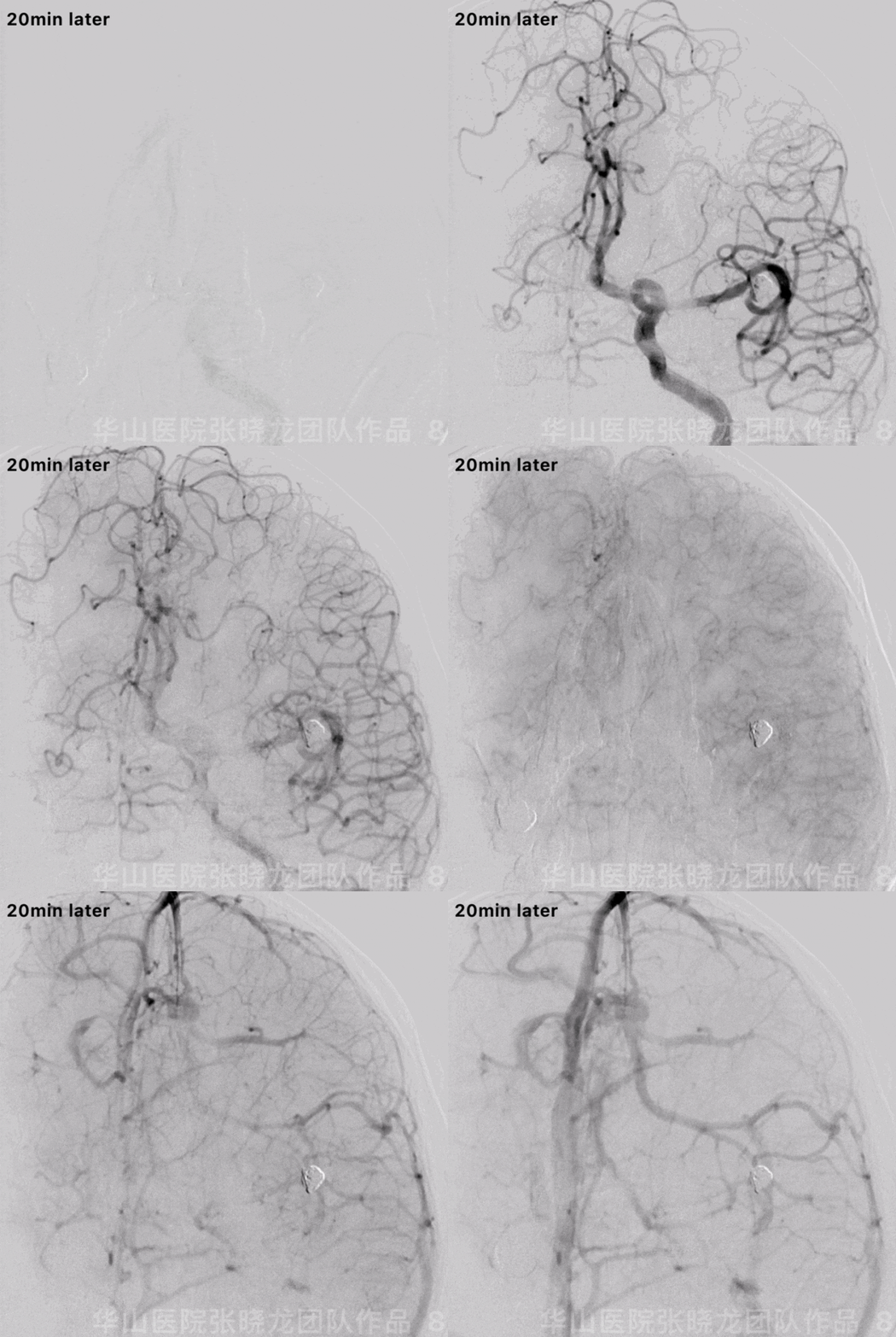
Figure 8 GIF. Parent artery was patent after waiting for 20 minutes.
图 8 GIF. 等待20min后复查造影,载瘤动脉通畅。
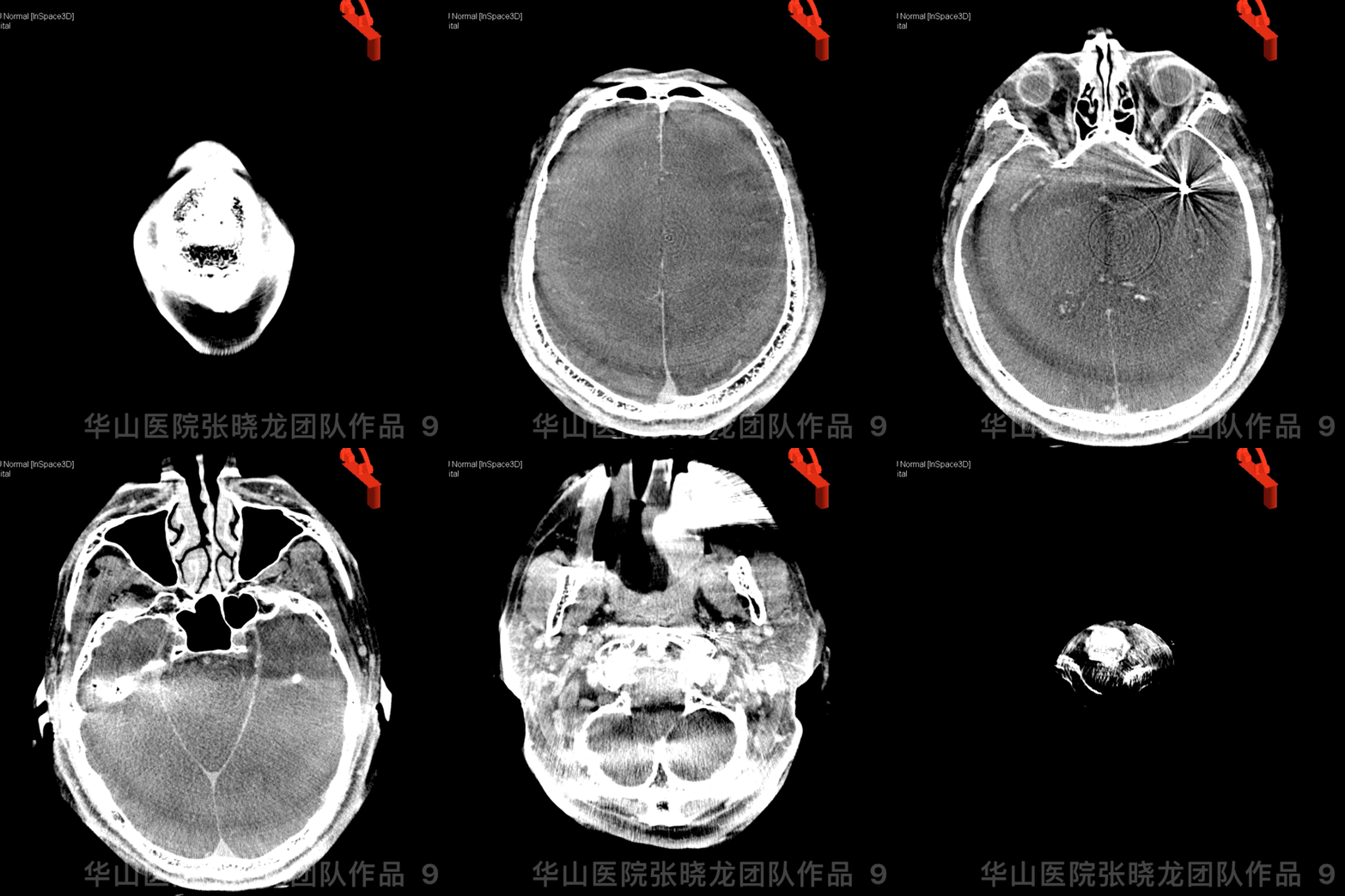
Figure 9 GIF. Post-operative dyna-CT did not demonstrate any hemorrhage.
图 9 GIF. 术后dyna-CT未见出血。
3
Post-Operation
Medication:
查体:双侧肌力正常,眼球各项运动佳,双侧巴氏征阴性,无新发神经功能缺损。

Figure 10. Solitaire straightened the superior trunk.
图 10. Solitaire拉直大脑中动脉上干。
Video 2. Intracranial vessels were intact by 7 month follow up.
4
Summary
1.A large left MCA inferior trunk initial segment dissecting aneurysm with daughter sacs indicating a high rupture risk should be treated.
4.Due to an acute curve of inferior trunk initial segment, Atlas stent was preferred instead of Solitaire stent with a relative high ischemic or occlusion risk.
5.Since Atlas stent was soft, this stent should be deployed early after the first soft frame coil embolized.
6.General heparinization was performed after inferior trunk super-selection (dangerous procedure).
7.If this aneurysm recurred in follow up period, pipeline can be deployed in the superior trunk. Pipeline was difficult to open in the inferior trunk.
8.Pipeline was not recommended in superior trunk at the first stage considering delayed stenosis or occlusion and delayed rupture due to daughter sac.
1.左侧大脑中动脉下干起始部大夹层动脉瘤伴子瘤,破裂风险高,建议治疗。
4.由于下干起始部急弯,选用Atlas支架,因为Solitaire支架相对缺血或血管闭塞事件风险较高。
5.Atlas支架比较软,所以支架在第一枚弹簧圈稳定成篮后再释放。
6.全身肝素化时机:由于下干超选风险高,下干超选成功后再行肝素化。
7.若随访时动脉瘤复发,考虑下干pipeline可能会打开困难,可在上干置入pipeline支架。
8.考虑到血流导向装置延迟狭窄或闭塞及延迟破裂风险,上干置入Pipeline支架不是一期治疗的首选方案。
![]()
点击或扫描上方二维码
查看更多“介入”内容


