

Review
Two cases
First case
• 40 y/o male.
• A large intracranial aneurysm was found accidentally.
病例 1
• 40岁,男性.
• 体检偶然发现左侧颈内动脉大动脉瘤。
• 神经查体:-。

Figure 1. A left large intracranial aneurysm was detected by local hospital.
Figure 2 GIF. Angiograms confirmed a left ICA terminal segment dissecting aneurysm mainly involving left M1 and A1 segment.
图 2 GIF. 造影证实左侧颈内动脉末端夹层动脉瘤,动脉瘤主要累及左侧大脑中M1及大脑前A1段。

图 3. 左侧颈内动脉压颈试验示前交通动脉发育良好。
1-1
Strategy
The left large ICA terminal segment aneurysm with irregular shape habouring a high rupture risk was suggested treatment.
Flow divertor device was unavailable at that time in our center.
Conventional stents assisted large coiling technique can be adopted for this type of aneurysms.
The Acom was well-developed, therefore the left A1 segment can be sacrificed if necessary.
Stents were planned to be deployed at the left middle cerebral artery.
左侧颈内动脉末端形态不规则大动脉瘤有破裂风险,建议治疗。
可以选择血流导向装置治疗该动脉瘤,但2015年时我中心没有血流导向装置。
我们采用传统支架辅助大圈栓塞技术治疗该动脉瘤。
前交通动脉发育良好,因此必要时可牺牲左侧A1段。
计划于左侧大脑中动脉释放支架。
1-2
Operation

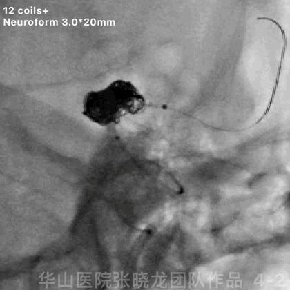
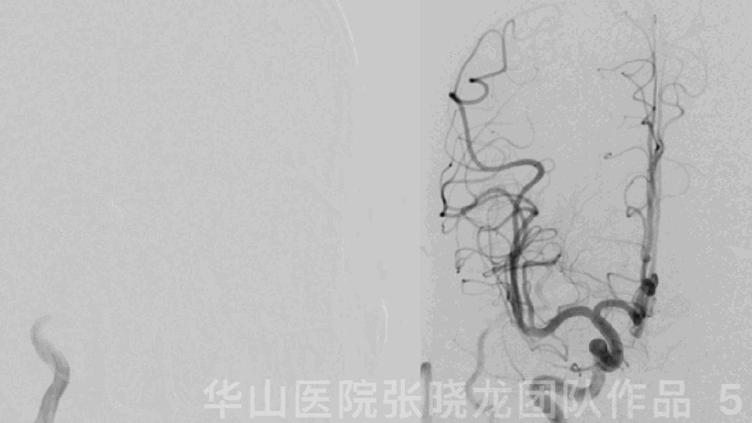
图 5 GIF. 复查造影动脉瘤颈少许残留,颅内血管通畅。
1-3
Post Operation
NE: GCS 15, bilateral muscle strength normal, no neurologic defect, bilateral Babinski negative.
Medication: Tirofiban maintained for 24 hours.
At discharge: Aspirin and Clopidogrel for long-term.
神经查体:GCS 15,四肢肌力正常,无新发神经功能缺损,双侧巴氏症阴性。
药物:替罗非班维持24h。
出院:阿司匹林氯及吡格雷长期口服。
Video 1. By 6 month follow up, angiograms depicted the aneurysm remained a bit relapsed. Stopped Aspirin and Clopidogrel. The residual neck was stable by 8 year follow up.
• 50 y/o fmale.
• suffering from sudden onset headache 2 weeks ago, and CT scan revealed SAH in local hospital.
• Medication: Olmesartan.
病例 2
• 50岁,女性.
• 2周前突发头痛,当地医院头颅CT提示蛛网膜下腔出血。
• 药物:奥美沙坦酯。


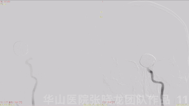
Figure 8. CTA depicted a left large irregular MCA dissecting aneurysm.


图 9 GIF. 造影证实左侧大脑中动脉大夹层动脉瘤伴左侧M1段局部管径扩张。当时由于机器原因,无法行3D旋转造影。前交通动脉发育良好。
2-1
Strategy
A large ruptured MCA dissecting aneurysm with a large irregular daughter sac haboured a high re-rupture risk, which should be treated.
Combined with CT scan, the daughter sac was thought of a ruptured point.
Staged embolization was planed
First stage - embolized the daughter sac to avoid re-bleeding.
second stage - After 7 days dual antiplatelet therapy, FD stent with coiling embolization was performed.
破裂出血的大脑中动脉大夹层动脉瘤伴不规则大子瘤,再次破裂出血风险高,建议治疗。
一期 – 为降低再次破裂出血风险,栓塞子瘤。
二期 – 一期治疗后口服双抗,7天后行血流导向支架。
2-2
First stage

Figure 10. Measurements: main aneurysm sac size 20.24*21.28mm, neck 9.74mm, proximal parent artery 3.88mm. A large C-curved Echelon-10 and a C-curved Prowler Plus microcatheters were navigated into the sac. Inserted 51 coils (TJWY-Perdenser 20mm*30cm (*6), 18mm*30cm (*6), 16mm*30cm (*3), 15mm*30cm (*8), 14mm*30cm (*8), 13mm*30cm (*8), 12mm*30cm (*6), 10mm*30cm (*6)) via a Prowler Plus. General heparization was performed.
视频 3. 术后即刻头颅CT未见出血。术后第一天复查头颅DWI提示左侧大脑半球散在新发梗死灶。
2-3
Post Operation
NE: GCS 15, bilateral eye movement and light reflux normal, speech normal, bilateral muscle strength normal, bilateral Babinski negative.
Medication:
Tirofiban 6ml/h maintained for 24h.
Aspirin 100mg and Clopidogrel 75mg were prescribed.
Methylprednisolone 120mg for one day, mannitol 100ml q8h.
查体:GCS 15,双侧眼球运动正常,对光反射灵敏,言语清,四肢肌力正常,双侧巴氏症阴性。
药物:
替罗非班6ml/h维持24小时。
予口服阿司匹林及氯吡格雷。
2-4
Second stage-7d later
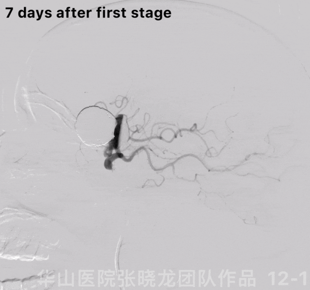


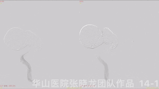
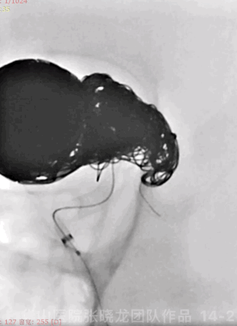
Figure 14 GIF. Angiograms showed the dissecting aneurysm almost invisible and parent artery patent. Massage the stent. Tirofiban 10ml was administered.
图 14 GIF. 复查造影动脉瘤基本不显影,载瘤动脉通常。按摩支架后予替罗非班10ml。
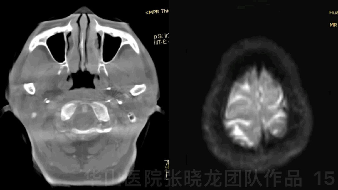
Figure 15 GIF. Dyna-CT did not depicted any hemorrhage and no new infarctions were detected by DWI.
图 15 GIF. 术后Dyna-CT未见出血,DWI未见新发脑梗死。
2-5
Post Operation
NE:GCS 15, bilateral eye movement and light reflux normal, speech normal, bilateral muscle strength normal, bilateral Babinski negative.
Medication:
Tirofiban 8ml/h maintained for 24h.
CYP2C19 PM.
Aspirin and Ticagrelor were prescribed.
查体:GCS 15,双侧眼球运动正常,对光反射灵敏,言语清,四肢肌力正常,双侧巴氏症阴性。
药物:
替罗非班8ml/h维持24小时。
氯吡格雷基因代谢慢代谢。
予口服阿司匹林及替格瑞洛。
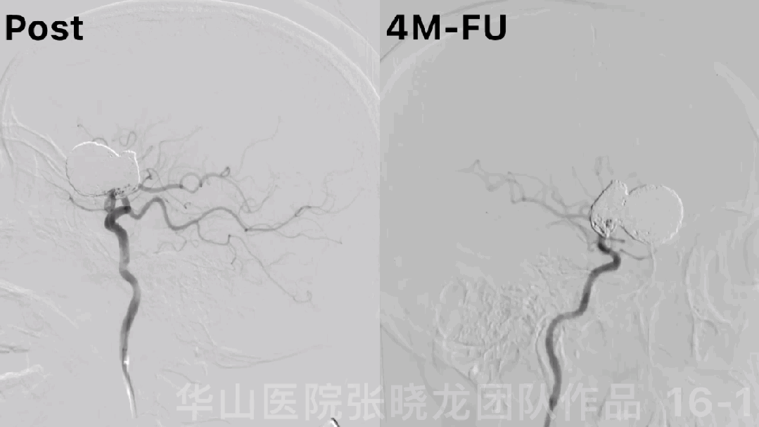
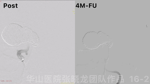
Figure 16 GIF. The aneurysm was embolized satisfactorily without relapse, while the parent artery was stenosis by 4 month follow up.

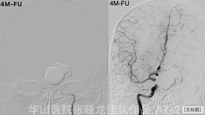
Figure 17 GIF. Angiograms showed the parent artery stenosis and the left A2 segment was compensated by anterior communicating artery via RICA. The patent did not suffer from any neurological deficit.

Figure 18. No new acute infarctions were observed from DWI by 4 month follow up.
3
Summary
Conventional stents assisted coiling technique can be adopted for ICA terminal segment dissecting aneurysms, which was not very giant nor significant expansion. The first case’s long-term follow up angiograms showed it was safe and effective. While flow divertors were preferable for giant dissecting aneurysms.
Embolizing the ruptured point during acute cerebral hemorrhage first. Flow divertors can be deployed at the second stage with enough dual antiplatelets preparation.
Parent artery stenosis after flow divertor stents deployment should be considered, which may be for the reason of vascular endothelialization. Extended the time of dual antiplatelets may work[1], while long-term follow up was still needed.
这种夹层动脉瘤需要支架辅助栓塞治疗,扩张不是非常严重的(非巨大动脉瘤),普通支架加弹簧圈的策略也可以有很好的结果,就像病例1。局部扩大明显,形成巨大动脉瘤的,首选FD支架治疗。
出血急性期,应首先止血,可在抗血小板药物准备后二期置入血流导向支架。
血流导向支架术后的载瘤动脉狭窄是应该考虑的,根据文献报道,考虑内膜增生过度导致的,延长双抗治疗的时间,可能会逆转血管狭窄,但仍需要长期随访证实。
[1] DC L, SJ C, JW O, AR C, CJ M, AP K. - Management of In-Stent Stenosis with Dual Antiplatelet Therapy Following Pipeline. D - 101528275. (- 1878-8769 (Electronic)):- e303-e9.
![]()

点击或扫描上方二维码
查看更多“介入”内容
