Review
History
• 65 y/o male.
• Suffered from transient blurred and hallucinate vision for one month. MRI revealed left temporal edema and DAVF.
• Med history: DM, HTN; no smoking, no alcohol consumption.
• PE: muscle strength was normal; No cognitive disorder, MMSE 29, MoCA 27.
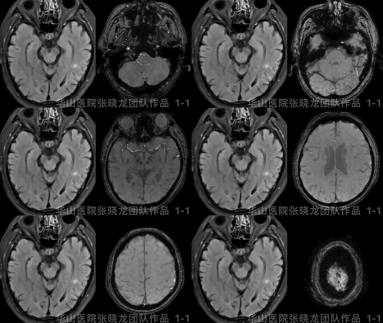
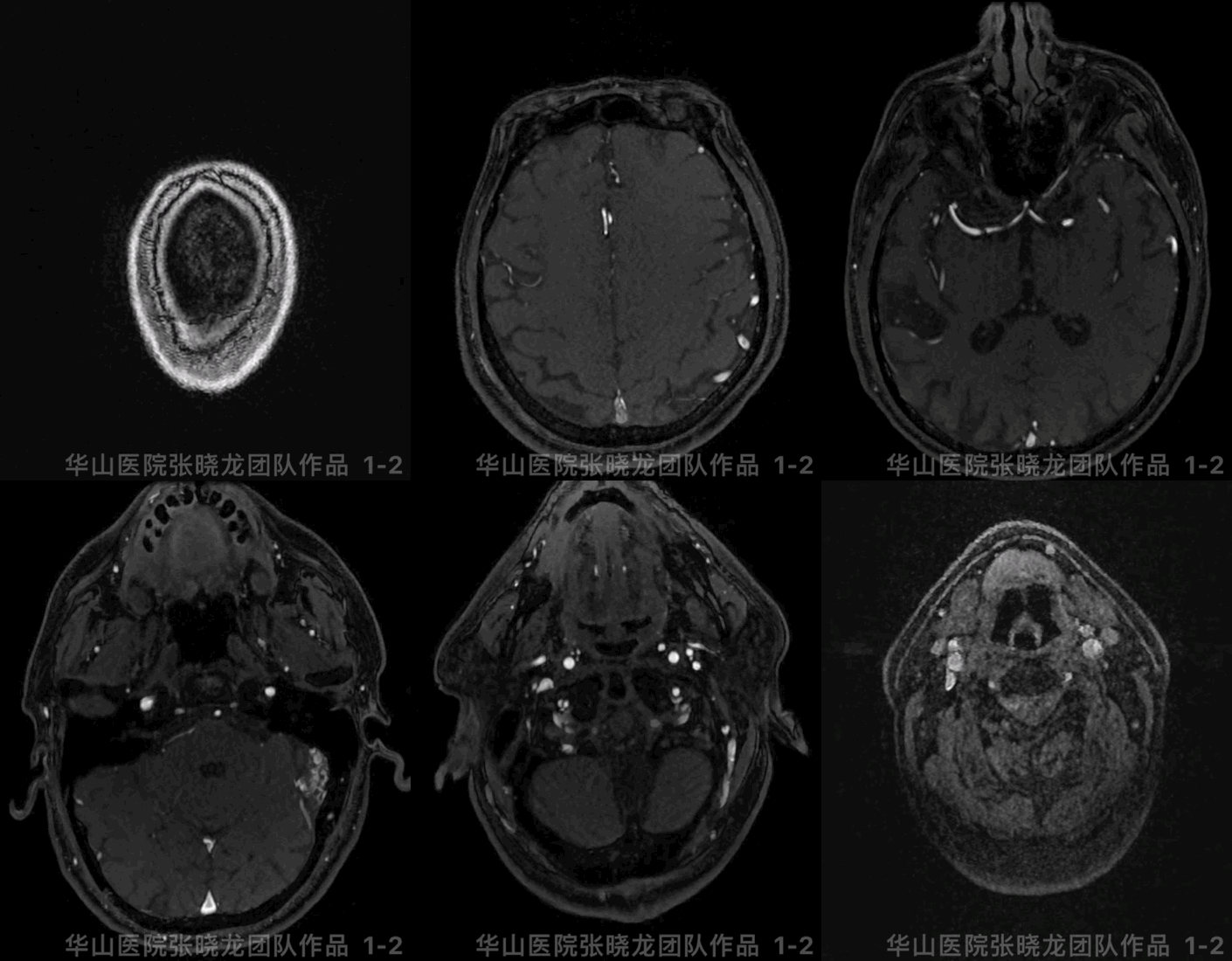
图 1 GIF. T2 flair示左侧颞叶异常信号,SWI示左侧大脑半球多发微出血灶,TOF示左侧乙状窦附近异常血管影。

Figure 2. Enhanced HR-MR revealed left transverse under-developed and sigmoid sinus occlusion with sinus septum formation in the left residue sinus.
图 2. 高分辨率增强MR示左侧横窦发育不良,乙状窦静脉间隔形成。
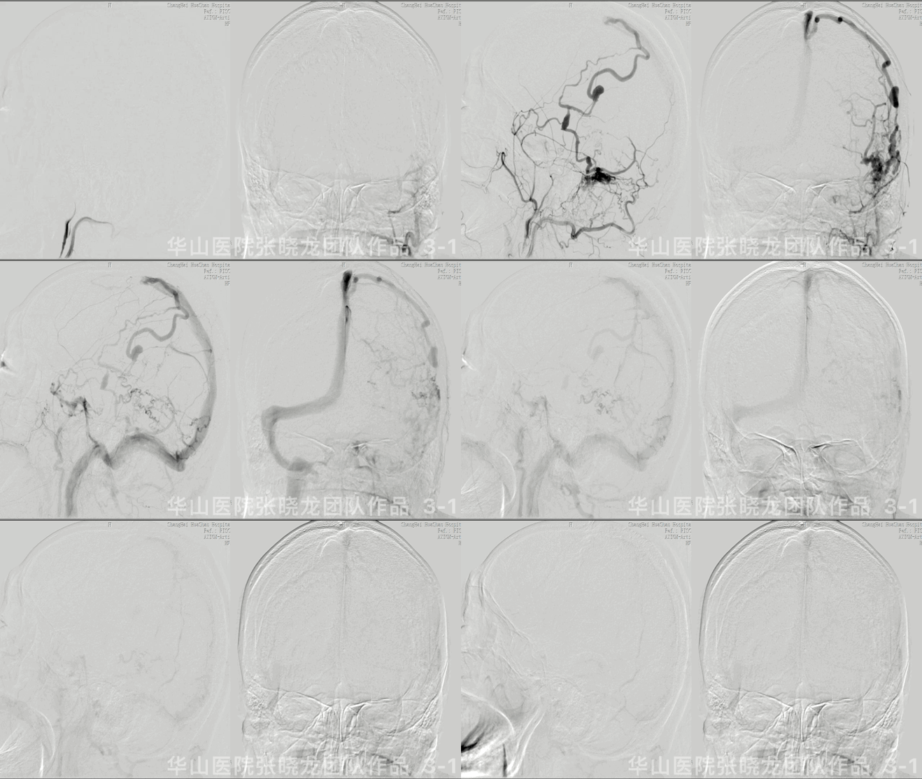
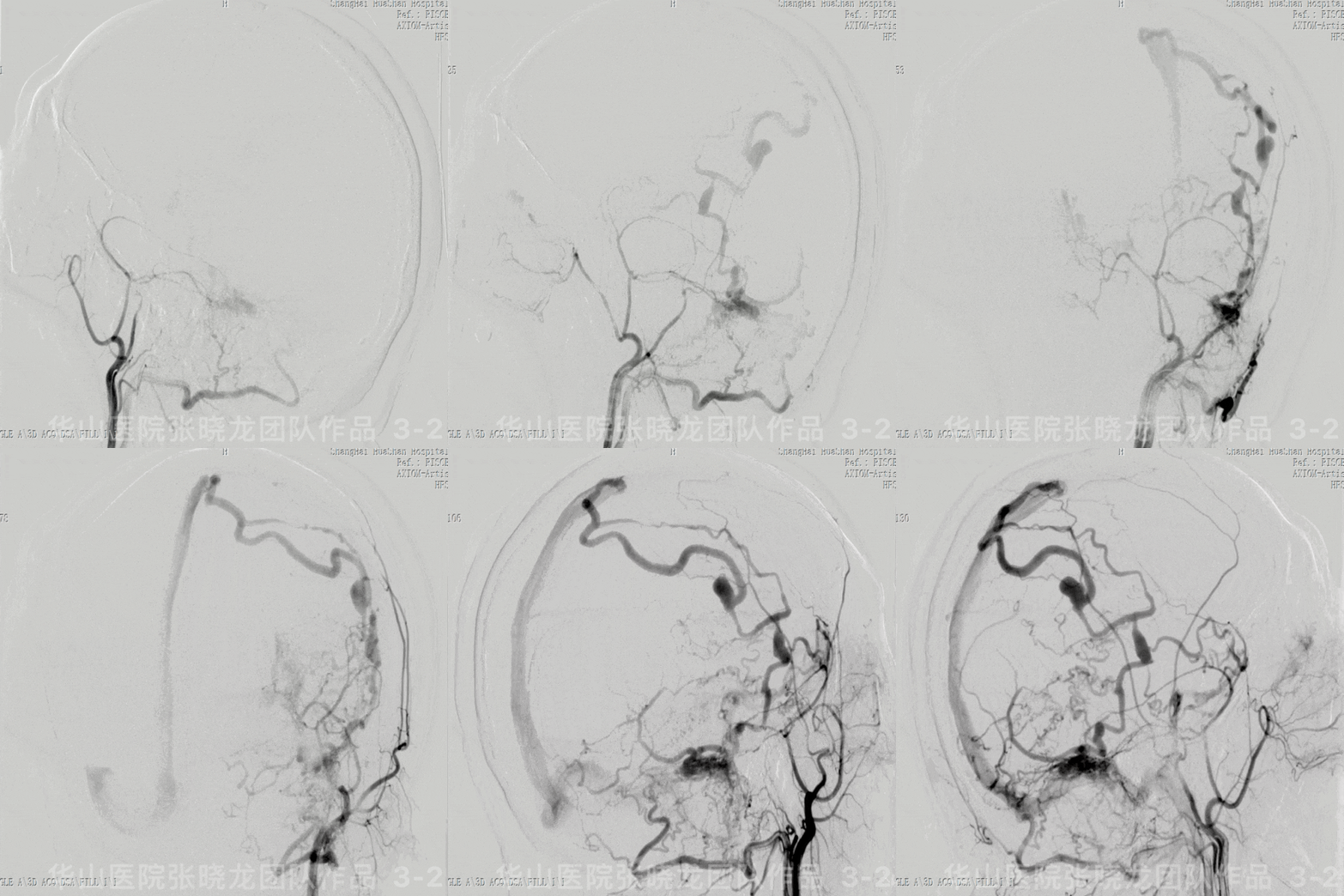
Figure 3 GIF. The fistula located at left lateral sinus and drained into an isolated sinus then refluxed to the cortical and deep vein, while venous aneurysms have formed (Cognard 2b).
1
Strategy
1.The drainage pattern of this left lateral sinus fistula originated from an isolated sinus to the cortical veins, which was related to the left sigmoid & transverse sinuses occlusion.
2.The left TS was underdeveloped, no significant venous edema or cognitive disorder, therefore we chose to occlude the residue sinus instead of the sinus recanalization.
3.The fistula refluxed to the occipital cortical vein and the vein of Labbe, therefore we planned to occlude the isolated sinus and preserved the bridge vein.
1.左侧侧窦区硬脑膜动静脉瘘,瘘口经孤立窦向皮层静脉逆流的引流方式,与乙状窦、横窦闭塞有关。
2


图 5. Marathon微导管超选入左侧脑膜中动脉后支,手推造影证实微导管到位,注入Onyx-18大约2.9ml,胶在残窦及供血动脉末端、共同静脉端铸型。

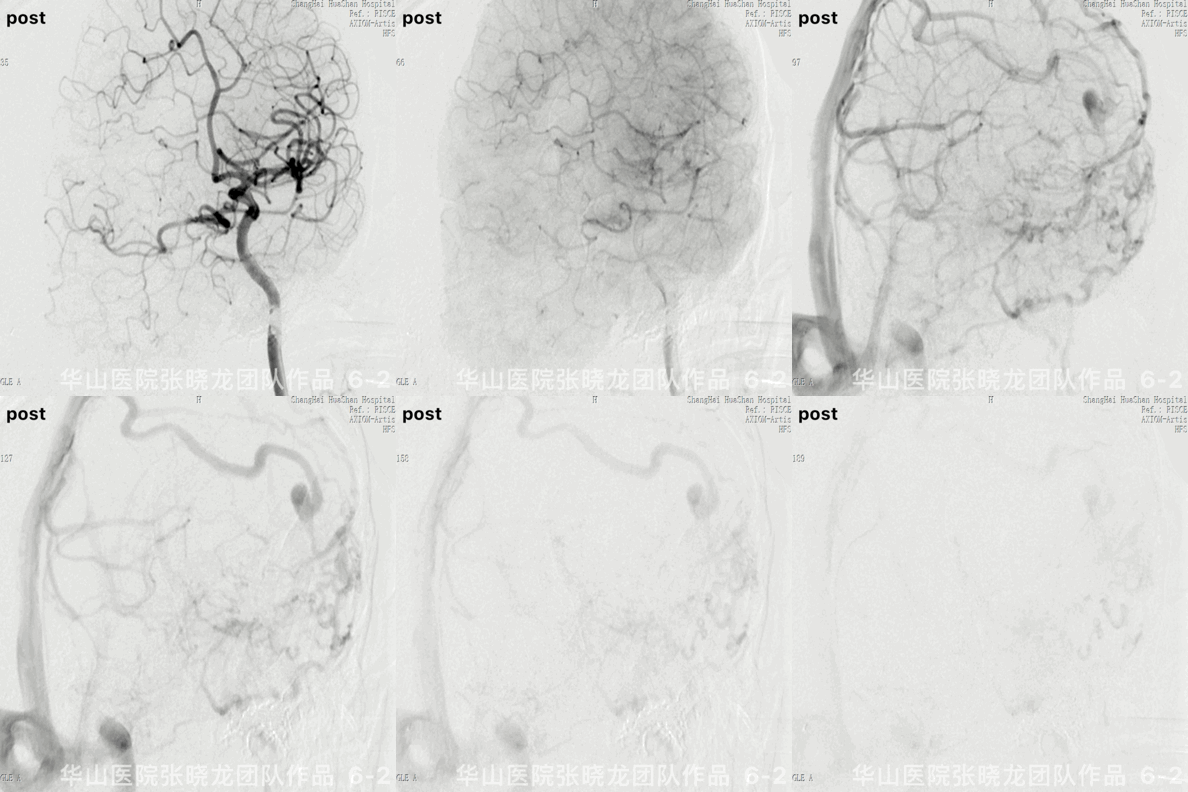
Figure 6 GIF. Angiograms showed the fistula was occluded and meanwhile the drainage veins were well preserved, cortical veins drained to superior sagittal sinus via Labbe vein and Trolard.
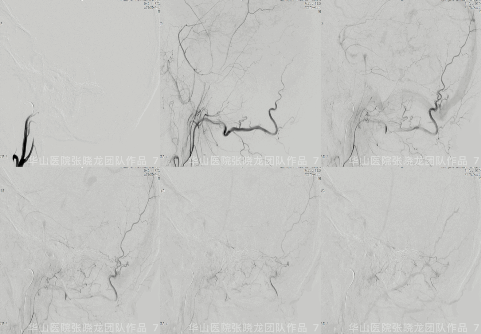
图 7 GIF. 复查造影瘘口消失。
3


Video 2. Left transverse and sigmoid sinus occluded and the cortical veins preserved well with normal drainage function.
视频 2. 复查左侧颈内动脉造影示左侧横窦乙状窦闭塞,皮层静脉保留完好,恢复正常引流功能。
4
Follow up
视频 3. 20个月随访原皮层静脉瘤样扩张管径基本恢复正常。

Figure 10. Edema on the left temporal lobe was not detected from T2 flair while micro-bleedings did not decrease by 20 month follow up.
5
Summary
1.The fistula was related to the left sigmoid and transverse sinuses occlusion.The fistula drained via the isolated occluded sinus to the cortical vein.
2.The left TS was underdeveloped, no significant venous edema or cognitive disorder, therefore we chose to occlude the residue sinus instead of the sinus recanalization.
3.Sinus recanalization was another option especially for patients with venous edema and severe cognitive disorder, indicating the sinus with important drainage function.
4.The fistula refluxed to the occipital cortical vein and the vein of Labbe, therefore we planned to occlude the isolated sinus and preserved the bridge vein. The drainage function of the cortical veins could be preserved.
5.After the embolization, the occipital cortical vein drained via the bridging vein then emptied into the vein of Labbe.
6.The venous aneurysms shrunk during the follow up after the hypertension was relieved.
1.左侧侧窦区硬脑膜动静脉瘘,瘘口经孤立窦向皮层静脉逆流的引流方式,与乙状窦、横窦闭塞有关。
2.磁共振显示静脉水肿范围小,临床上无认知障碍,考虑左侧横窦非优势侧,所以治疗上首选更为高效的闭塞残窦而非开通静脉窦,随访swi仍提示多发微出血灶,提示局部仍存在静脉高压。
3.开通静脉窦也是可选方案,尤其使用静脉水肿及认知障碍严重的患者,提示该侧静脉窦功能重要,有保留、开通、重建的必要性。
4.精准栓塞应当闭塞残窦及Labbe桥静脉与静脉窦结合部的“桥静脉硬膜内段”,而不应扩大损毁范围累及至软膜静脉端。该病例中保留了远心端蛛网膜下的Labbe静脉,使得皮层软膜静脉的侧枝代偿得以保留。
5.瘘口栓塞后,枕叶皮层静脉经桥静脉向Labbe静脉引流。
6.20个月随访原静脉瘤样扩张消失,皮层静脉管径恢复正常。
张晓龙
复旦大学附属华山医院
复旦大学附属华山医院放射科主任医师,博士、教授、博士生导师;
斯坦福大学医学院客座临床教授;
主持国家自然科学基金3项,第一作者或通讯作者发表国内外权威期刊文章50余篇;
中华医学会、放射学会、卫生部医政司等组织中担任副主任委员、组长等职务.《中国名医百强榜》神经介入专业中国十强(2012年度、2013年度、2014年度、2015-16年度、2017-18年度);
擅长复杂和疑难脑血管疾病的介入治疗,如复杂脑动脉瘤的栓塞,硬脑膜动静脉瘘栓塞,脑动静脉畸形栓塞,脑梗死的支架,脊髓血管畸形治疗;
自1995年开始从事脑血管疾病介入诊治工作和研究,师从黄祥龙教授、沈天真教授和凌锋教授,是我国最早从事神经介入的专家之一。2010年9月至今连续介入治疗颅内动脉瘤1500余例,无操作致死.
点击或扫描上方二维码
查看更多“介入”内容


