Review
History
35 y/o male.
Suffering from sudden onset of severe headache with vomiting 20 days ago.
Subarachnoid hemorrhage was detected from local hospital CT scan. A right ACA dissecting aneurysm was found by CTA and DSA in local hospital.
Past medical history: HTN for 6 years and DM for 4 years.
Medication: Perindopril, metformin, dapagliflozin.
NE: (-)
35岁,男性。
20天前突发剧烈头痛伴呕吐。
当地医院头颅CT平扫提示蛛网膜下腔出血。当地医院CTA和DSA示右侧大脑前动脉夹层动脉瘤。
既往史:高血压6年,糖尿病4年。
药物:培哚普利,二甲双胍,达格列净。
神经查体:-

Figure 1. High density of interhemispheric fissure cistern were detected by CT scan.
图 1. CT平扫示纵裂池高密度影。

Figure 2. CTA revealed an irregular right anterior cerebral artery dissecting aneurysm.
图 2. CTA示右侧大脑前动脉不规则夹层动脉瘤。

Figure 3. HR-MR demonstrated an irregular anterior cerebral artery with partial wall enhancement.
图 3. 高分辨磁共振示右侧大脑前不规则动脉瘤伴瘤壁部分强化。
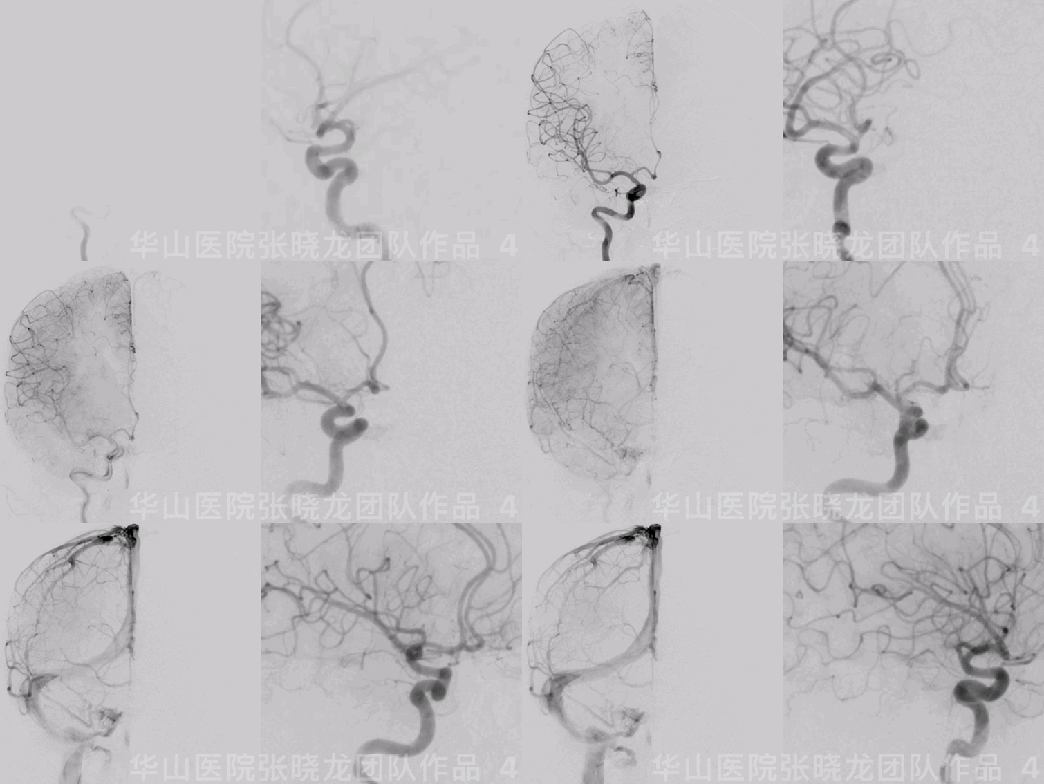
Figure 4 GIF. DSA confirmed a small right anterior cerebral artery dissecting aneurysm.
图 4 GIF. DSA证实右侧大脑前动脉小夹层动脉瘤。

Figure 5 GIF. 3D reconstruction depicted an anterior cerebral artery dissecting aneurysm with an irregular sac and two perforators originating from distal part of the dissecting aneurysm and proximal segment of parent artery respectively.
图 5 GIF. 3D重建示大脑前动脉瘤伴瘤腔不规则扩张,动脉瘤瘤体远端及邻近载瘤动脉发出2支穿支。
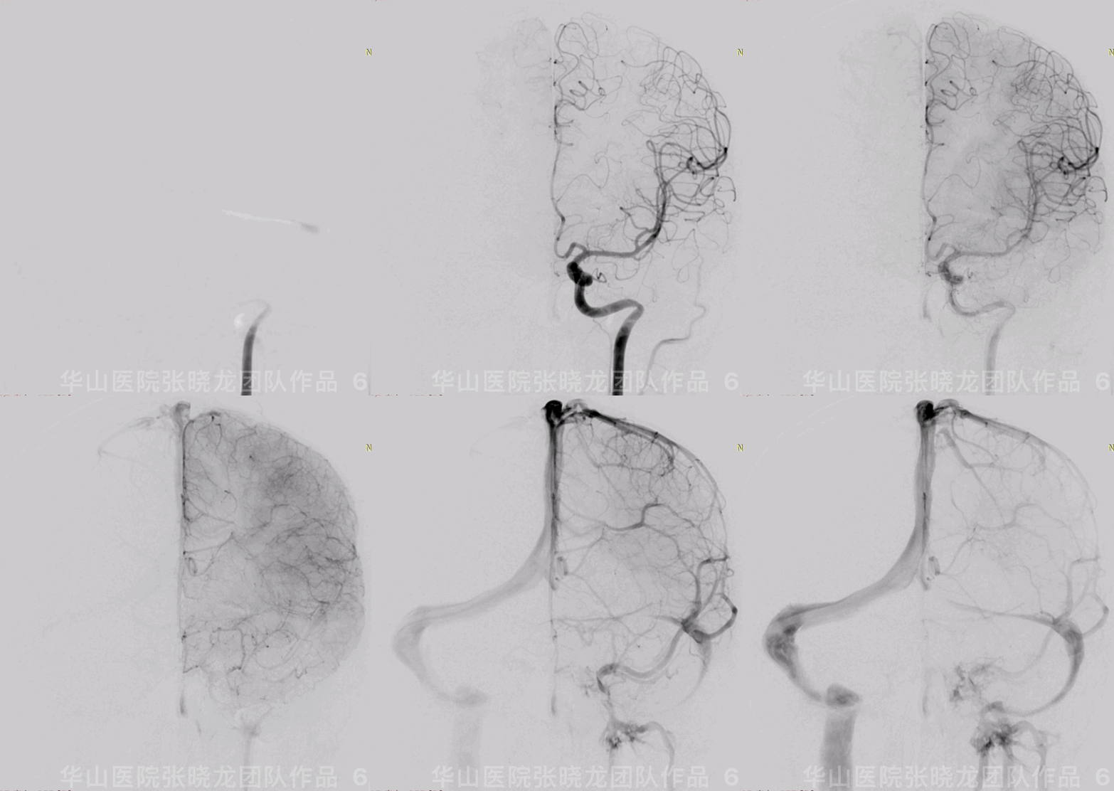
Figure 6 GIF. Left ICA angiogram showed the anterior communicating artery well developed.
图 6 GIF. 左侧颈内动脉造影示前交通动脉发育良好。
1
Strategy
Anterior longitudinal fissure SAH was detected by CT scan, and an irregular right A1 segment dissecting aneurysm was found simultaneously. Therefore the ruptured dissecting aneurysm should be treated as soon as possible.
Contralateral ACA and anterior communicating artery developed well.
Two branches arose from the distal part of the aneurysm, that should be preserved.
Dissecting aneurysm dorsal side presented irregular sacs with enough space to insert coils.
Three different strategies can be considered.
• FD stent can preserve the perforating branches. However for an acute ruptured aneurysm, post stenting hemorrhage should be considered.
• Dissecting aneurysm parent artery proximal segment occluded was another option, which was not the first choice in our center.
• Post large coils stenting technique can embolize the dissecting aneurysm and preserve the parent artery simultaneously. But it may be difficult to preserve the perforating branches.
CT平扫提示前纵裂蛛网膜下腔出血,同时CTA和DSA发现右侧A1段不规则夹层动脉瘤,出血位置与夹层动脉瘤相符,所以考虑夹层动脉瘤破裂导致蛛网膜下腔出血。该破裂夹层动脉瘤建议尽快治疗。
左侧大脑前动脉和前交通动脉发育良好。
夹层动脉瘤远端有两支穿支血管发出,栓塞时需要保护。
夹层动脉瘤背侧有扩张囊腔,有足够空间填入弹簧圈。
3种不同介入治疗策略可以考虑。
• 血流导向装置可以保护穿支,但急性期破裂夹层动脉瘤,血流导向装置有支架后再出血风险。
• 闭塞夹层动脉瘤近端载瘤动脉,但该策略在我们中心不是首选。
• 大圈支架后释放技术栓塞夹层动脉瘤的同时可以保护载瘤动脉,但可能影响动脉瘤上发出的穿支。

Figure 7. Two different working projections were selected, one for microcatheter navigation and the other for coil insertion. Measurements: An size 2.05*2.1mm, proximal parent artery 2.09mm and distal parent artery 1.8mm.
图 7. 选择了两个不同的工作角度,一个用来填圈,另一个微导管到位。测量:动脉瘤大小2.05*2.1mm,近端载瘤动脉直径2.09mm,远端载瘤动脉直径1.8mm。
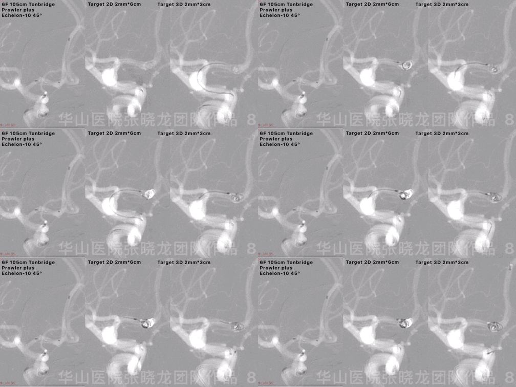
Figure 8 GIF. General heparinization was conducted. 6F 105cm Tonbridge guiding catheter was placed into the right cavernous sinus segment. A Prowler Plus microcatheter was navigated into the right A2 segment and Echelon-10 into the aneurysm sac. Inserted a first coil Target 2D 2mm*6cm from working projection I, then shifted to working projection II showing the first coil protruded into the distal part of the aneurysm. Retrieved the first coil and re-inserted the first coil from working projection II. Continued inserting a second coil Target 3D 2mm*3cm (undetached).
图 8 GIF. 行全身肝素化。将6F 105cm Tonbridge 导引导管置于右侧颈内在动脉海绵窦段,Prowler Plus微导管在微导丝导引下至于右侧大脑前动脉A2段,Echelon-10微导管至于动脉瘤腔内。微导管到位后在工作角度I(微导管工作角度)上填入第一枚弹簧圈Target 2D 2mm*6cm,更换至工作角度II显示动脉瘤远端清晰,发现弹簧圈填入动脉瘤远端,弹簧圈可影响穿支,遂在工作角度II上重新填入第一枚弹簧圈,反复确认弹簧圈不影响动脉瘤远端。由于微导管稳定,继续填入另一枚弹簧圈Target 3D 2mm*3cm(暂不解脱)。

Figure 9 GIF. Deployed Solitaire 4*29mm stent. Angiogram showed the dissecting aneurysm densely packed and the two perforators remained. Then detached the second coil. Tirofiban 10ml was administered.
图 9 GIF. 瘤颈部释放Solitaire 4*29mm支架。复查造影示夹层动脉瘤致密栓塞,两小穿支显影。解脱第2枚支架。经导引导管给予替罗非班10ml。
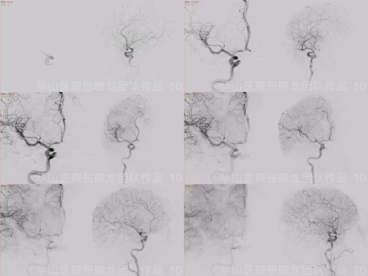
Figure 10 GIF. Waiting for 10 minutes. Angiograms showed the two perforators patent.
图 10 GIF. 等待10分钟,再次造影复查,旋转示原动脉瘤远端两穿支显影良好。

Figure 11. 3D reconstructions confirmed the two perforators existed. Vessels colored in orange referred to pre-operation and green to post-operation. Black dotted line and purple dotted line demonstrated two branched pre/post operation.
图 11. 3D重建确认两穿支存在。橘黄色是术前血管形态,绿色为术后。黑色和紫色虚线分别代表术前术后两穿支血管。

Figure 12 GIF. Retrieved the microcatheter ( avoid microcatheter tip pushing coils). Right M3 segment distal branches filling defects were observed and the aneurysm was embolized densely with parent artery patent. Tirofiban 10ml was administered.
图 12 GIF. 撤出微导管(回撤微导管时避免微导管头段推弹簧圈致弹簧圈移位)。复查造影右侧大脑中动脉M3以远分支血管可见充盈缺损,夹层动脉瘤致密栓塞,载瘤动脉通畅。经导引导管给予替罗非班10ml。
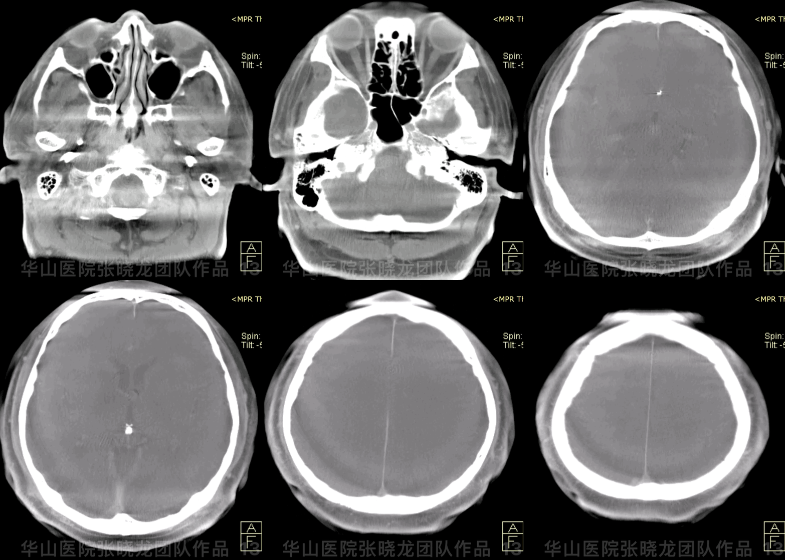
Figure 13 GIF. Dyna-CT showed no hemorrhage.
图 13 GIF. Dyna-CT未见出血。

Figure 14. DWI depicted dotted acute infarctions on the right frontal and temporal lobes.
图 14. DWI示右侧额颞叶少许点状急性脑梗死。
2
Post Operation
NE: GCS 15, slight central facial paralysis (48h recovered), bilateral eye movement normal, bilateral muscle strength and sensibility normal, bilateral Babinski negative.
Medication:
Tirofiban 9ml/h maintained 48 hours.
Aspirin 100mg for long term and Clopidogrel 75mg for 3 months were prescribed (AA 100% and ADP 100%; CYP2C19 NM).
查体:患者GCS 15,轻度中枢性面瘫(48h后恢复),双侧眼球运动正常,四肢肌力、感觉正常,双侧巴氏症阴性。
药物:
替罗非班9ml/h维持48h。
阿司匹林100mg长期口服,氯吡格雷75mg口服3月后停药(阿司匹林抑制率100%,氯吡格雷抑制率100%;氯吡格雷基因代谢正常)。
3
Summary
Anterior longitudinal fissure SAH was detected by CT scan, and an irregular right A1 segment dissecting aneurysm was found simultaneously. Therefore the ruptured dissecting aneurysm should be treated as soon as possible.
Contralateral ACA and anterior communicating artery developed well.
Two branches arose from the distal part of the aneurysm, that should be preserved.
Dissecting aneurysm dorsal side presented irregular sacs with enough space to insert coils.
Working projections and rotational DSA as well as 3D reconstructions confirmed the two perforators patent.
Three different strategies can be considered.
• FD stent can preserve the perforating branches. However for an acute ruptured aneurysm, post stenting hemorrhage should be considered.
• Dissecting aneurysm parent artery proximal segment occluded was another option, which was not the first choice in our center.
• Post large coils stenting technique can embolize the dissecting aneurysm and preserve the parent artery simultaneously. But it may be difficult to preserve the perforating branches.
For the dissecting aneurysm, the parent artery was very fragile which may rupture or occlude during navigating a microcatheter.
Long- term follow-up is needed.
CT平扫提示前纵裂蛛网膜下腔出血,同时CTA和DSA发现右侧A1段不规则夹层动脉瘤,出血位置与夹层动脉瘤相符,所以考虑夹层动脉瘤破裂导致蛛网膜下腔出血。该破裂夹层动脉瘤建议尽快治疗。
左侧大脑前动脉和前交通动脉发育良好。
夹层动脉瘤远端有两支穿支血管发出,栓塞时需要保护。
夹层动脉瘤背侧有扩张囊腔,有足够空间填入弹簧圈。
弹簧圈填入后,需要在工作角度、旋转造影和3D重建上反复确认两穿支血流通畅。
3种不同介入治疗策略可以考虑。
• 血流导向装置可以保护穿支,但急性期破裂夹层动脉瘤,血流导向装置有支架后再出血风险。
• 闭塞夹层动脉瘤近端载瘤动脉,但该策略在我们中心不是首选。
• 大圈支架后释放技术栓塞夹层动脉瘤的同时可以保护载瘤动脉,但可能影响动脉瘤上发出的穿支。
夹层动脉瘤的载瘤动脉往往很脆弱,微导管超选时可能导致载瘤动脉破裂或闭塞。
该病例需要长期随访。
点击或扫描上方二维码
查看更多“介入”内容


