Review
History
• 58 y/o female.
• Suffering from right transient headache for 10 days. MRI revealed a right ICA giant dissecting aneurysm with thrombus formation.
• Past medical history: HTN for 2 years.
• Medication: Felodipine.
• NE: -.
• 58岁,女性。
• 发作性头痛10天。MRI提示右侧颈内动脉巨大夹层动脉瘤伴血栓形成。
• 既往史:高血压2年。.
• 药物:非洛地平。
• 神经查体:-。

Figure 1. MRI depicted a right ICA giant dissecting aneurysm and thrombus existed in the sac.
图 1. MRI提示右侧颈内动脉颅内巨大夹层动脉瘤,瘤腔内血栓形成。

Figure 2. CTA revealed a right anterior cerebral artery giant dissecting aneurysm.
图 2. CTA示右侧大脑前动脉巨大夹层动脉瘤。
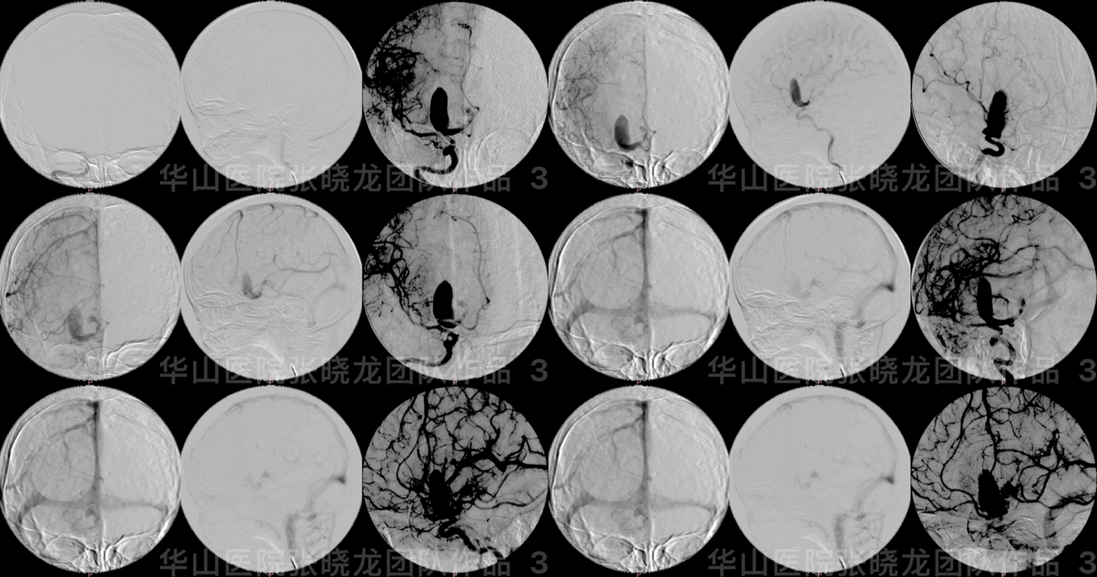
Figure 3 GIF. DSA confirmed a right A1 initial segment giant dissecting aneurysm with irregular shape and thrombus formation.
图 3 GIF. DSA证实右侧A1起始段巨大夹层动脉瘤,动脉瘤形态不规则,瘤腔内血栓形成。
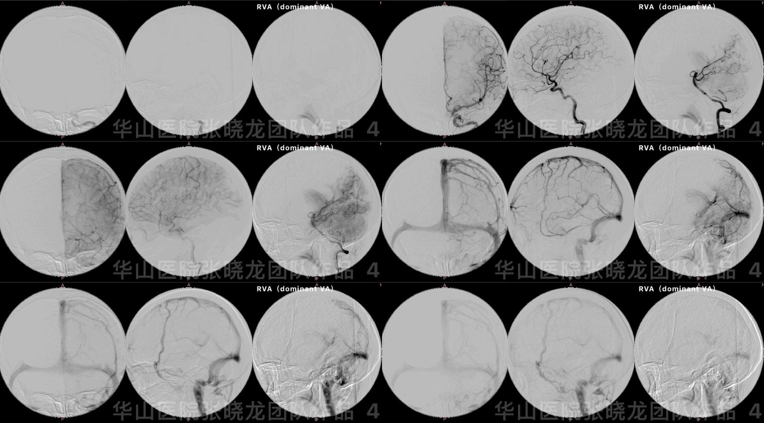
Figure 4 GIF. Anterior communicating artery and left anterior cerebral artery did not reveal from right ICA angiogram. Meanwhile right dominant vertebrate artery did not compensate the right anterior cerebral artery region.
图 4 GIF. 右侧颈内动脉造影前交通动脉及左侧大脑前动脉未见显影。右侧优势椎动脉造影未见后循环代偿右侧大脑前动脉供血区。
1
Strategy
A right A1 initial segment irregular giant dissecting aneurysm harboured obvious mass effect and a high rupture risk, which should be treated.
The aneurysm on un-enhanced MRI mismatched with on angiogram because of thrombus formation.
BOT test will be conducted.
If BOT test was negative, right A1 segment can be sacrificed.
If BOT test was positive, stent assisted coiling technique will be adopted to preserve the A1 segment.
FD was not available in 2009.
右侧A1起始段不规则巨大夹层动脉瘤,占位效应明显,且有破裂风险,建议治疗。
动脉瘤在非增强MRI上与造影不匹配,考虑动脉瘤腔内部分血栓形成。
栓塞前行BOT试验。
若BOT试验阴性,右侧A1段可以闭塞。
若BOT试验阳性,建议采用支架辅助栓塞保护A1段血管。
2009年的病例,那时我中心还不能用FD支架。
2
Operation
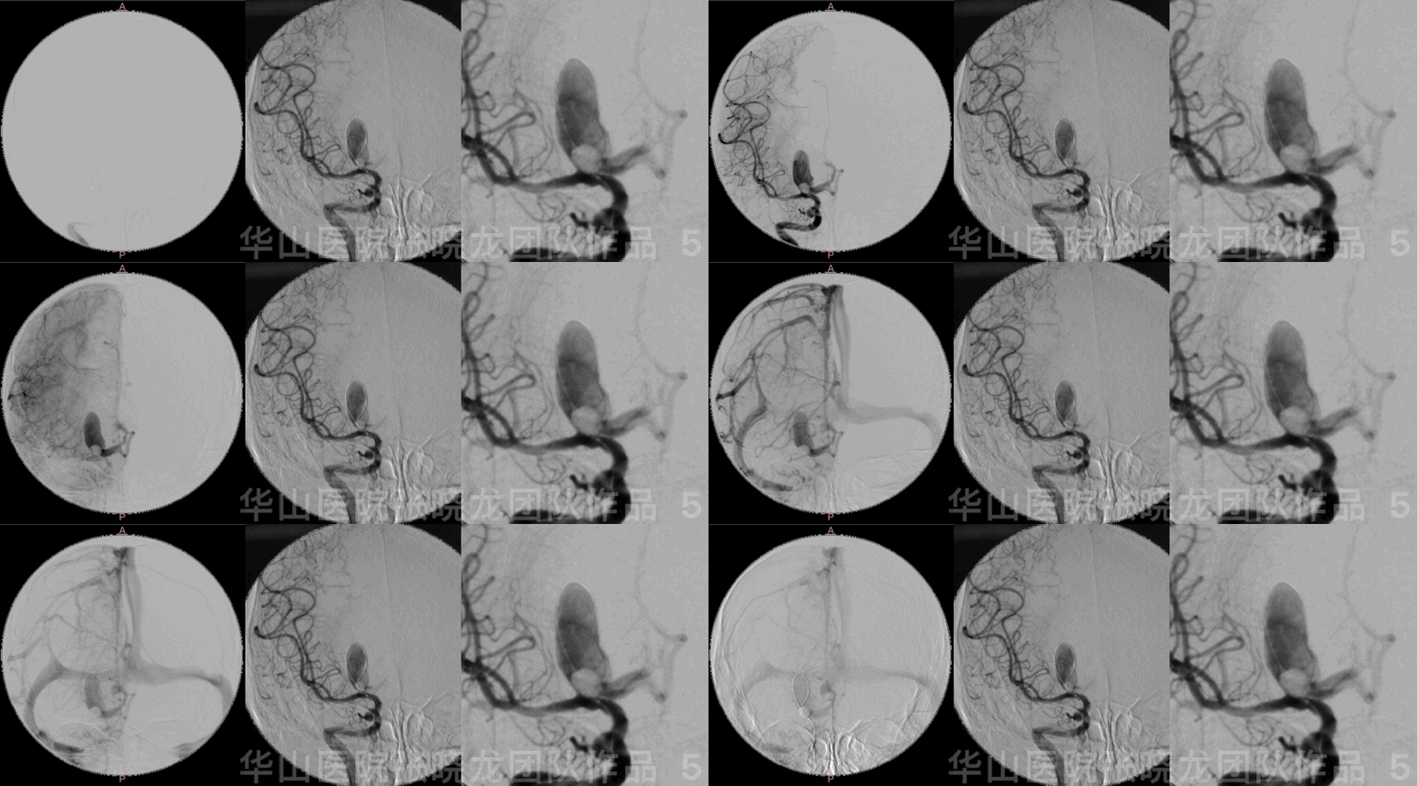
Figure 5 GIF. BOT test was conducted. Dilated balloon at the A1 initial segment but failed to occluded the right A1 artery.
图 5 GIF. 右侧A1起始部动脉瘤颈处反复尝试BOT试验,无法完全闭塞A1段动脉。
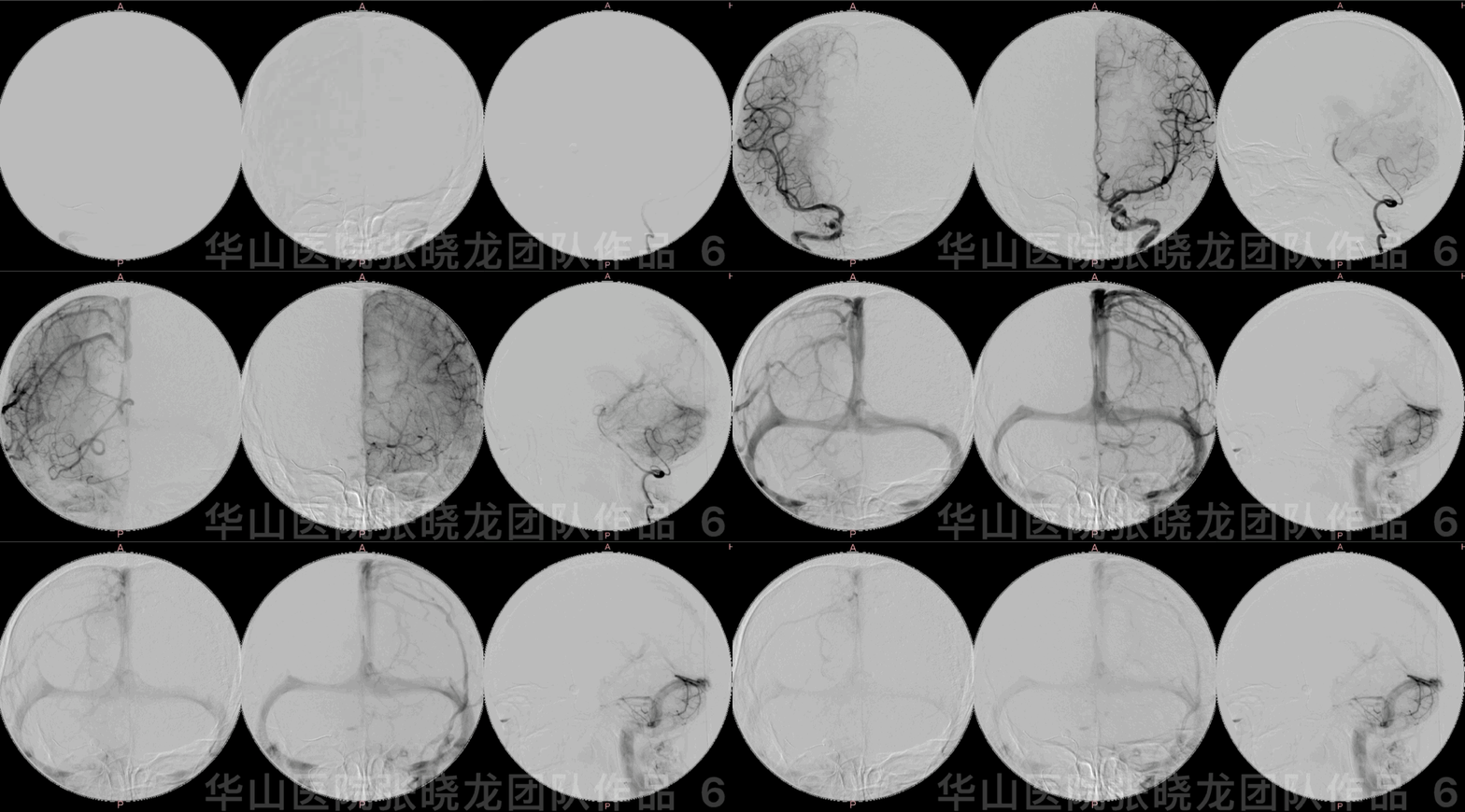
Figure 6 GIF. A Hyperform 4*7mm balloon was placed at the right M1 segment, then dilated the balloon herniation into the aneurysm sac. BOT test succeeded. Right A1 segment can be revealed from left ICA angiogram while right A2 segment did not visualize clearly. Dominant vertebrate artery did not compensate the right anterior cerebral artery region.
图 6 GIF. Hyperform 4*7mm球囊放置在右侧大脑中动脉M1段,部分充盈球囊,球囊疝入动脉瘤内,达到BOT的目的。遂行左侧颈内动脉造影,右侧A1段显影,右侧A2段以远显影差。椎动脉造影未见后循环代偿右侧大脑前动脉供血区。
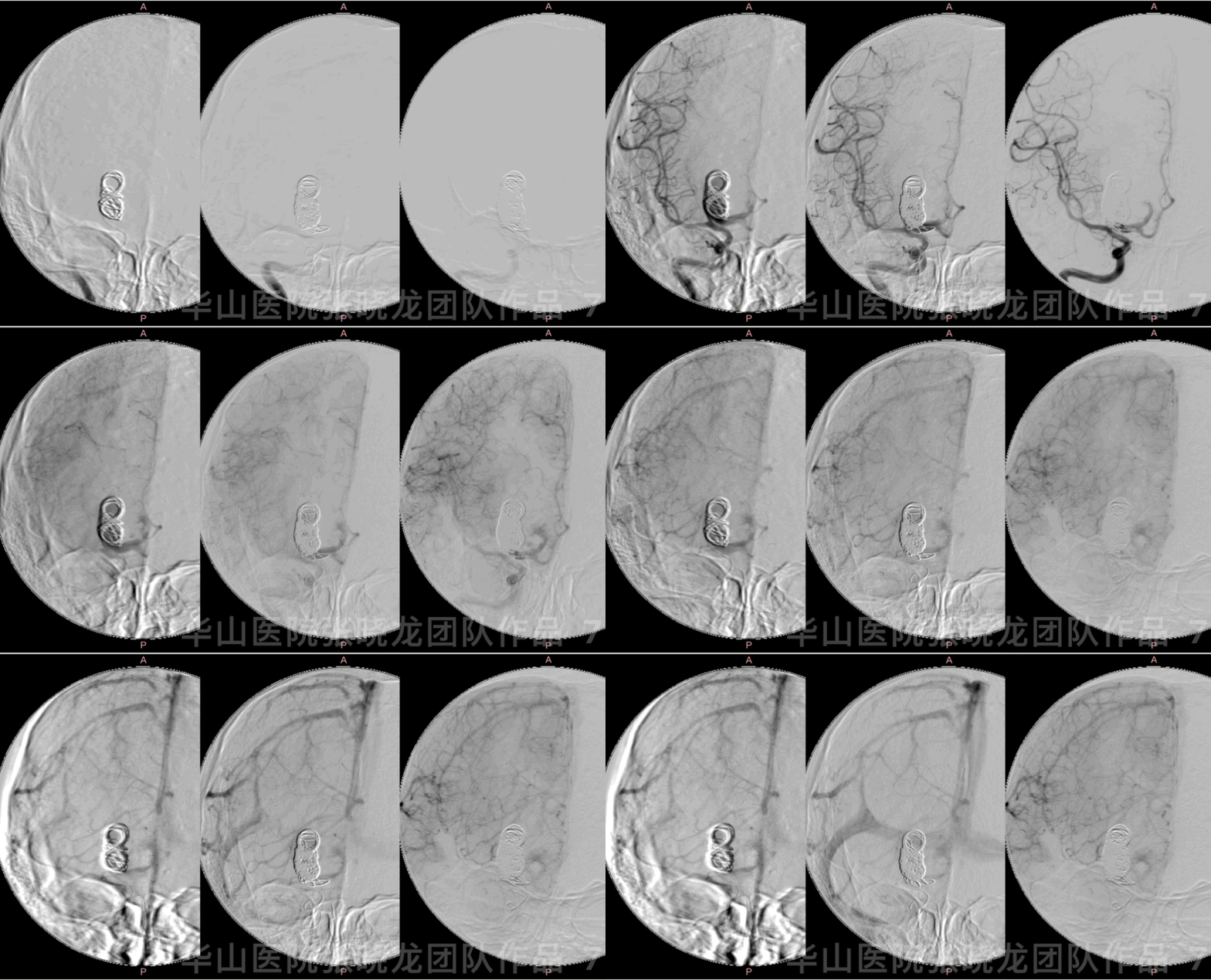
Figure 7 GIF. Insert the following 14 coils (Microplex 20mm*60cm, 20mm*50cm, 10mm*30cm, 8*25, Hydrosoft 5mm*20cm (2), 6mm*20cm (2), 4mm*10cm (4), 2mm*8cm (2)) in sequence.
图 7 GIF. 依次填入以下14枚弹簧圈(Microplex 20mm*60cm, 20mm*50cm, 10mm*30cm, 8*25, Hydrosoft 5mm*20cm (2), 6mm*20cm (2), 4mm*10cm (4), 2mm*8cm (2)) 。
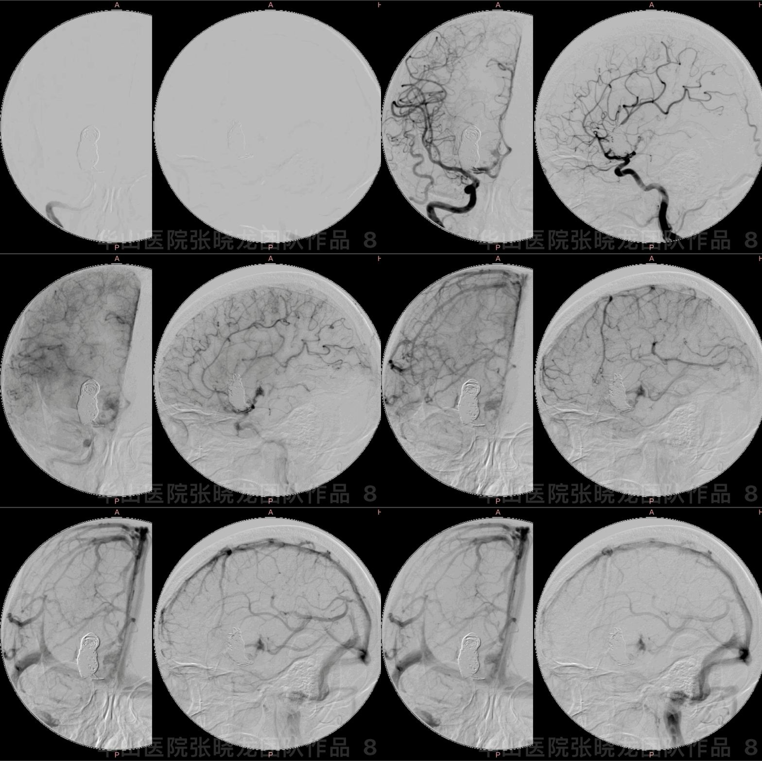
Figure 8 GIF. Right ICA angiogram showed a bit remnant of the aneurysm and A1 segment was not occluded completely to avoid post-operative large area cerebral infarctions.
图 8 GIF. 右侧颈内动脉造影显示动脉瘤仍有残余,右侧A1段未完全闭塞,期待A1段缓慢闭塞,以防术后大面积脑梗死。
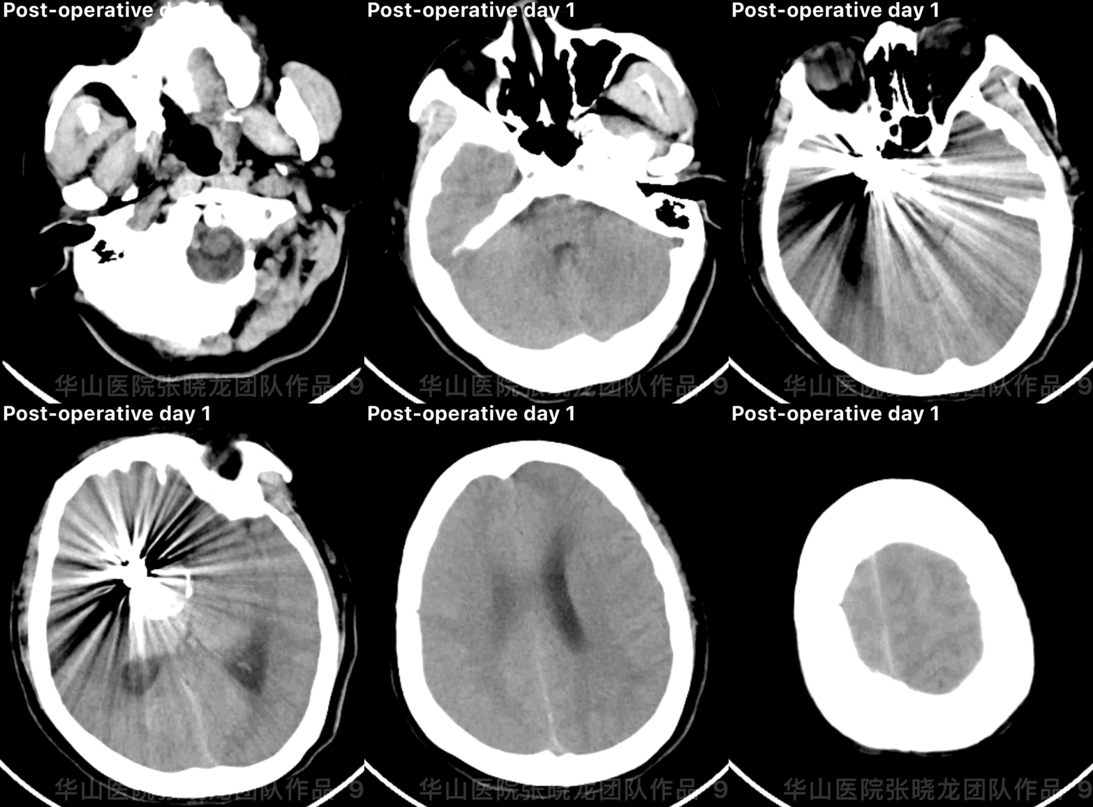
Figure 9 GIF. CT did not demonstrate any hemorrhage one day after operation.
图 9 GIF. 术后第一天复查头颅CT未见出血。
3
Post Operation
NE: GCS 15, headache and vomiting (2 days later improved), eye movement normal, bilateral pupils light reflux normal, muscle strength normal, bilateral Babinski negative.
查体:GCS 15分,头痛伴呕吐(2天后好转),双侧眼球运动正常,双侧对光反射灵敏,四肢肌力正常,双侧巴氏征阴性。
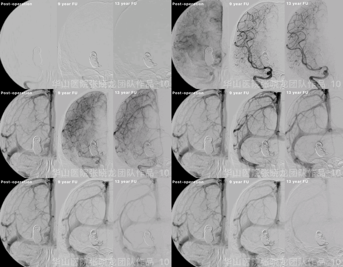
Figure 10 GIF. By 9 years follow up, the aneurysm was totally embolized while the right A1 initial part could be observed. By 13 years follow up, the right A1 segment was occluded and the aneurysm was not relapsed.
图 10 GIF. 9年随访,动脉瘤完全栓塞,右侧A1起始部少许显影。13年随访,右侧A1段闭塞,动脉瘤未见复发。
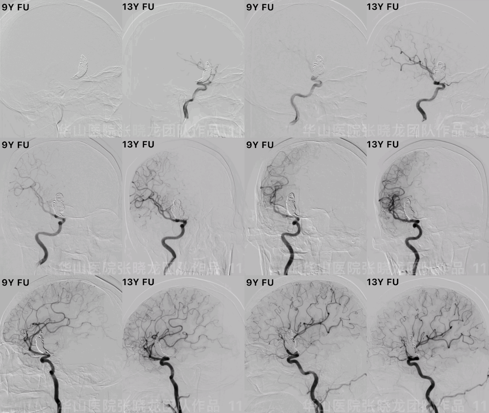
Figure 11 GIF. No recurrence of the aneurysm was observed by follow up.
图 11 GIF. 随访旋转DSA证实动脉瘤无残余或复发。
Video 1. Left anterior cerebral artery and right posterior artery compensated the right anterior cerebral artery region.
视频 1. 左侧大脑前动脉及右侧大脑后动脉供血右侧大脑前动脉供血区。

Figure 12. No acute infarctions or increased mass effect were detected by 9 years follow up.
图 12. 9年随访头颅MRI未见急性脑梗塞或占位效应增加。
4
Summary
A right A1 initial segment irregular giant dissecting aneurysm indicating mass effect and a high rupture risk should be treated.
The aneurysm on un-enhanced MRI mismatched with on angiogram because of thrombus formation, which was not visualized on angiogram and not inserted coils. By 13 years follow up, the aneurysm was not relapsed.
The aneurysm located at the A1 artery initial segment and BOT test failed when balloon dilated at the A1 segment. While BOT test succeeded when a Hyperform balloon was placed at the right M1 segment and the balloon protruded into the aneurysm sac after dilating, which made full use of super-compliance of Hyperform.
BOT test showed the anterior communicating artery can only compensate the right side partially, therefore right A1 segment was occluded chronically to lower post-operative cerebral infarctions risk. Simple large coiling was adopted. No acute infarction occurred while the aneurysm was totally embolized by follow up.
FD was not available in 2009 in our center while Pipeline stent can be chosen nowadays.
右侧A1起始段不规则巨大夹层动脉瘤,占位效应明显,且有破裂风险,建议治疗。
动脉瘤在非增强MRI上与造影不匹配,考虑动脉瘤腔内部分血栓形成。血栓栓塞部分造影上大部分不显影,该部分瘤腔未填入弹簧圈,随访时动脉瘤未见残余或复发。
动脉瘤位于A1段起始部,球囊置于A1段行BOT试验无法成功。因此充分运用了Hyperform球囊的超顺应性,球囊放置在中动脉,部分充盈的球囊疝入动脉瘤内,闭塞了右侧A1起始部,达到了BOT的目的。
BOT试验表明前交通动脉只能部分代偿右侧大脑前动脉供血区(A2段以远显影不良),因此采用了单纯大圈栓塞技术,右侧A1段慢性闭塞降低术后脑梗发生率。随访时无新发脑梗同时动脉瘤致密栓塞。
2009年时我中心还不能使用FD支架,现在治疗可以选择Pipeline支架。
点击或扫描上方二维码
查看更多“介入”内容


