

Review
History
• 55 y/o male.
• Suffered from transient dizziness for 6 months. Local hospital DSA revealed a left middle cerebral artery aneurysm.
• Past medical history: HTN for 20+ years and DM for 15 years with blood sugar unsatisfactory management; no drinking or smoking.
• Medication: Valsartan, Sitagliptin, Metformin.
• NE: -.
• 55岁,男性。
• 发作性头晕6月。当地医院脑血管造影提示左侧大脑中动脉瘤。
• 既往史:高血压20余年,糖尿病15年,血糖控制不佳;否认饮酒和吸烟。
• 药物:缬沙坦,西格列汀,二甲双胍。
• 神经查体:-。

Figure 1. DSA revealed an irregular left middle cerebral artery aneurysm.
图 1. DSA示左侧大脑中不规则动脉瘤。

Figure 2. Bilateral vertebrate arteries and right internal carotid artery were normal.
图 2. 双侧椎动脉及右侧颈内动脉未见异常。
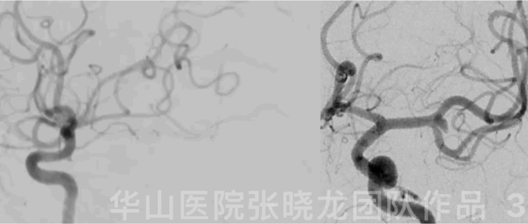
Figure 3 GIF. Rotational DSA confirmed an irregular left middle cerebral artery aneurysm incorporating the superior trunk and lenticulostriate arteries arose from the M2 segment superior trunk.
图 3 GIF. 旋转DSA证实左侧大脑中动脉不规则动脉瘤,动脉瘤累及大脑中动脉上干,豆纹动脉发自左侧大脑中动脉M2段上干。
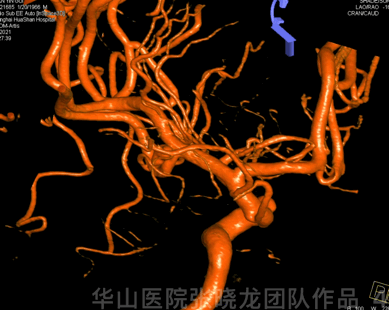
Figure 4 GIF. 3D reconstruction showed a lobular left middle cerebral artery aneurysm.
图 4 GIF. 3D重建提示左侧大脑中动脉分叶状动脉瘤。
1
Strategy
A lobular left middle cerebral artery aneurysm indicating a high rupture risk should be treated.
The aneurysm incorporated the large superior trunk and lenticulostriate arteries arose from the superior trunk, which should be preserved.
A Solitaire stent will be selected to deploy at the MCA superior trunk to straighten the parent artery in order to decrease the recurrence rate.
Small and soft coils were chosen to densely pack the irregular small aneurysm sacs.
左侧大脑中分叉部分叶状动脉瘤,破裂风险高,建议治疗。
动脉瘤累及大脑中动脉粗大上干,且豆纹动脉发自上干,所以上干需要保护。
计划Solitaire支架在大脑中动脉上干释放,拉直载瘤动脉,降低复发风险。
选用小、软圈致密栓塞不规则动脉瘤瘤腔。

Figure 5. Measurements: aneurysm size 2.1*2.67mm; parent artery diameter 2.5mm.
图 5. 测量:动脉瘤大小2.1*2.67mm;载瘤动脉直径2.5mm。
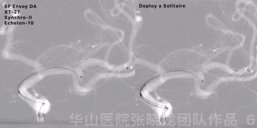
Figure 6 GIF. 6F Envoy DA guiding catheter was placed into the left internal carotid artery cavernous sinus segment. A XT-27 microcatheter was positioned into the left middle cerebral artery superior trunk via a Synchro-II microwire. A straight-tipped Echelon-10 microcatheter was advanced into the aneurysm sac. Deploy a Solitaire stent 4*20mm at the superior trunk covering the aneurysm neck.
图 6 GIF. 6F Envoy DA导引导管置于左侧颈内动脉海绵窦段,XT-27微导管在Synchro-II微导丝导引下至于左侧大脑中动脉M2段上干。直头Echelon-10微导管置于瘤腔。于上干释放Solitaire支架覆盖瘤颈。
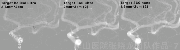
Figure 7 GIF. Inserted the following coils (Target helical ultra 2.5mm*4cm, Target 360 ultra 2mm*3cm (2), Target 360 nano 1.5mm*2cm (2)) in sequence and packed the aneurysm densely.
图 7 GIF. 依次填入以下弹簧圈(Target helical ultra 2.5mm*4cm, Target 360 ultra 2mm*3cm (2), Target 360 nano 1.5mm*2cm (2)),将动脉瘤致密栓塞。
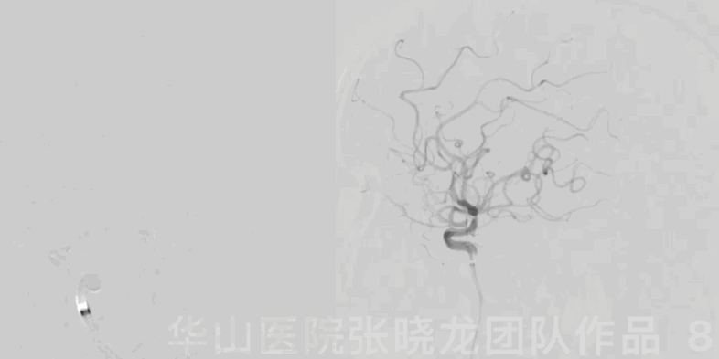
Figure 8 GIF. General heparinization was performed. Angiogram showed the aneurysm packed satisfactorily and no hemorrhage was observed. Tirofiban 15ml was administered.
图 8 GIF. 行全身肝素化。复查血管造影示动脉瘤栓塞满意,未见出血或栓塞。经导管给予替罗非班15ml。
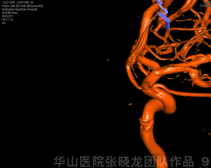
Figure 9 GIF. Aneurysm was not observed from 3D reconstruction and the parent artery was patent.
图 9 GIF. 3维重建未见动脉瘤,载瘤动脉通畅。
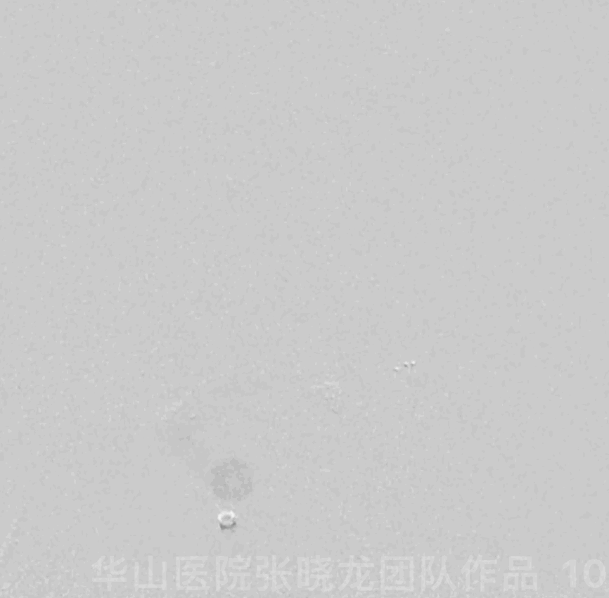
Figure 10 GIF. The aneurysm was embolized densely and the parent artery was patent.
图 10 GIF. 复查造影动脉瘤栓塞满意,载瘤动脉通畅。
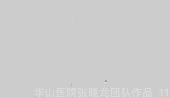
Figure 11 GIF. The intracranial vessels were intact.
图 11 GIF. 颅内血管完好。
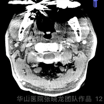
Figure 12 GIF. Dyna-CT did not depict hemorrhage.
图 12 GIF. Dyna-CT未见出血。
2
Post-operation
NE: GCS 15, no headache or dizziness, normal eyeball movement, bilateral pupil light reflex normal, bilateral muscle strength normal, bilateral Babinski negative.
Medication: Tirofiban 15ml/h maintained 48 hours.
AA 82.6%, ADP 70%.
At discharge: Clopidogrel 75mg for 3 month and Aspirin 100mg for long term.
查体:GCS 15,无头痛头晕,眼球各项运动佳,双侧瞳孔对光反射灵敏,双侧肌力正常,双侧巴氏征阴性。
药物:替罗非班15ml/h维持48h。
血栓弹力图:阿司匹林抑制率82.6%,氯吡格雷抑制率70%。
出院:氯吡格雷75mg口服3月,阿司匹林长期口服。
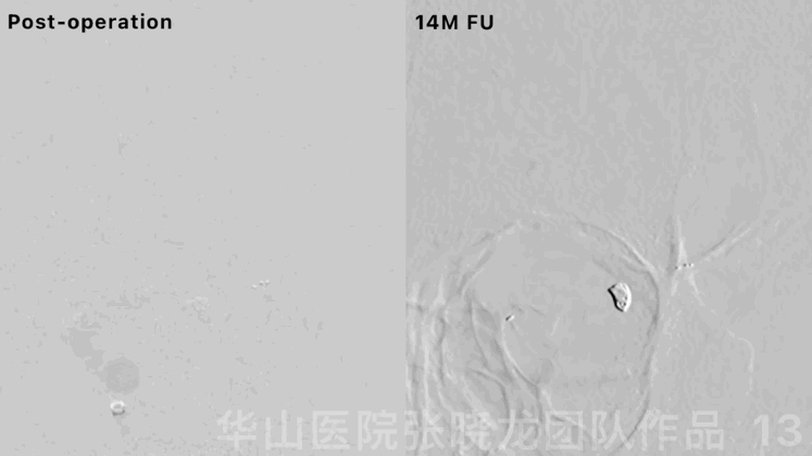
Figure 13 GIF. No recurrence was visualized by 14 month follow up angiogram.
图 13 GIF. 14个月复查脑血管造影未见动脉瘤复发。

Figure 14. The parent artery straightened significantly.
图 14. 载瘤动脉成角明显拉直。

Figure 15 GIF. Rotational DSA demonstrated the aneurysm dense packing by 14 month follow up.
图 15 GIF. 14个月复查旋转DSA示动脉瘤致密栓塞。
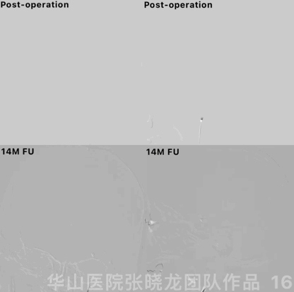
Figure 16 GIF. The intracranial vessels remained intact by 14 month follow up.
图 16 GIF. 14个月复查颅内血管完好,未见狭窄或出血。
3
Summary
A lobular left middle cerebral artery aneurysm indicating a high rupture risk should be treated.
The aneurysm incorporated the large superior trunk and lenticulostriate arteries arose from the superior trunk. Meanwhile no antiplatelets were prescribed before operation with microcatheters not difficult to navigate, a stent was deployed at the superior trunk and general heparinization can be performed. While general heparinization was conducted after the aneurysm was densely packed considering the aneurysm was small and irregular.
Solitaire stent with the merit of parent artery angle significant increasement and low in-stent restenosis risk, was selected to deploy at the MCA superior trunk in order to decrease the recurrence rate.
Working projection selection should visualize super-selection route and lobular morphology.
Small and soft coils were chosen to pack the irregular small aneurysm sacs, while a relative large first coil was selected for a stable basket.
左侧大脑中分叉部分叶状动脉瘤,破裂风险高,建议治疗。
动脉瘤累及大脑中动脉粗大上干,且豆纹动脉发自上干,所以支架在上干释放。同时该患者术前无抗血小板准备,术中微导管到位不困难,术中可行肝素化。但考虑小动脉瘤形态不规则,所以动脉瘤致密栓塞后再行肝素化。
Solitaire支架具有载瘤动脉成角拉直明显、支架内狭窄风险低的优势,于大脑中动脉上干释放,降低复发风险。
工作角度需要清楚显示上干超选路径及动脉瘤形态。
选用小、软圈栓塞不规则动脉瘤瘤腔,但应避免成篮首圈过小。

点击或扫描上方二维码
查看更多“介入”内容
