Case Review
• 64 y/o, female.
• Suffering from paroxysmal unsteady gait one month. Local hospital MRA revealed a right vertebrate aneurysm.
• Medical history: Chronic hepatitis B, HTN, asthma and rheumatoid arthritis.
• Medication: Amlodipine.
• NE: (-)
• 64岁,女性。
• 发作性行走不稳1月。外院MRA提示右侧椎动脉瘤。
• 既往史:慢性乙肝,高血压,哮喘和类风湿关节炎。
• 药物:氨氯地平。
• 神经查体:-。

图 1. MRA提示右侧椎动脉 V4段夹层动脉瘤。

图 2. 高分辨率MRI提示右侧椎动脉夹层动脉瘤,瘤壁强化,局部血栓形成。
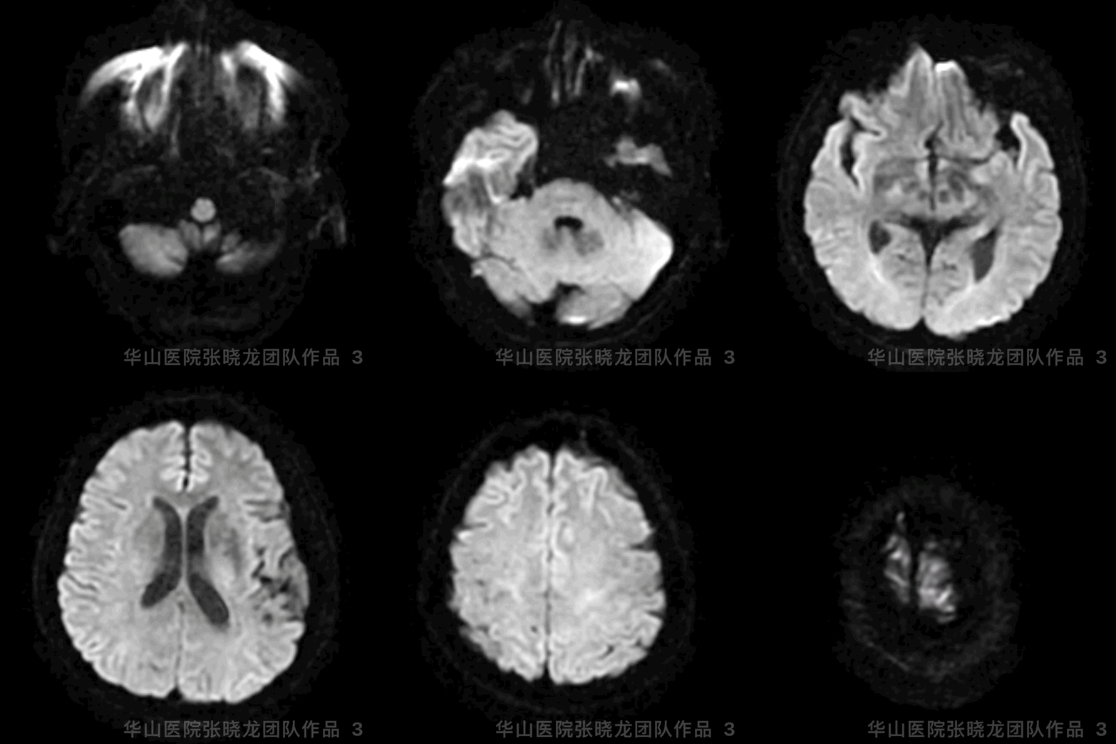
图 3 GIF. DWI未见急性脑梗死。

图 4. DSA显示左侧劣势椎动脉。

图 5. DSA证实右侧椎动脉V4段夹层动脉瘤。
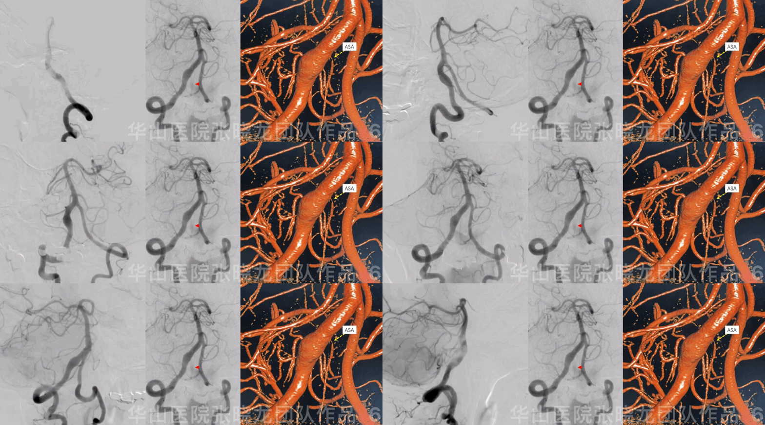
图 6 GIF. 右侧优势椎动脉,脊髓前动脉发自夹层动脉瘤远端。
1
Strategy
• A right dominant vertebrate artery V4 segment dissecting aneurysm with focal thrombosis and wall enhancement should be treated.
• Post large coiling stenting technique was recommended for the dissecting aneurysm.
• The important branch (ASA) arising from distal part of the aneurysm must be preserved.
• A proper working projection displayed the important artery ASA meanwhile attention coils effecting the branch.
• 右侧优势椎动脉V4段夹层动脉瘤局部血栓形成、瘤壁强化,建议栓塞治疗。
• 该夹层动脉瘤采用大圈辅助支架后释放技术。
• 脊髓前动脉发自夹层动脉瘤远端,该分支必须保护。
• 治疗时选择合适的工作角度显示脊髓前动脉,同时警惕弹簧圈填塞时影响该分支。
2
Operation
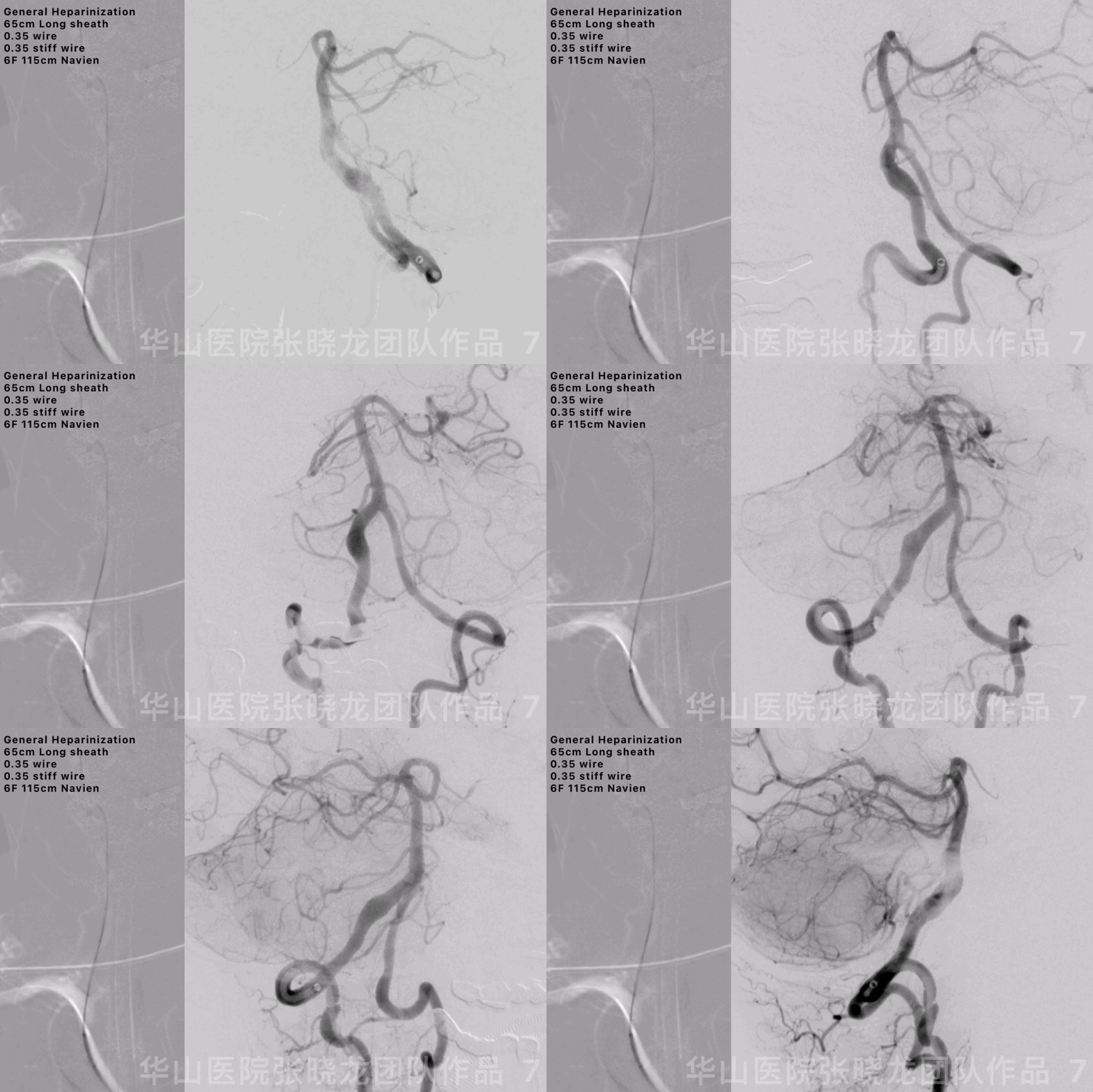
图 7 GIF. 全身肝素化。将6F 115cm Navein和65cm长鞘在双0.35导丝导引下置于右侧锁骨下动脉。然后Navein中间导管在双0.35导丝(一根正常导丝一根硬导丝)导引下置于右侧椎动脉V4段。造影清楚显示右侧椎动脉夹层动脉瘤及脊髓前动脉。

图 8. 选择合适的工作角度。测量:夹层动脉瘤长径6.8mm,近端载瘤动脉4.2mm,远端载瘤动脉4.1mm。
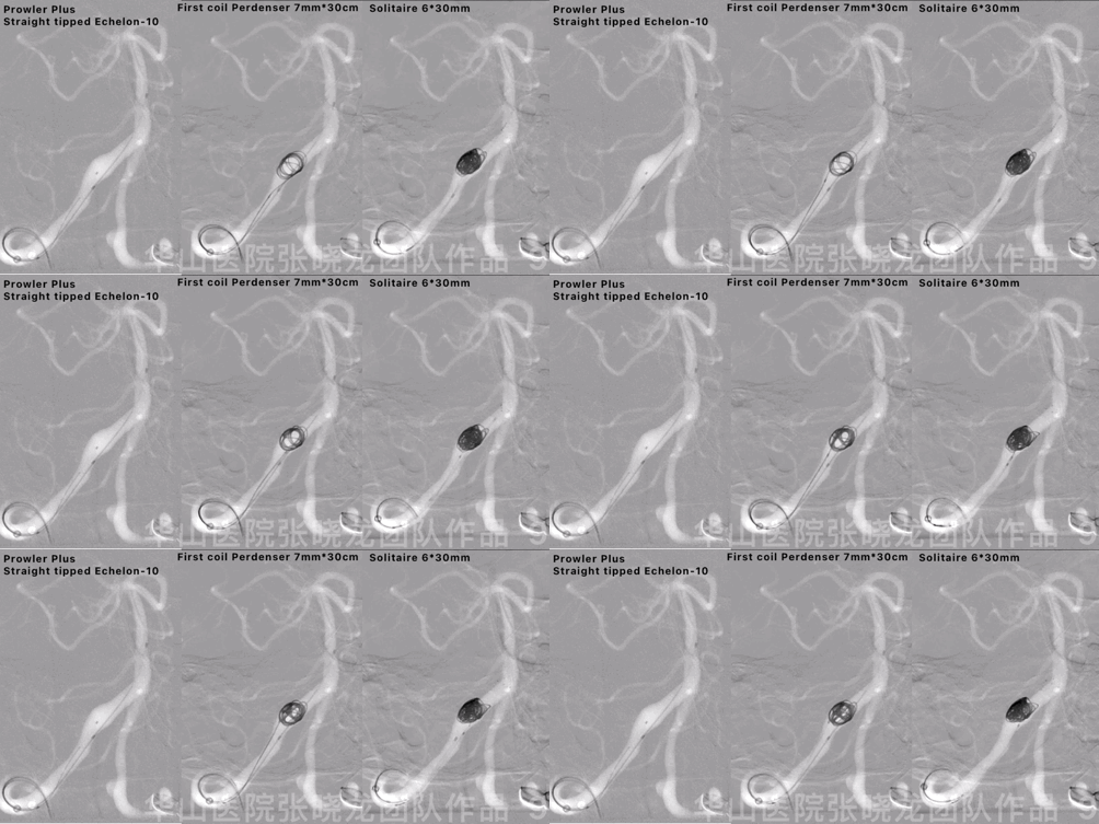
图 9 GIF. 将Prowler Plus微导管置于基底动脉近端,将直头微导管置于瘤腔。经微导管填入Perdenser 7mm*30cm和6mm*20cm两枚弹簧圈,然后瘤颈部释放Solitaire 6*30mm支架。
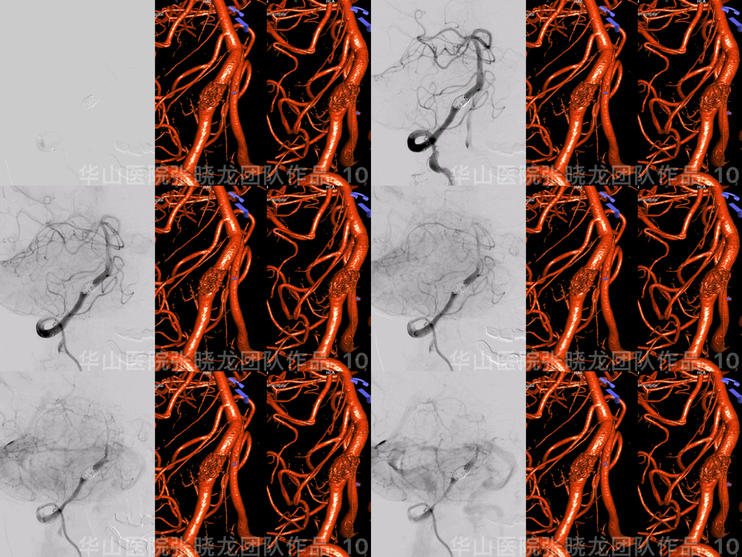
图 10 GIF. 复查造影证实动脉瘤不显影,载瘤动脉及脊髓前动脉显示良好。
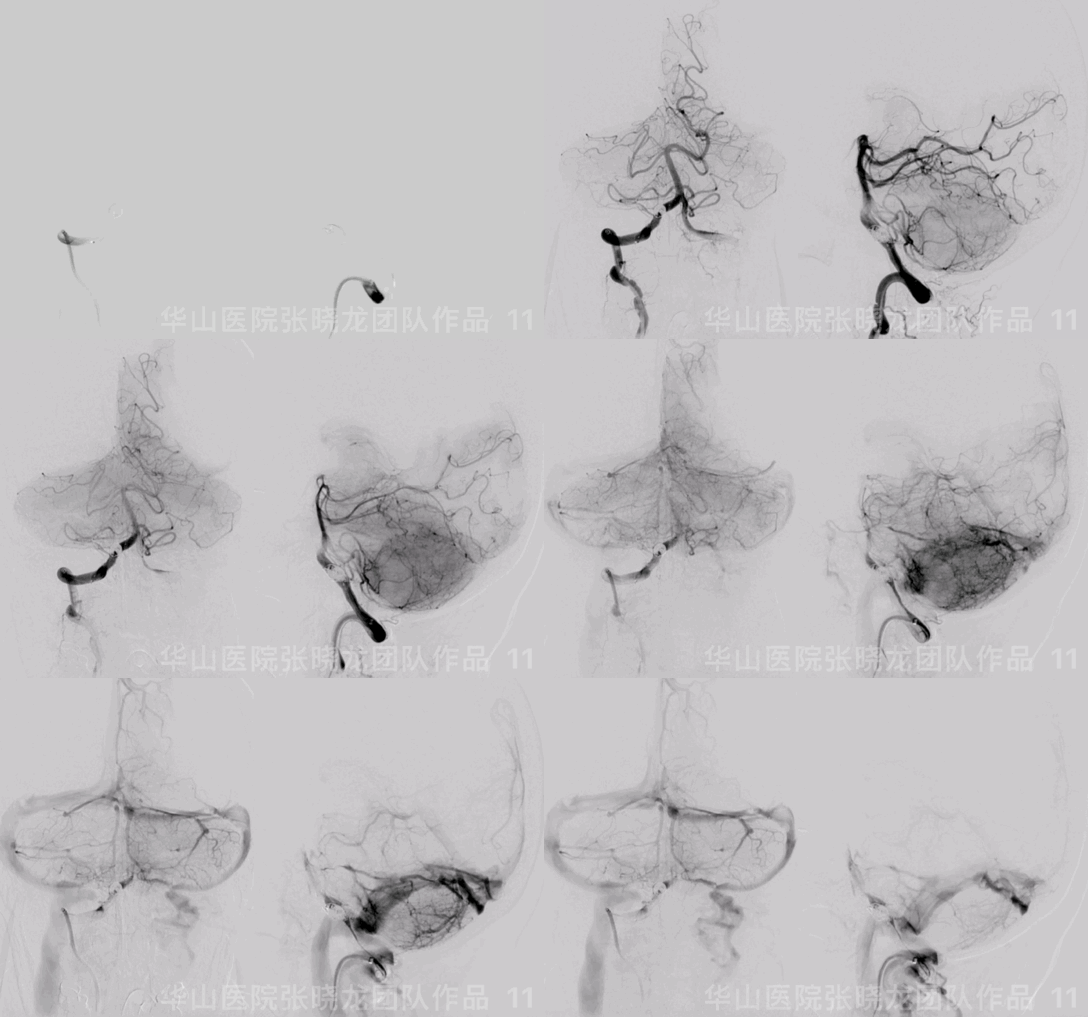
图 11 GIF. 造影示颅内血管完好,动脉瘤致密栓塞。经导引导管给予替罗非班9ml。
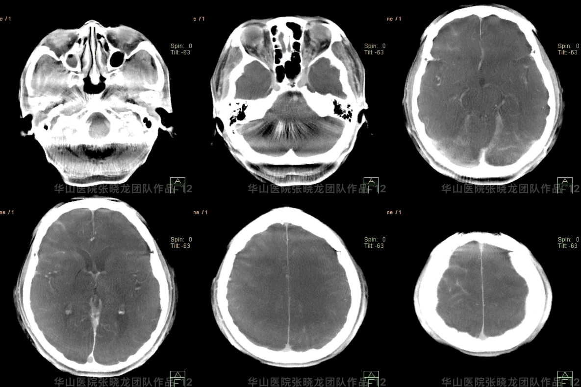
图 12 GIF. Dyna-CT未见出血。
3
Post Operation
• Tirofiban 8ml/h maintained 24h considering the patient took Cilostazol and Clopidogrel 2 days before the operation.
• NE: GCS 15, no swallowing difficulty, no dizziness, eye movement normal, bilateral normal strength, no new neurologic defect.
• At discharge: Clopidogrel for 3 month and Cilostazol for long term. ADP 48%.
• 患者术前2天开始口服西洛他唑及氯吡格雷,术后替罗非班8ml/h维持24h。
• 神经查体:GCS 15,无吞咽困难,无头晕,眼球运动正常,四肢肌力正常,未见新发神经功能缺损。
• 出院:氯吡格雷3月,西洛他唑长期口服。氯吡格雷抑制率48%。
Video 1. Angiography did not show any relapsed of the dissecting aneurysm and the ASA patency by 16 month follow-up.
Video 2. Rotational DSA exhibited the parent artery and branches patent.

Figure 13. Intracranial vessels were intact.
图 13. 颅内血管完好。
Summary
• A right dominant vertebrate artery V4 segment dissecting aneurysm with focal thrombosis and wall enhancement should be treated.
• Post large coiling stenting technique working as a focal flow divertor could lower the recurrence of for the dissecting aneurysms.
• The important branch (ASA) arising from distal part of the aneurysm must be preserved.
• A proper working projection displayed the important artery ASA meanwhile attention coils effecting the branch.
• Due to severe arteriosclerosis, the guiding catheter was difficult to navigate into the distal V2 segment. So two wires (one regular wire and one stiff wire) were used to provide enough support.
• Continue single antiplatelet and next follow up was scheduled in 2-3 years.
• Pipeline was not the preferred choice for the sort of dissecting aneurysms in our center, which presented with mild expansion plus anterior spinal artey originating from.
• 右侧优势椎动脉V4段夹层动脉瘤局部血栓形成、瘤壁强化,建议栓塞治疗。
• 大圈辅助支架后释放技术有局部密网作用,能降低该类动脉瘤复发风险。
• 脊髓前动脉发自该夹层动脉瘤远端,该分支必须保护。
• 治疗时选择合适的工作角度显示脊髓前动脉,同时警惕弹簧圈填塞时影响该分支。
• 由于动脉粥样硬化严重,导引导管很难超选至V2段以远,所以用一根常规导丝和一根硬导丝来提供足够支撑力。
• 继续单抗,2-3年后再次随访。
• 这种扩张不明显的夹层动脉瘤,合并远端脊髓前动脉发出,在我们中心血流导向支架不是首选治疗方案。
张晓龙
复旦大学附属华山医院
复旦大学附属华山医院放射科主任医师,博士、教授、博士生导师;
斯坦福大学医学院客座临床教授;
主持国家自然科学基金3项,第一作者或通讯作者发表国内外权威期刊文章50余篇;
中华医学会、放射学会、卫生部医政司等组织中担任副主任委员、组长等职务.《中国名医百强榜》神经介入专业中国十强(2012年度、2013年度、2014年度、2015-16年度、2017-18年度);
擅长复杂和疑难脑血管疾病的介入治疗,如复杂脑动脉瘤的栓塞,硬脑膜动静脉瘘栓塞,脑动静脉畸形栓塞,脑梗死的支架,脊髓血管畸形治疗;
自1995年开始从事脑血管疾病介入诊治工作和研究,师从黄祥龙教授、沈天真教授和凌锋教授,是我国最早从事神经介入的专家之一。2010年9月至今连续介入治疗颅内动脉瘤1500余例,无操作致死.
点击或扫描上方二维码,
前往 张晓龙教授 学术主页
查看更多精彩内容
点击或扫描上方二维码
查看更多“介入”内容


