


Case Review
• 27 y/o female.
• CC: Suffered from repeated headache for one year.
• Local hospital MRA showed a giant basilar artery dissecting aneurysm with partial sac thrombosis.
• Past medical history: mild anemia (pre-operational hemoglobin (HGB) 112g/L). No HTN nor DM.
• NE:(-).
• 27岁,女性。
• 主诉:反复头痛1年。
• 当地医院MRA提示基底动脉巨大夹层动脉瘤伴部分瘤腔血栓形成。
• 既往史:轻度贫血(术前血红蛋白112g/L),否认高血压及糖尿病。
• 神经查体:-。

图 2 GIF. 双侧椎动脉造影证实基底动脉主干巨大夹层动脉瘤伴部分瘤腔血栓形成,残腔内造影剂滞留明显。
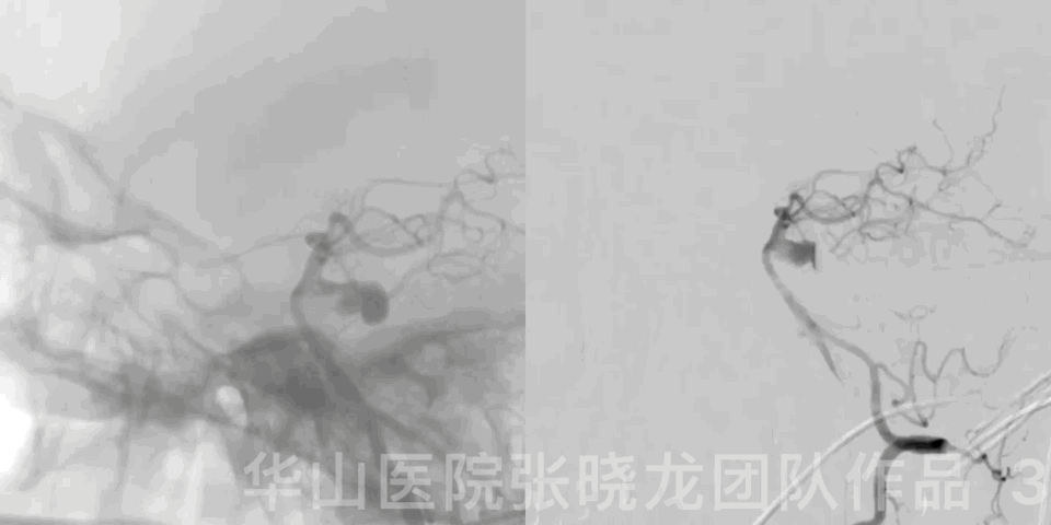
Figure 3 GIF. Rotational DSA depicted a wide neck dissecting aneurysm of upper-middle segmental basilar artery.
图 3 GIF.旋转DSA证实基底动脉中上段相对宽颈夹层动脉瘤。

Figure 4. Bilateral under-developed posterior communicating arteries.
1
Strategy
A giant dissecting aneurysm of upper-middle segmental basilar artery tended to have a risk of parent occlusion, ischemic events and rupture, which should be treated.
Pipeline for upper-middle segmental basilar artery aneurysms was prone to cause perforating events. Once the perforating branches affected, severe complications would happen.
LVIS stent-assisted coiling (rivet technique) was adopted for this type aneurysm to lower the recurrence risk.
Dense packing of neck region aimed the recurrence while loose packing of aneurysm dome purposed to relieve the mass effect.
基底动脉中上段夹层大动脉瘤有载瘤动脉闭塞、缺血及出血风险,该动脉瘤建议治疗。
基底动脉中上段动脉瘤用Pipeline支架容易影响穿支,可导致严重的并发症。
为降低动脉瘤复发风险,该动脉瘤采用LVIS支架辅助弹簧圈栓塞(铆钉技术)。
动脉瘤瘤颈部致密栓塞降低复发风险,而动脉瘤体疏松填塞降低占位效应。
2
Operation

图 5. 动脉瘤测量:动脉瘤大小:5.43*9.56mm 动脉瘤瘤颈:5.20mm 近端载瘤动脉直径:1.78mm 远端载瘤动脉直径:2.89mm。
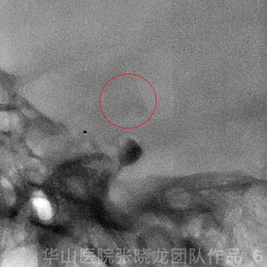
Figure 6 GIF. General heparinization. 6F guiding catheter was navigated into left vertebrate artery C3. Fluoroscopy showed contrast medium stagnation at the thalamus region when trying to placing Headway-21 microcatheter into left posterior cerebral artery.
图 6 GIF. 行全身肝素化。将6F导引导管置于左侧椎动脉C3水平。尝试将Headway-21微导管置于左侧大脑后动脉时透视下发现丘脑造影剂滞留。
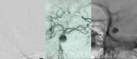
图 7 GIF. 反复造影及透视未见明确出血点。
Figure 8 GIF. Headway-21 microcatheter (steam shaped) was navigated into left P2 segment and Echelon-10 45° (steam shaped) to aneurysm sac.
图 8 GIF. 将Headway-21微导管塑形后超选入左侧P2段,Echelon-10 45° 头端塑形后置于动脉瘤腔。
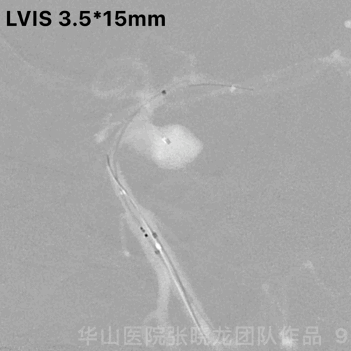
Figure 9 GIF. LVIS stent was deployed.
图 9 GIF. 释放LVIS支架。

Figure 10 GIF. Insert the first coil (Microplex-10 7mm*30cm) satisfactorily. Then continue insert the following coils: Microplex-10 6mm*20cm (2), Microplex-10 5mm*20cm.
图 10 GIF. 填入Microplex-10 7mm*30cm第一枚弹簧圈。然后依次填入2枚Microplex-10 6mm*20cm和1枚Microplex-10 5mm*20cm弹簧圈。

Figure 11 GIF. Rivet technique at the neck region. Microplex-10 5mm*20cm, Microplex-10 3mm*10cm, Microplex-10 2mm*8cm (2) were inserted.
图 11 GIF. 瘤颈部采用铆钉技术。瘤颈部填入以下弹簧圈:Microplex-10 5mm*20cm, Microplex-10 3mm*10cm, Microplex-10 2mm*8cm (2)
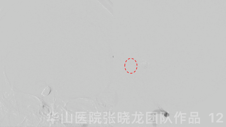
Figure 12. Angiography showed the aneurysm was densely packed but a small amount of thrombosis formed intra-stent.
图 12. 复查造影动脉瘤致密栓塞,支架内少许血栓形成。
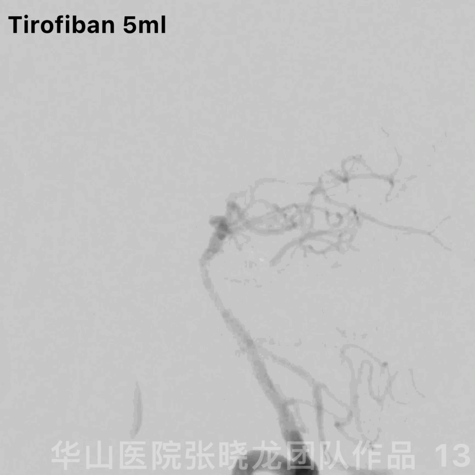
Figure 13 GIF. Tirofiban 5ml was administered. Rotational DSA demonstrated the parent artery patent.
图 13 GIF. 给予替罗非班5ml。旋转DSA证实载瘤动脉通畅。
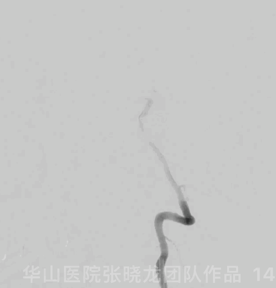
Figure 14. Redo angiography after waiting for 30 minutes and no hemorrhage was found.
图 14. 等待30分钟后再次复查造影,仍未发现出血。
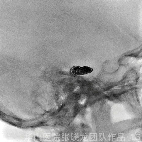
Figure 15 GIF. Coiling configuration.
图 15 GIF. 弹簧圈铸形。

Figure 16. Post-operational instant CT (2019.02.15 13:42) depicted high density in the sulci and cistern.
图 16. 术后即刻CT (2019.02.15 13:42) 提示部分脑沟及脑池内高密度影。

Figure 17. High density remained in the interpeduncular cistern on the Post-operative day 1 CT (2019.02.16 10:30). Therefore subarachnoid hemorrhage was considered.
图 17. 术后第1天(2019.02.16 10:30)复查头颅CT,脚间池内仍有高密度影,考虑蛛网膜下腔出血。
3
Post Operation
Tirofiban 5ml/h maintained 48 hours.
Dual antiplatelets were prescribed.
NE: Mild headache, bilateral pupillary light reflex normal, bilateral normal strength, no dizziness, normal swallowing, bilateral Babinski -.
替罗非班5ml/h微泵维持48h。
口服双抗。
神经查体:轻度头痛,双侧瞳孔对光反射灵敏,四肢肌力正常,无头晕及吞咽困难,双侧巴氏征阴性。

Figure 18. Left side of the pons was compressed and mass effect did not increase by the follow up.
图 18. 磁共振随访可见桥脑左侧受压,随访较术前占位效应无加重。
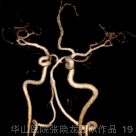
Figure 19 GIF. 3D construction by 42 month follow up.
图 19 GIF. 42月随访时3D重建。
Video 1. The aneurysm was no relapsed and parent artery was patent by 42 month follow up.
视频 1. 42个月随访未见动脉瘤复发,载瘤动脉通畅。
Video 2. Angiography confirmed the aneurysm was densely packed by 42-month follow up. The intracranial vessels were intact.
视频 2. 42个月随访造影证实动脉瘤致密栓塞。颅内血管完好。
Video 3. Patient status.
视频 3. 病例近况。
Summary
Aneurysm rupture should be considered first because of contrast medium delayed absorption on post-operational CT and limited manipulation.
Although fluoroscopy and angiography were conducted repeatedly, no definite hemorrhage was found during the operation. However, Dyna-CT was not performed peri-operation, which was the limits.
A giant dissecting aneurysm of upper-middle segmental basilar artery tended to have a risk of parent occlusion, ischemic events and rupture, which should be treated.
Pipeline for upper-middle segmental basilar artery aneurysms was prone to cause perforating events. Once the perforating branches affected, severe complications would happen.
LVIS stent-assisted neck dense coiling (rivet technique) was adopted for this type aneurysm to lower the recurrence risk. Long term follow up demonstrated LVIS assisted large coiling (rivet technique) for upper-middle segmental basilar artery aneurysms was safe and efficient.
Avoiding too large-stiff coils and loose packing of the aneurysm dome purposed to relieve the mass effect.
Stop Aspirin and next follow up was scheduled in 5 years.
术后CT造影剂延迟吸收及微导管微导丝无过多操作,所以首先考虑动脉瘤破裂出血。
尽管我们反复透视及造影,术中均未发现明确的出血。但不足之处是我们术中未行Dyna-CT。出血位于子囊上方,考虑动脉瘤瘤顶部出血,反复观察出血未增加,因此未中和肝素,计划快速动脉瘤栓塞止血。
基底动脉中上段夹层大动脉瘤有载瘤动脉闭塞、缺血及出血风险,故该动脉瘤建议治疗。
基底动脉中上段动脉瘤用Pipeline支架容易影响穿支,可导致严重的并发症。
为降低动脉瘤复发风险,该动脉瘤采用LVIS支架辅助加瘤颈部弹簧圈致密栓塞(铆钉技术),长期随访表明该方案是安全有效的。
避免使用过大弹簧圈以及远端子囊未致密栓塞可以防止占位效应进一步加重。
42个月随访动脉瘤无复发,建议停阿司匹林,5年后再次复查脑血管造影。

张晓龙
复旦大学附属华山医院
复旦大学附属华山医院放射科主任医师,博士、教授、博士生导师;
斯坦福大学医学院客座临床教授;
主持国家自然科学基金3项,第一作者或通讯作者发表国内外权威期刊文章50余篇;
中华医学会、放射学会、卫生部医政司等组织中担任副主任委员、组长等职务.《中国名医百强榜》神经介入专业中国十强(2012年度、2013年度、2014年度、2015-16年度、2017-18年度);
擅长复杂和疑难脑血管疾病的介入治疗,如复杂脑动脉瘤的栓塞,硬脑膜动静脉瘘栓塞,脑动静脉畸形栓塞,脑梗死的支架,脊髓血管畸形治疗;
自1995年开始从事脑血管疾病介入诊治工作和研究,师从黄祥龙教授、沈天真教授和凌锋教授,是我国最早从事神经介入的专家之一。2010年9月至今连续介入治疗颅内动脉瘤1500余例,无操作致死.
.png")
点击或扫描上方二维码,
前往 张晓龙教授 学术主页
查看更多精彩内容

点击或扫描上方二维码
查看更多“介入”内容
