


Case Review
• 34 y/o male.
• Suffered from intracranial pulsatile tinnitus for 5 months.
• Past medical history: Left cavernous sinus dual arteriovenous fistula embolization was performed 2 months ago in other hospital. Paroxysmal headache accompanied with orbit pain 10 years ago, relieved in one year.
• PE: No eye movement deficit; no conjunctival congestion, no relative infection.
• 34岁男性。
• 颅内搏动性耳鸣5个月。
• 既往史:2月前于外院行左侧海绵窦区硬脑膜动静脉瘘栓塞术。10年前有发作性头痛伴眼眶痛,一年后该症状自行缓解。
• 查体:无眼球运动障碍;无结膜充血,无相关感染。

图 1. MRI提示左侧海绵窦外侧流空影。
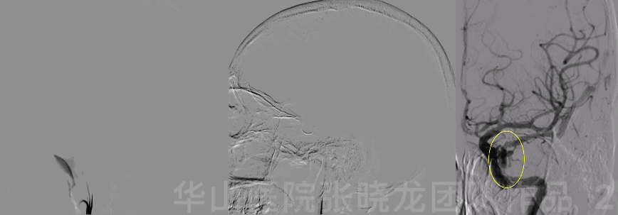
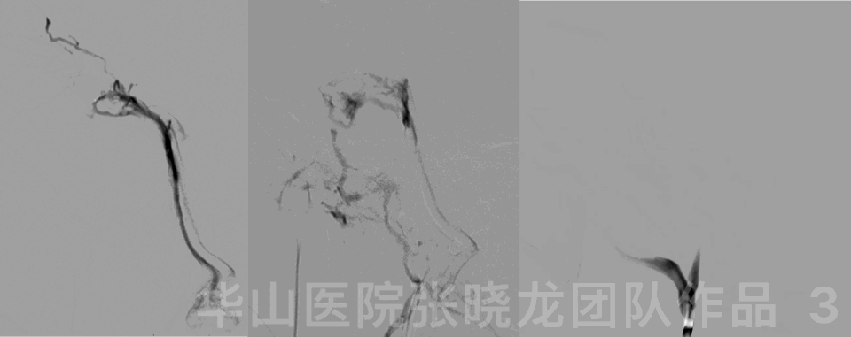
图 3 GIF. 尝试经静脉途径栓塞失败,经静脉途径的微导管进入不同的间隔,但难以进入外侧海绵窦。遂经脑膜副动脉超选至瘘口,注入Onyx-18约0.6ml。外院术中血压或心率的变化没有记录。
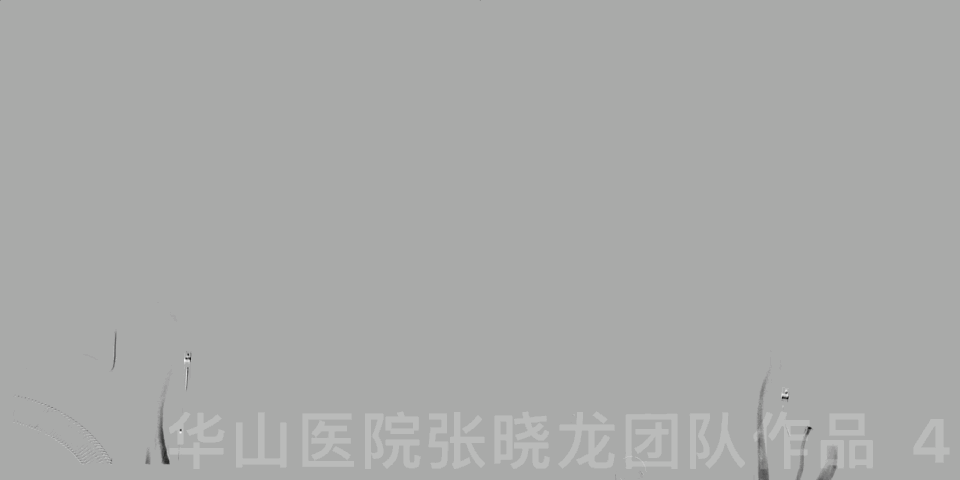
图 4 GIF. 术后颈总动脉造影未见瘘口残余。
1
Post Operation
After the TAE, the tinnitus disappeared but severe headache occurred. Then headache relived but pulsatile tinnitus recurred 10 days later. Tinnitus Handicap Inventory (THI) scored 14 on admission to our department.
术后患者耳鸣消失但出现严重头痛,影响睡眠。10天后,头痛好转但耳鸣出现。再次入院时耳鸣妨碍量表(THI)14分。
2
2nd Angio
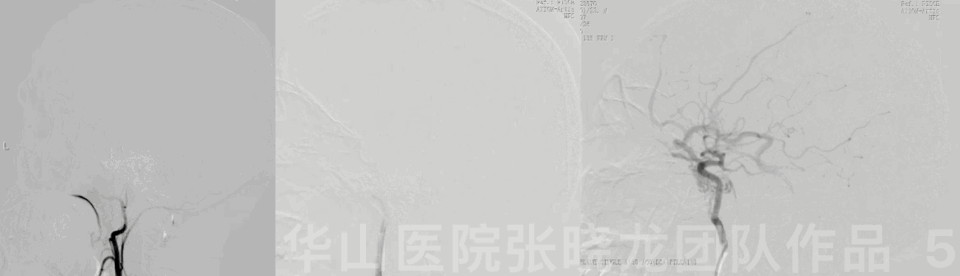
图 5 GIF. DSA (2021.11.24)造影示残余瘘口由左侧颌内动脉和颈内动脉分支供血,经大脑中浅静脉和岩下窦引流。

图 6. 对侧的正常蝶顶窦和海绵窦。

图 7. 瘘口首先进入外侧海绵窦 (红色区域),再向侧裂静脉(橙色虚线箭头)和海绵窦后上间隙-岩下窦(蓝色虚线)引流。

图 8. 红色区域(海绵窦前外侧间隔)是瘘口的受体间隔,蓝色虚线(海绵窦后内侧间隔)是引流间隔。
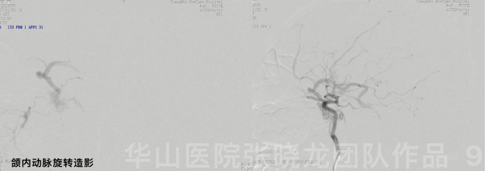
图 9 GIF. 选择造影显示了海绵窦外侧间隔与海绵窦后部的关系。
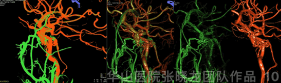
Figure 10 GIF. 3D reconstruction.
图 10 GIF. 3D重建。
3
Strategy
The inferior petrosal sinus (IPS) was still revealed from fistulous drainage, therefore transvenous embolization from IPS to the lateral cavernous sinus was theoretically accessible.
Transarterial embolization via ECA was an alternative method.
There existed cranial nerves and endocranium between lateral-cavernous sinus and cavernous sinus. Hence transvenous embolization could serve as an attempt because it was difficult for microcatheter to pass through.
However, Onyx injection in the lateral-cavernous sinus via transarterial route had a risk of cranial nerve palsy.
少量瘘口沿颈内动脉后部进入海绵窦内侧间隔,并经岩下窦引流,因此经静脉途径从岩下窦至海绵窦外侧间隔栓塞瘘口理论上是可行的。
经颈外动脉栓塞瘘口也是另一可行的治疗方案。
外侧海绵窦与海绵窦之间解剖上存在颅神经及硬膜阻隔,经静脉途径微导管可能非常难以通过,所以经静脉途径治疗只能作为尝试。
而经动脉途径在外侧间隔注胶有颅神经麻痹风险。
4
2nd Operation
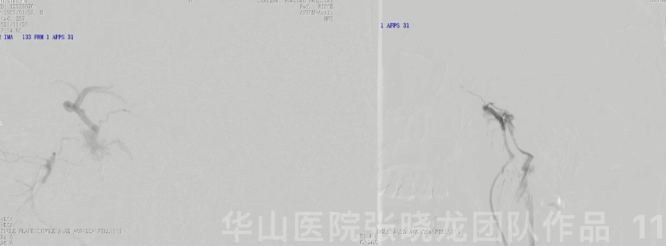
图 11 GIF. 颌内动脉超选择造影(左)见瘘口通过大脑中浅静脉逆流。岩下窦的直接静脉造影(右)见海绵窦内侧间隔及眼静脉显影,但未见大脑中浅静脉逆流,说明内侧间隔与外侧间隔之间沟通不充分。
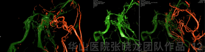
图 12 GIF. 颈外动脉与岩下窦双融合重建显示海绵窦瘘口间隔(外侧)和引流间隔(后内侧)的相互关系。
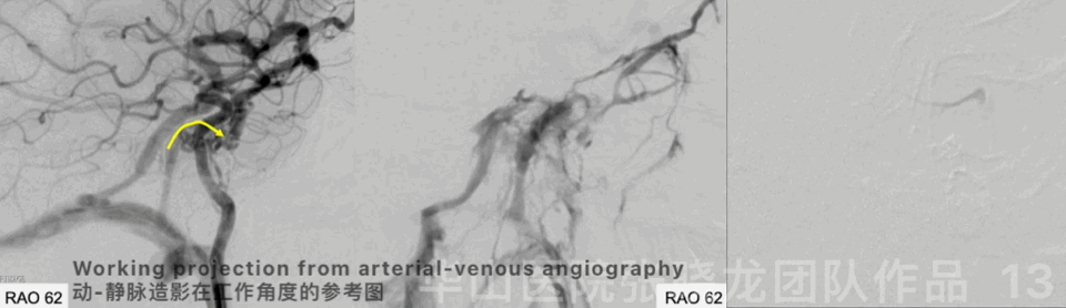
图 13 GIF. 微导管经岩下窦进入海绵窦。经静脉系统造影未见大脑中浅静脉显影,说明所进入的窦腔并非责任间隔。

图 14. Echelon-10 尝试经静脉途径超选外侧间隔,两根微导管均不能成功到位。正位上微导管均在海绵窦的内侧间隔,而首次治疗的胶位于相对外侧(蓝色区)。
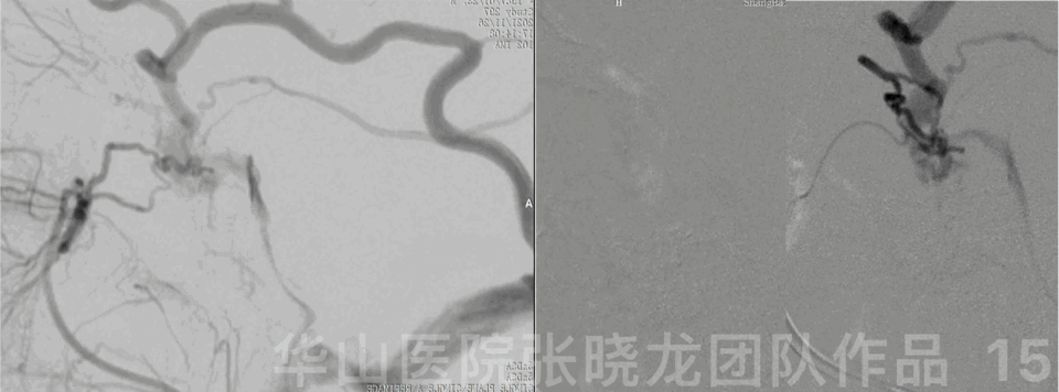
图 15 GIF. 经静脉途径失败后,改为经动脉栓塞。
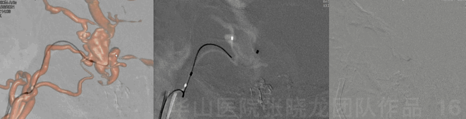
图 16 GIF. 微导管经颌内动脉超选至瘘口终末端。
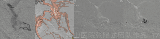
图 17 GIF. 经微导管注入onyx-18约1.6ml。在注射DMSO 期间患者心率下降。
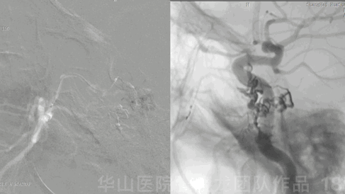
图 18 GIF. Onyx的搏动性弥散提示其他供血动脉的竞争性血流。

图 19. 胶少量弥散至眼动脉硬膜回返支供血的硬膜静脉端。
图 20 GIF. 术后DSA显示瘘口消失。眼动脉硬膜回返支内造影剂停滞。大脑中浅静脉恢复正向血流。

Figure 21. Dyna-CT
showed the onyx cast.
5
Post Operation
PE: increased left pupil diameter, normal light reflex, left eye abduction deficit, left periorbita and forehead hypoesthesia. Pulsatile tinnitus disappeared, bilateral muscle strength normal.
Medication: Dehydration, corticosteroid, antiplatelet therapy were administered.
At discharge: Aspirin for 1 month and rehabilitation therapy.
查体:患者左侧瞳孔略增大,左侧对光反射正常,左眼外展受限,左眶周及额部感觉减退。耳鸣消失,四肢肌力正常。
药物:予脱水、激素、抗血小板治疗。
出院:阿司匹林口服1月,当地康复医院左眼外展训练。

The Pic cite from: Gailloud P. Angiographic anatomy of the laterocavernous sinus. AJNR Am J Neuroradiol. 2000
6

图 23. 患者出院后行20天高压氧治疗。9个月随访时,左眼外展功能恢复,左侧眶周及额部感觉减退明显好转。
Video 1. 9 month follow-up angiography showed no recurrence of DAVF. Ophthalmic artery stagnation disappeared. Venous drainage returned to normal.
视频 1. 9个月造影复查,未见瘘口残余及复发。眼动脉阻滞消失,颅内静脉引流恢复正常。
Video 2. 9 month follow-up DSA demonstrated the left cavernous sinus drainage function preserved and the inferior petrosal sinus was patent.
视频 2. 9个月DSA随访证实左侧海绵窦功能保留,左侧岩下窦引流通畅。
Summary
In this case the venous thrombosis and septum devided the CS into two devisions: the fistula was located in the lateral cavernous sinus and mainly refluxed to the SMCV rather than antegrade drainage to the inferior petrous sinus. The medial septum was connected to the ophthalmic vein and IPS.
The lateral CS to medial CS was obstructed, therefore the fistula refluxed from lateral CS to the SMCV.
Due to the IPS was still revealed from fistulous drainage, our primary strategy is access to the lateral CS via transvenous route. However the anatomic septum might obstruct the microcatheter access route. Therefore we changed to the TAE therapy.
The injection of DMSO via IMA or AMA can induced HR or BP decrease. The DMSO should be injected within minimal dosage and slower rate.
The toxic effect of the DMSO can induced nerve paralysis. Abduction deficit should be awared in TAE of the lateral CS DAVF. Dehydration, corticosteroids and hyperbaric oxygen therapy could alleviate the symptom.
DSA follow up should be performed 2-3 years later.
该病例中静脉血栓和纤维间隔将海绵窦的引流主要分成两个部分:瘘口位于海绵窦外侧间隔,经大脑中浅静脉逆流为主,少量向岩下窦引流。内侧间隔向前连接眼静脉,向后连接岩下窦。
海绵窦外侧至内侧引流通路阻塞,因此瘘口经海绵窦外侧向大脑中浅静脉逆流。
由于瘘口仍有少量经岩下窦引流,所以我们最先的策略是经静脉途径到达海绵窦外侧间隔栓塞瘘口。但由于海绵窦内外间隔之间解剖上存在硬膜、颅神经阻隔,经静脉途径微导管可能难以通过,所以我们最终采用了经动脉途径栓塞。
经颌内动脉或脑膜副动脉注射DMSO时,由于三叉-心反射会引起心率和血压下降。DMSO注射应控制剂量及速率。
DMSO的毒性可引起神经麻痹。外展受限是Onyx栓塞治疗海绵窦外侧间隔瘘口常见的并发症。术后的脱水、激素冲击治疗、高压氧等可改善症状。
建议2-3年后行DSA随访。

点击或扫描上方二维码
查看更多“介入”内容
