Review
• 68 y/o, female.
• CC: suffered from bilateral limb static tremor for 2 years. The symptom aggravated for 1 month. A left Pcom and MCA aneurysms were detected for one month.
• Medical history: HTN for 2 months, well-controlled.
• Medication: Aspirin; Atorvastatin.
• PE: (-)
• 68岁,女性。
• 主诉:四肢静止性震颤2年,加重1月。检查发现左侧后交通动脉瘤及左侧大脑中动脉瘤1月。
• 既往史:高血压2月,血压控制良好。
• 药物:阿司匹林;阿托伐他汀。
• 神经查体:阴性。

图 1. 当地医院头颅CTA提示左侧后交通及大脑中动脉瘤。
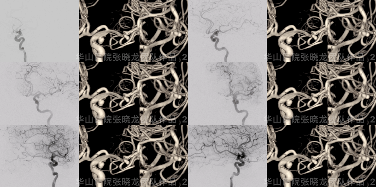
图 2 GIF. 近端入路扭曲。后交通窄颈动脉瘤,大脑中动脉瘤分叶伴2枚子瘤。
1
Strategy
• Since both the Pcom and the irregular MCA aneurysm have a high rupture risk, treatment is necessary.
• For the narrow-necked PcomA aneurysm, simple coiling with dual microcatheter technique is preferred.
• For the MCA aneurysm, stent-assisted coiling is a back-up if simple coiling fails. Two sacs should be densely packed.
• 后交通动脉瘤及不规则大脑中动脉瘤破裂风险高,建议治疗。
• 后交通窄颈动脉瘤,首选双微导管单纯弹簧圈栓塞技术。
• 大脑中动脉瘤先采用单纯栓塞,若单纯栓塞失败,采用支架辅助栓塞。大脑中动脉瘤2枚子瘤须致密栓塞。
2
Step 1
Embolize the left MCA aneurysm with two daughter sacs.
栓塞左侧大脑中动脉瘤及其子瘤
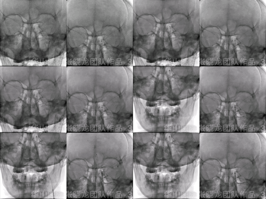
Figure 3 GIF. The guiding catheter was pushed to confirm the proximal tortuous vessel. Fluoroscopy shows no contrast medium stagnation. 125cm MP + 6F Tonbridge guiding catheter+ 0.35 guiding wire General heparinization.
图 3 GIF. 向前推动导引导管,顺应性良好。透视未见造影剂滞留。 使用125cm MP导管+6F通桥导引导管+0.35导引导丝。 全身肝素化。
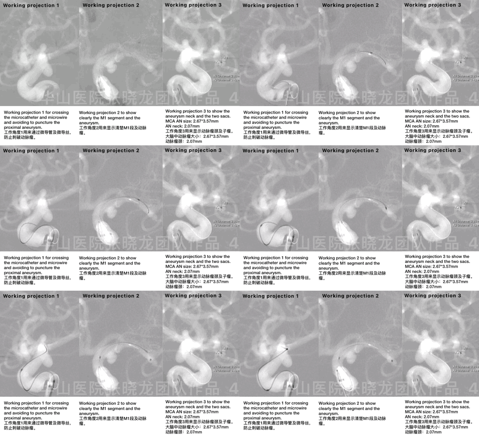
图 4 GIF. 采用多个工作角度。
Video 1. Working projection 3. Echelon-10 45° unshaped was navigated into the MCA aneurysm by the support of Synchro II. Insert a helical 3mm*8cm coil with “8” framing in the two sacs.
视频 1. 在工作角度3下操作。将未塑形的Echelon-10 45° 微导管在Synchro II微导丝支撑下超选至大脑中动脉瘤腔内。然后在2个瘤腔内以“8”字成篮方式填入1枚helical 3mm*8cm弹簧圈。
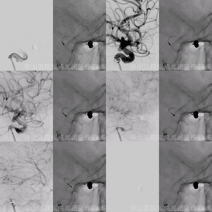
Figure 5 GIF. Continue to insert two coils (Prime helix 2mm*6cm and 1.5mm*2cm). The MAC aneurysm was packed densely by angiography. Retrieving the microcatheter and pushing the coil loop with the microcatheter.
图 5 GIF. 继续填入Prime helix 2mm*6cm和1.5mm*2cm两枚弹簧圈。复查造影证实大脑中动脉瘤致密栓塞。遂撤回微导管,撤回微导管时用微导管头端推压弹簧圈袢。
3
Step 2
Embolize the left Pcom aneurysm
栓塞左侧后交通动脉瘤
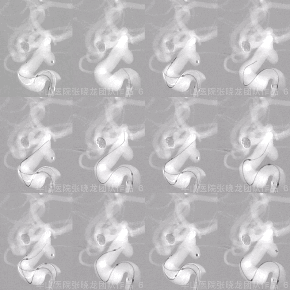
图 6 GIF. 将2根Echelon-10 45°微导管头端塑“C”弯后置于动脉瘤腔内。
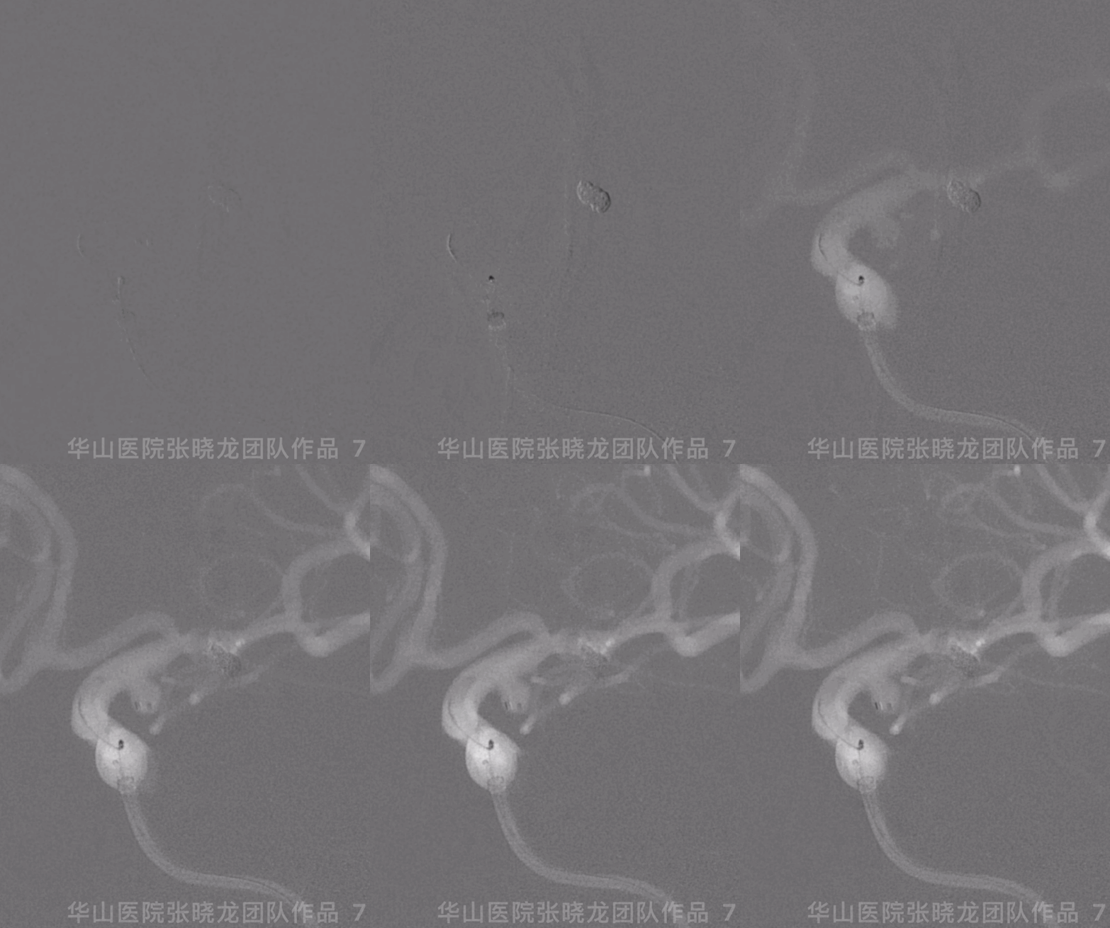
Figure 7 GIF. Changed to another working projection for aneurysm embolization.
AN size: 4*4.04mm
AN neck: 2.24mm
图 7 GIF. 更换工作角度栓塞动脉瘤。
动脉瘤大小:4*4.04mm.
动脉瘤颈:2.24mm
Video 2. Perdenser 4.5mm*12cm via the 1st microcatheter, undetached. Target-360 3mm*4cm via the 2nd microcatheter.
视频 2. 经过第一根微导管填入Perdenser 4.5mm*12cm弹簧圈后暂不解脱。然后经第二根微导管填入Target-360 3mm*4cm弹簧圈。
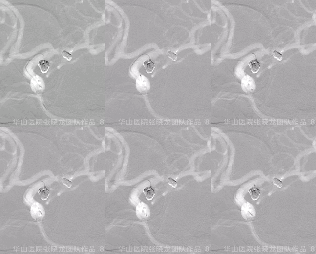
图 8 GIF. 继续填入Target-360 3mm*4cm和Target-360 1.5mm*3cm弹簧圈。
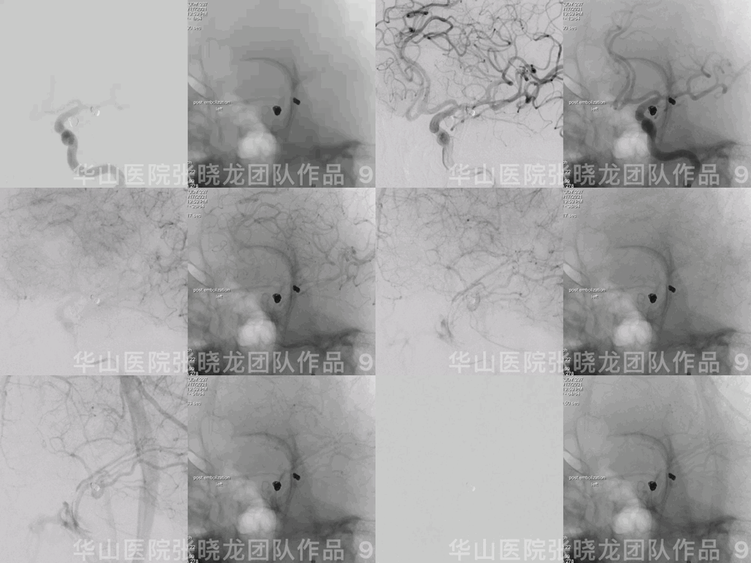
图 9 GIF.造影显示动脉瘤致密栓塞。
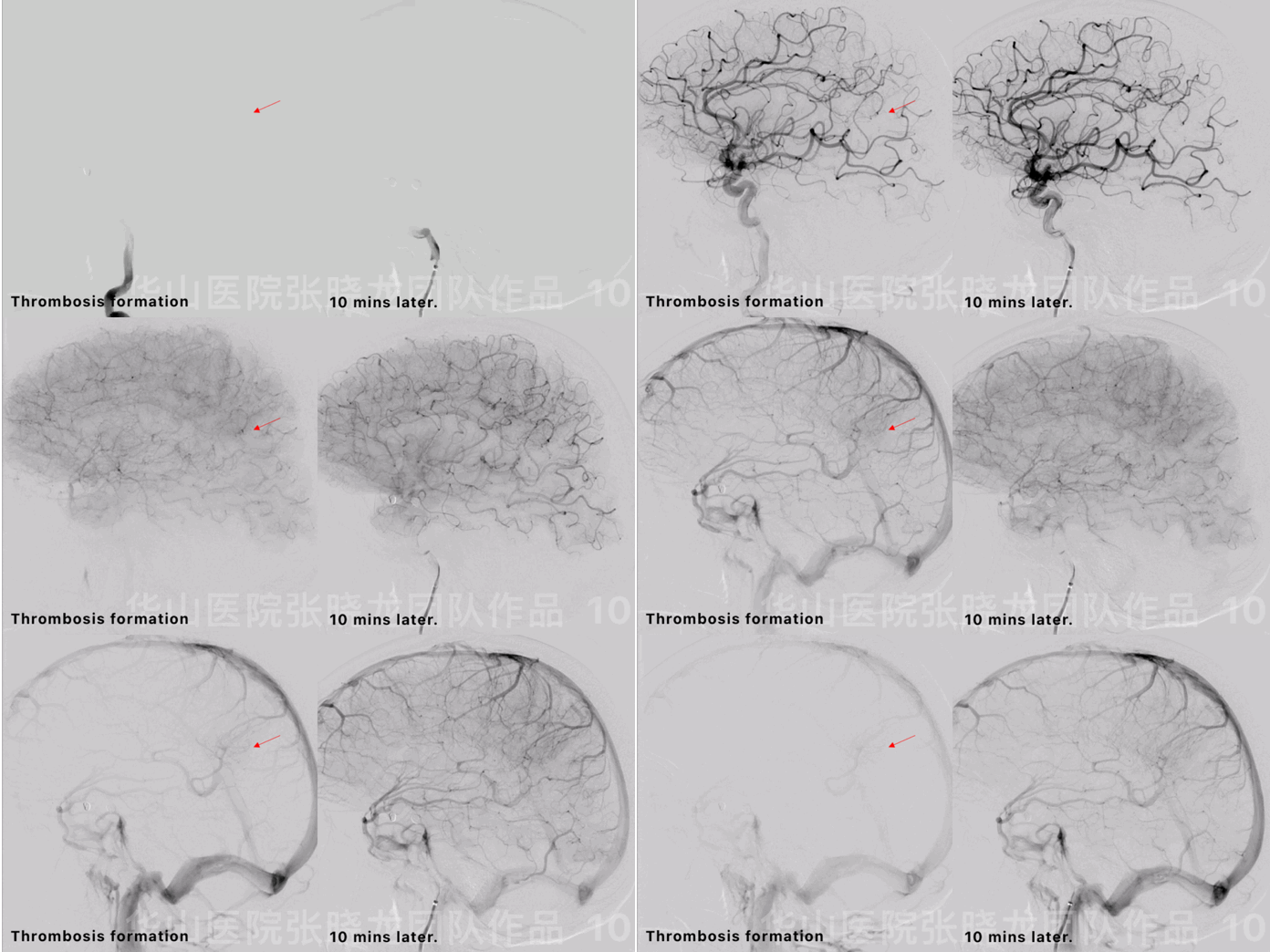
Figure 10 GIF. Thrombosis formed in a small left MCA branch. Tirofiban 15ml was administered and the thrombus resolved 10 minutes later.
图 10 GIF. 左侧大脑中动脉小分支内局部血栓形成。经导引导管注入替罗非班15ml,等待10min后复查造影血栓基本消失。
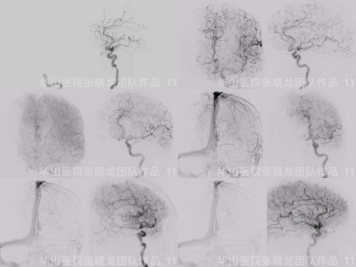
Figure 11 GIF. AP view and rotational DSA both confirmed aneurysms packed densely and intracranial artery intact.
图 11 GIF. 前后位及旋转造影均证实动脉瘤致密栓塞,颅内血管完好。
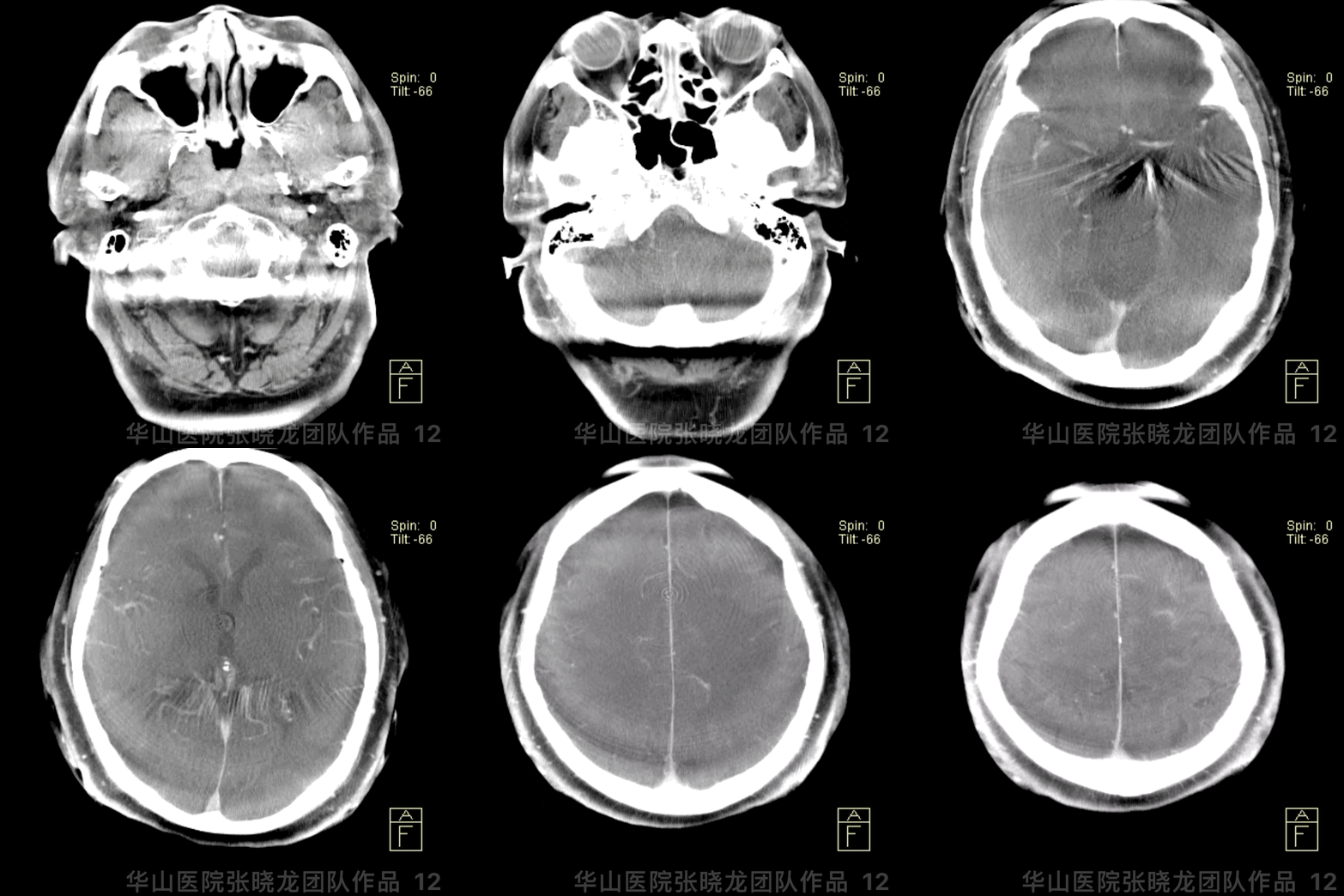
图 12 GIF. Dyna-CT未见出血。
4
Follow-up (8-month)
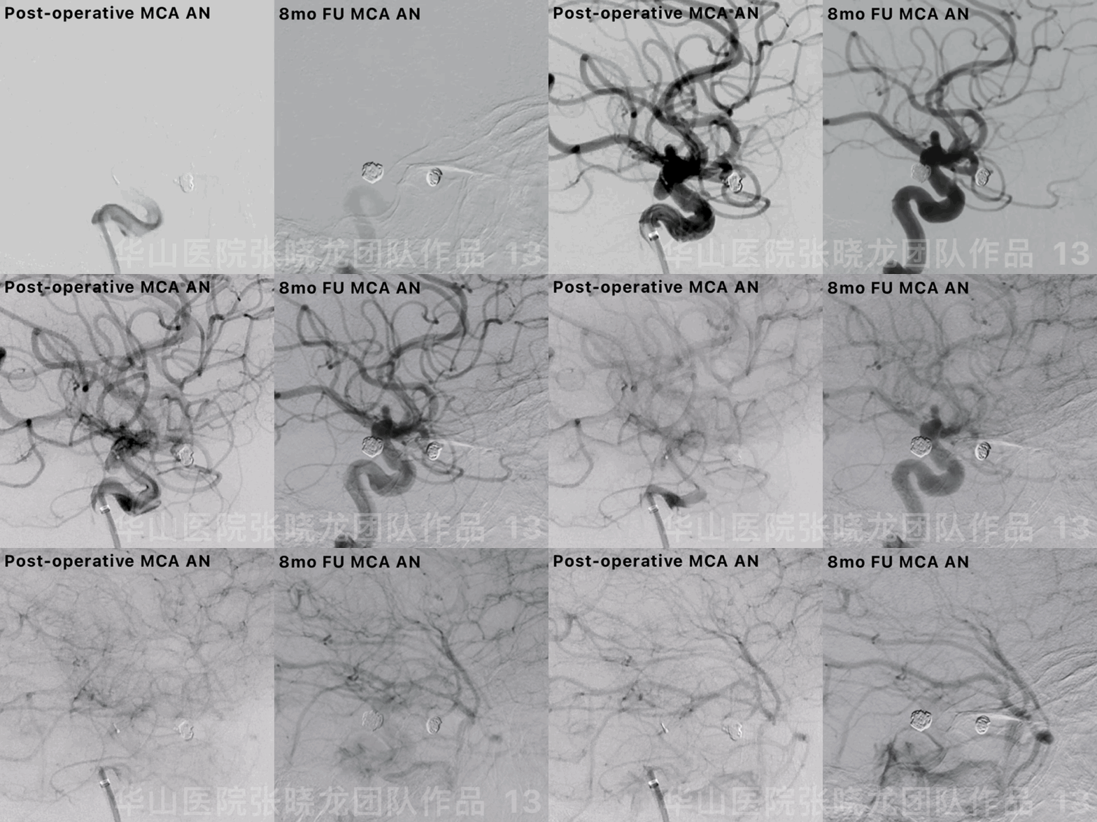
图 13 GIF. 8个月随访DSA提示左侧大脑中动脉瘤致密栓塞。
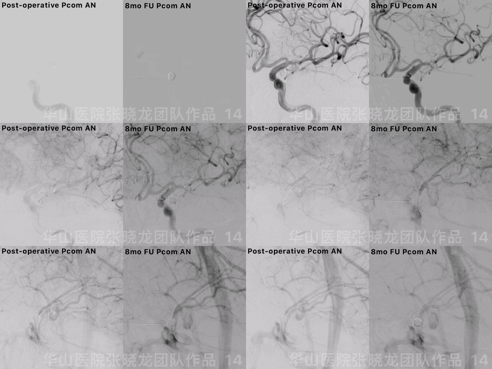
图 14 GIF. 8个月随访DSA提示左侧后交通动脉瘤致密栓塞。
Video 3. 8 month follow up rotational DSA showed the dense packing of aneurysms with parent artery patency.
视频 3. 8个月旋转DSA证实动脉瘤均致密栓塞,无残留,载瘤动脉通畅。

图 15. 颅内血管完好。
Summary
• Since both the Pcom and the irregular MCA aneurysms have a high rupture risk, endovascular treatment is performed.
• For the relatively narrow-necked PcomA aneurysm, simple coiling with dual microcatheter technique is preferred.
• We select more stiff and thicker perdenser coil to form a basket frame for the Pcom AN and that may decrease the recurrence rate.
• For the MCA aneurysm, the two sacs were densely packed. The first large coil was selected because of the stable microcatheter. Long framing coil with a “8” configuration in the two sacs is stable which can decrease the recurrence rate.
Three working projections were selected:
Working projection 1 for crossing the microcatheter and microwire and can avoid puncturing the proximal aneurysm.
Working projection 2 to show clearly the M1 and aneurysm.
Working projection 3 to show the aneurysm neck and the two sacs.
• Tonbridge guiding catheter tip is softer than Navien which is suitable for tortuous route. The guiding catheter was pushed to conform the proximal tortuous vessel to prevent vasospasm or thrombus.
• The case need long term follow up.
• 后交通动脉瘤及不规则大脑中动脉瘤破裂风险高,建议治疗。
• 后交通窄颈动脉瘤,首选双微导管单纯弹簧圈栓塞技术。
• 后交通动脉瘤使用更粗更硬的perdenser弹簧圈成框,可能会降低复发率。
大脑中动脉瘤采用单纯栓塞,2个瘤腔均需致密栓塞。由于微导管稳定,第一枚弹簧圈可以选用尺寸相对更大、更长的。弹簧圈在2个瘤腔内“8”字成篮,这种成篮更稳定,动脉瘤复发风险低。
栓塞大脑中动脉瘤时选用3个不同的工作角度:
工作角度1用来通过微导管及微导丝,防止刺破动脉瘤。
工作角度2用来显示清楚M1段及动脉瘤。
工作角度3用来显示动脉瘤颈及2个子瘤。
• 通桥导引导管比Navien更软,所以更适合这种迂曲的通路。向前推动导引导管,顺应性良好,防止血管痉挛及血栓形成。
• 该病例仍需要长期随访。


