本文为Rhoton解剖视频中《Fiber Pathways of the Cerebrum》这一章节,主要讲解了大脑联络纤维、投射纤维、联合纤维、基底节、额叶、颞叶、边缘系统等传导通路,由Kaan Yagmurlu教授讲解。共截取270张图片。
笔者水平所限,错误之处请批评指正!


大脑纤维通路
Fiber Pathways of the Cerebrum
▼大脑由灰质和白质构成。灰质(也称为皮层),是大脑的外层结构,可根据表面解剖予以划分。
从功能性来说,额叶(下图)为意识和思维中枢,位于中央沟前方。
The cerebrum has grey and white matter. The grey matter, which is outer layer of the cerebrum, also called as the cortex, is classified according to surface anatomy.Basically, the frontal lobe is located in front of the central sulcus. Functionally, the frontal lobe is the conscious and thought center,
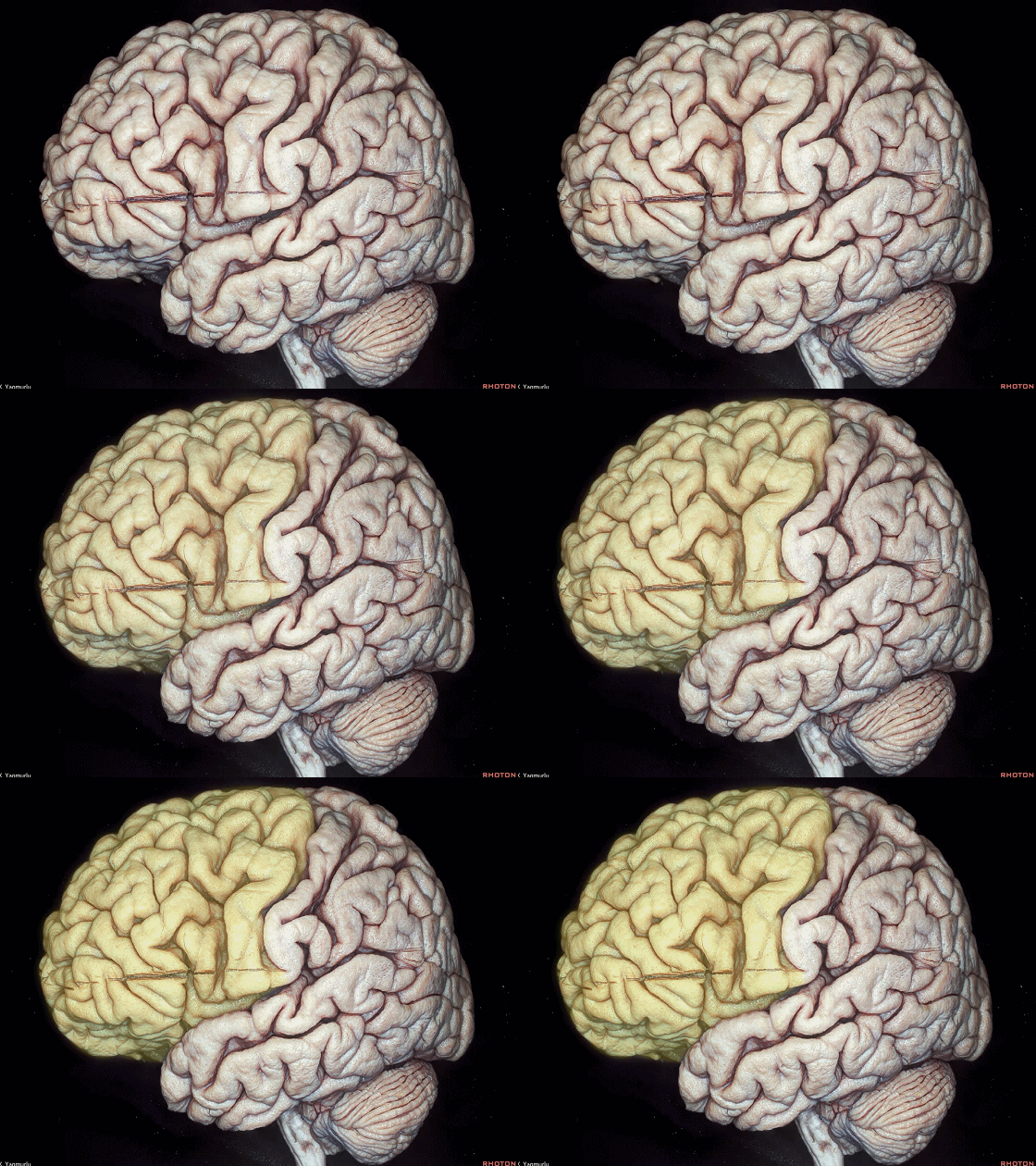
▼顶叶为定位中枢,位于额叶与枕叶之间。
The parietal lobe is the navigation center,which is located between the frontal and occipital lobes.
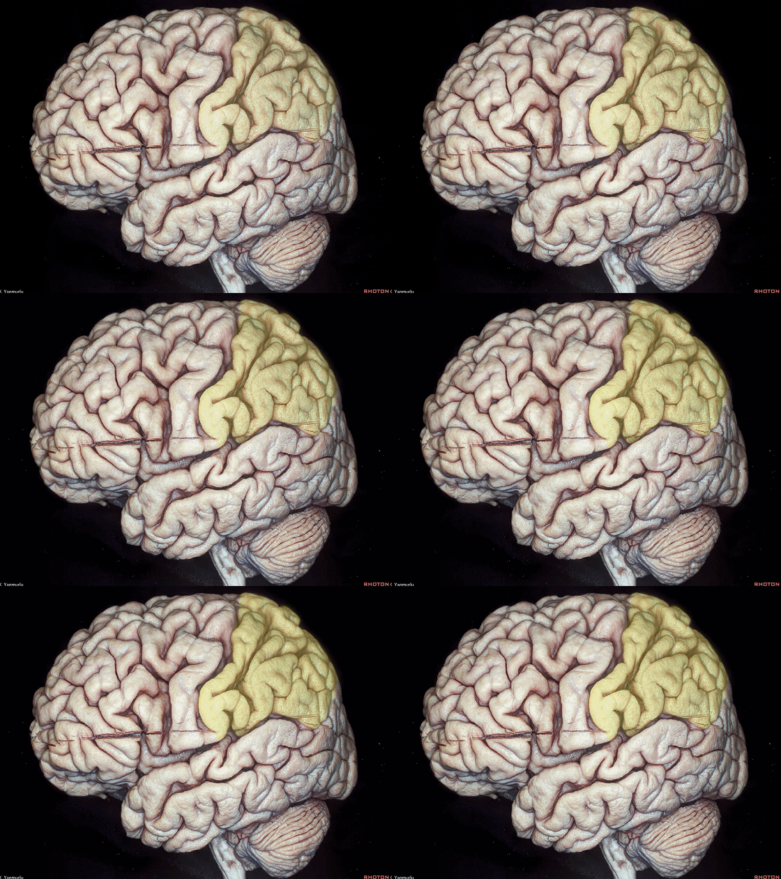
▼枕叶为视觉中枢,位于 枕前切迹 与顶枕沟上端 连线后方。
The occipital lobe is the vision center. The occipital lobe is located behind an imaginary line drawn between the preoccipital notch and the top of the parieto-occipital sulcus.
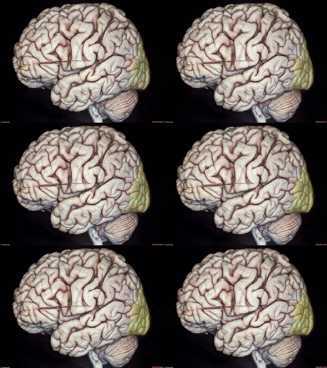
▼颞叶是记忆和听觉中枢,居于大脑的下部。
The temporal lobe is the inferior part of the cerebrum.the temporal lobe is the memory and sound center of the cerebrum.
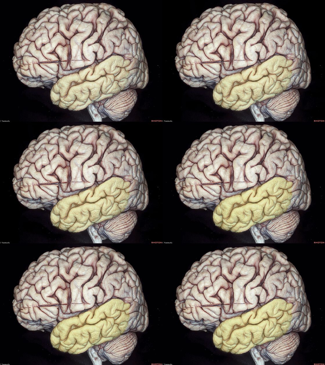

▼大脑的白质,介于皮层灰质和基底节之间,包含三种纤维:联络纤维、投射纤维、联合纤维。
联络纤维(下图)连接同侧大脑半球内的不同皮层区域。
The white matter of the cerebrum, which underlies the grey matter, and intervenes between cortical grey matter and the basal ganglia, contains three types of fibers.The association fibers connect the different regions in the same hemisphere.
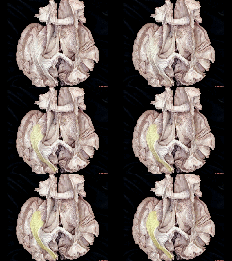
▼投射纤维,例如放射冠(下图),连接皮层至脑的尾端和脊髓。
The projection fibers, such as the corona radiata, connect the cortex with caudal parts of the brain and spinal cord.
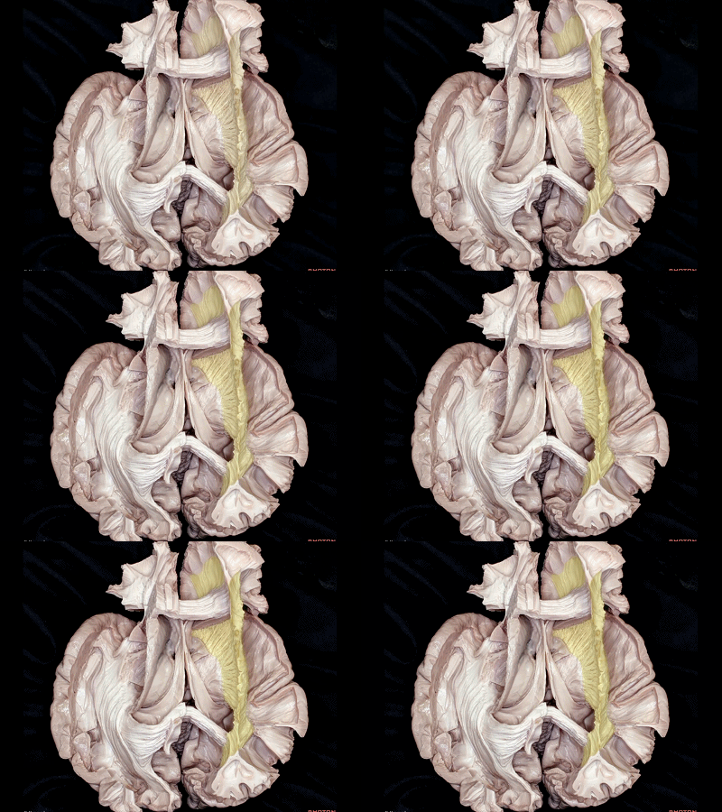
▼联合纤维,例如胼胝体(下图),跨过中线连接两侧大脑半球。
The commissural fibers, such as the corpus callosum, connect the two hemispheres across the median plane.
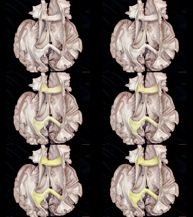
▼首先来看联络纤维,其分为 短联络纤维 和 长联络纤维。
短纤维 又称为 U状纤维,位于浅部,恰位于皮层灰质的深面。长纤维位于较深处。
弓状束(下图)为额顶颞长联络纤维。
Let's start from the association fibers, which has the short and long types. The short fibers, also called as U fibers, are located superficially, just underlying the cortical grey matter. And the long fibers are located a deeper position. The arcuate fasciculus is the frontoparietotemporal long association fibers
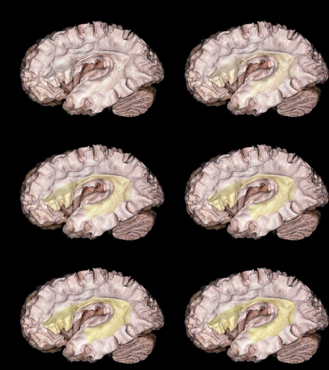
▼弓状束 连接 Wernicke区 至 Broca区。
Wernicke区(下图)包括 颞上回 和 颞中回 的中后部
The arcuate fasciculus that connects Wernicke's area, to Broca's area which consists of the middle and posterior parts of the superior and middle temporal gyri,
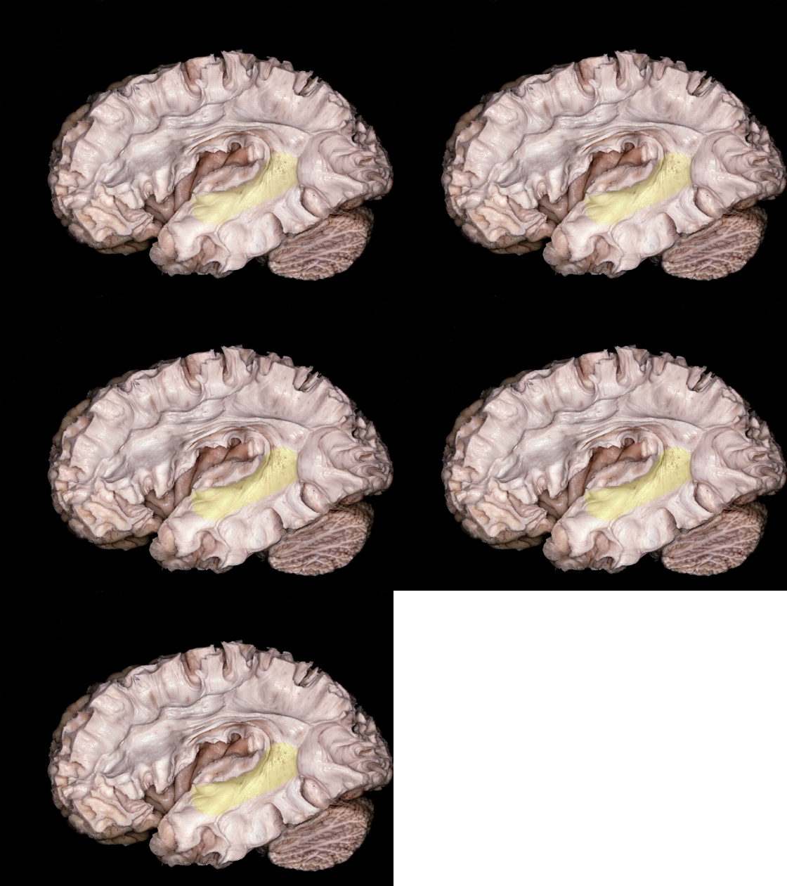
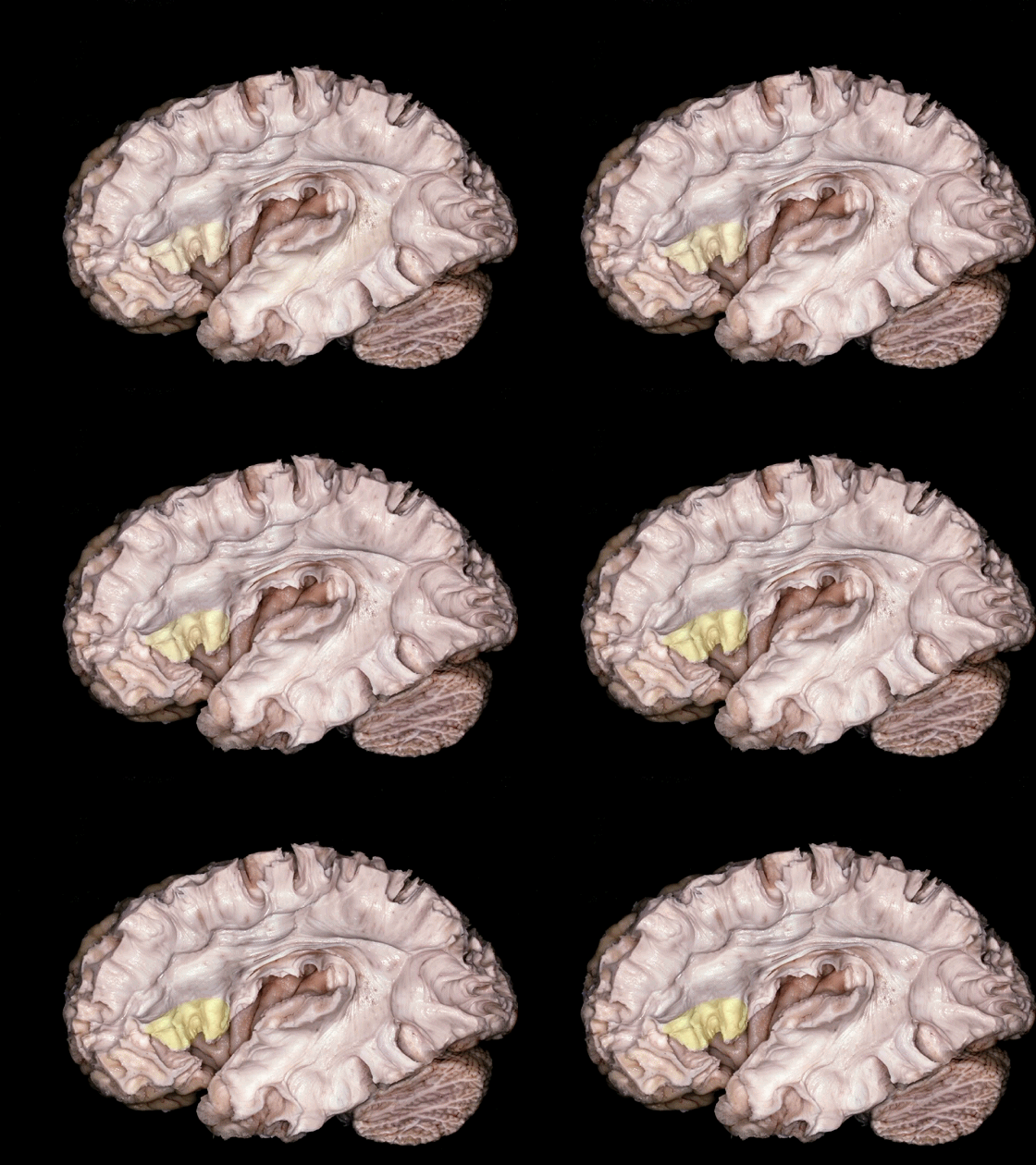
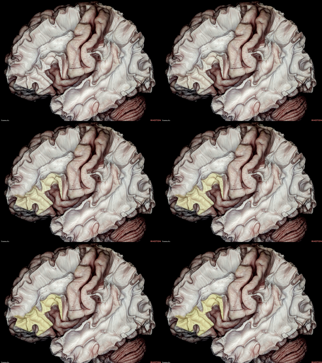
▼Broca区(下图)位于 额下回 的 后三分之二。
Broca's area located in the posterior two thirds of the inferior frontal gyrus.
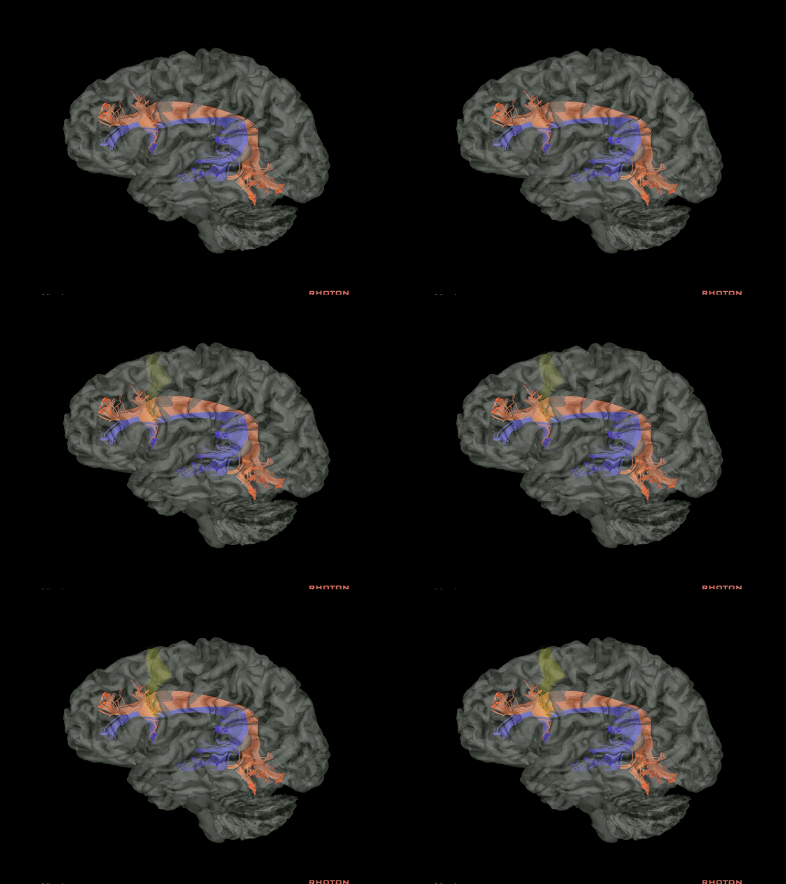
▼DTI成像 表明 弓状束 终于中央前回(下图) 以及Broca区
This DTI demonstrates that the arcuate fasciculus terminates in the precentral gyrus as well as Broca's area
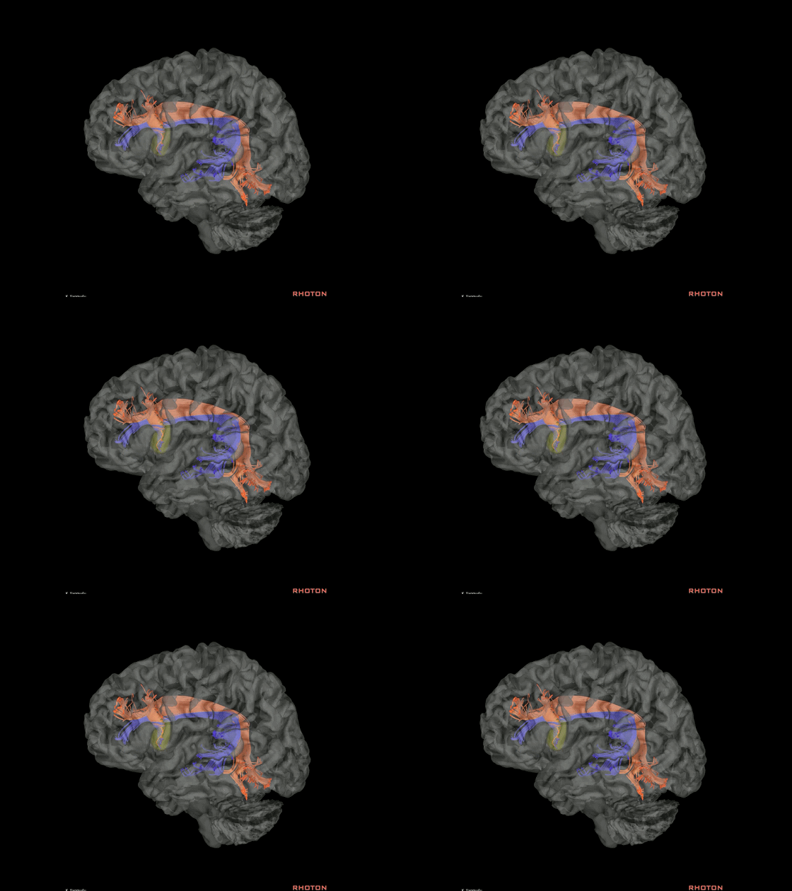
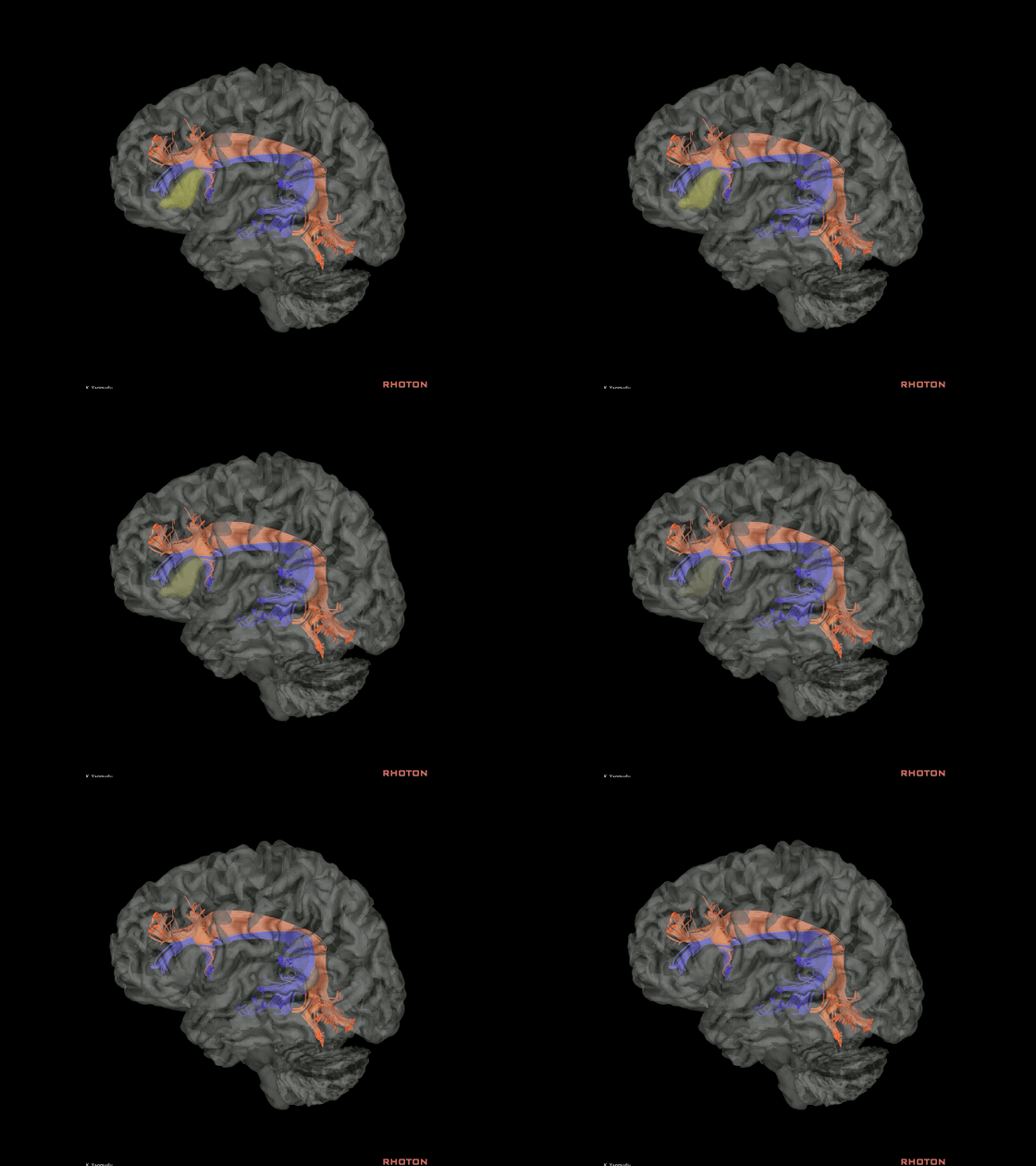
▼Broca区由额下回的 盖部 和 三角部 构成。
Broca's area, which is composed of the pars opercularis and triangularis in the inferior frontal gyrus.
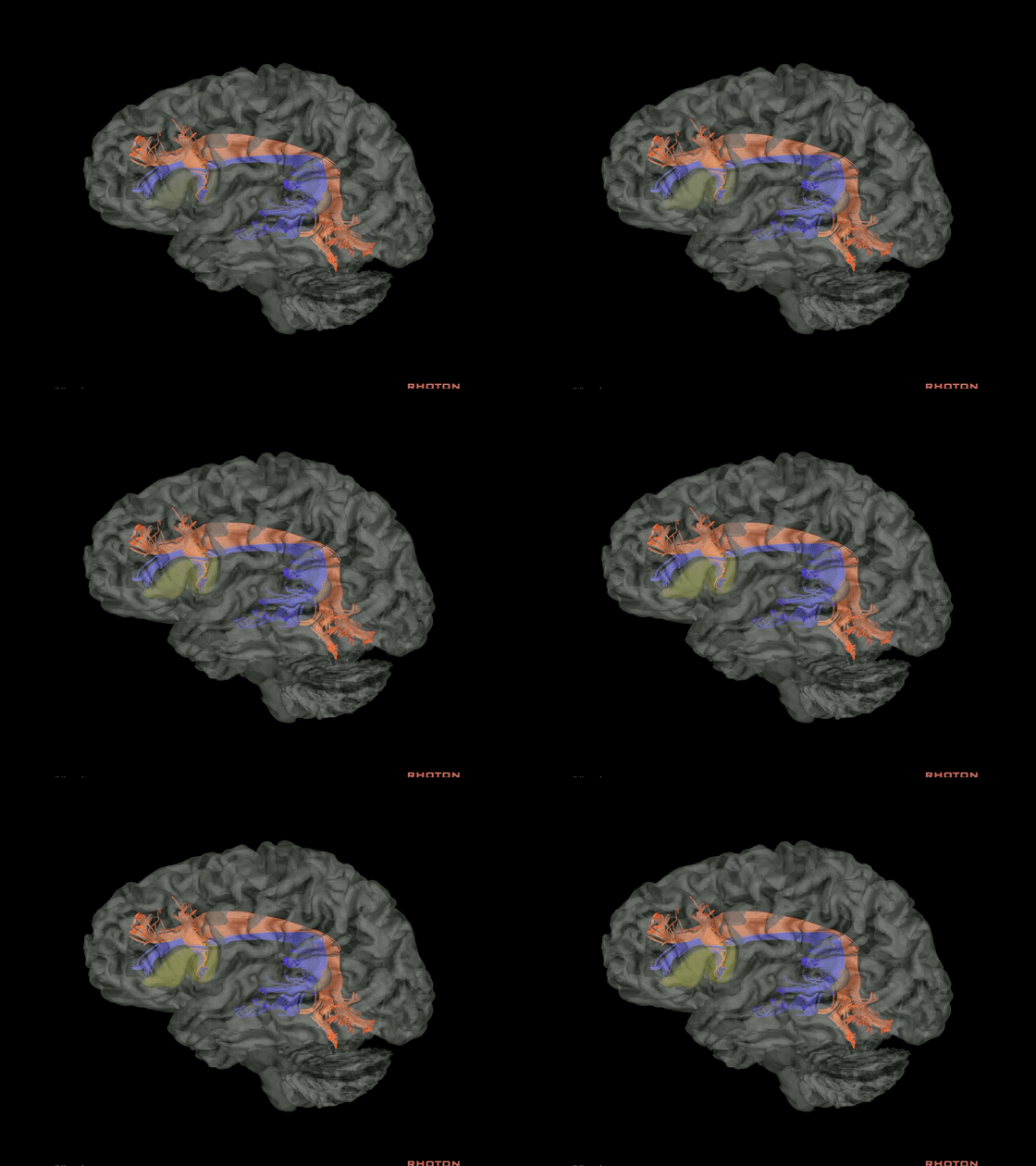
▼这是 盖部
pars opercularis
▼这是 三角部
triangularis
▼弓状束进一步分为 腹侧部(蓝色部分) 和 背侧部(橙色部分)。
The arcuate fasciculus is subdivided into a ventral part, which is blue one, and a dorsal part, which is orange one

▼在外侧裂上区,主要的长联络纤维是 上纵束
其包含三个部分:上纵束 I(背侧通路),上纵束 II(中间通路),上纵束 III(腹侧通路)。
In the suprasylvian area, the main long association fiber pathway is the superior longitudinal fasciculus (SLF), which has three parts: SLF I (dorsal pathway), SLF II (middle pathway), and SLF III (ventral pathway).
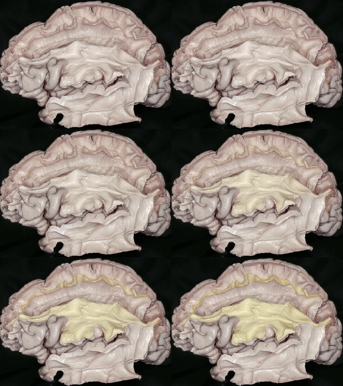
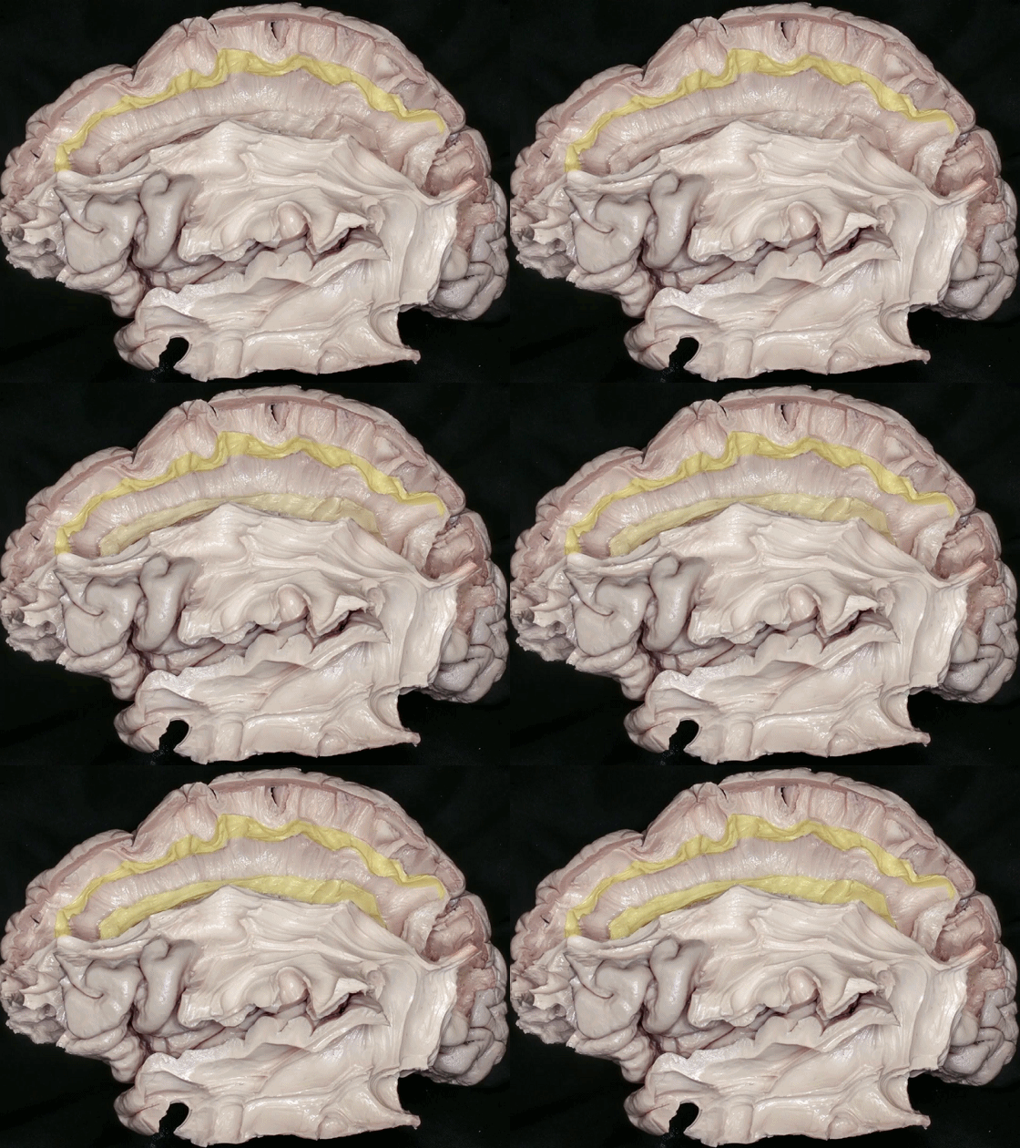
▼这是上纵束 I ,位于额上回内
The SLF I is positioned within the superior frontal gyrus,
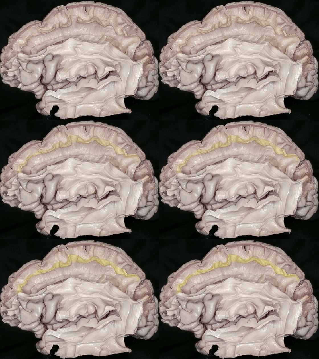
▼上纵束 I 位于扣带沟上岸内,行于扣带(下图)上方,连接顶叶上部与扣带回前部。
The SLF I lies within the superior bank of the cingulate sulcus and travels just above the cingulum to connect the superior parietal lobe and anterior cingulate cortex.
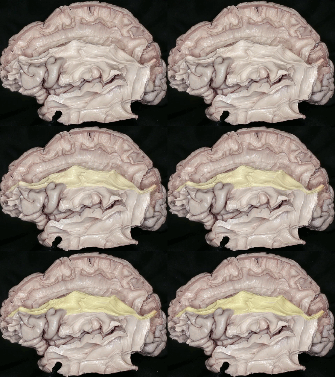
▼这是上纵束 II ,位于额中回内。
上纵束 II 连接 顶叶下部 与 额中回。
the SLF II within the middle frontal gyrus, The SLF II extends from the inferior parietal lobe to the middle frontal gyrus.
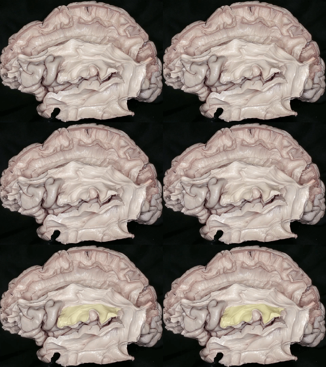
▼这是上纵束 III, 位于额下回内。
上纵束 III 行于额顶叶盖部,连接 顶叶下部 与 额下回。
and the SLF III within the inferior frontal gyrus. The SLF III courses within the frontoparietal operculum and extends from the inferior parietal lobe to the inferior frontal gyrus.
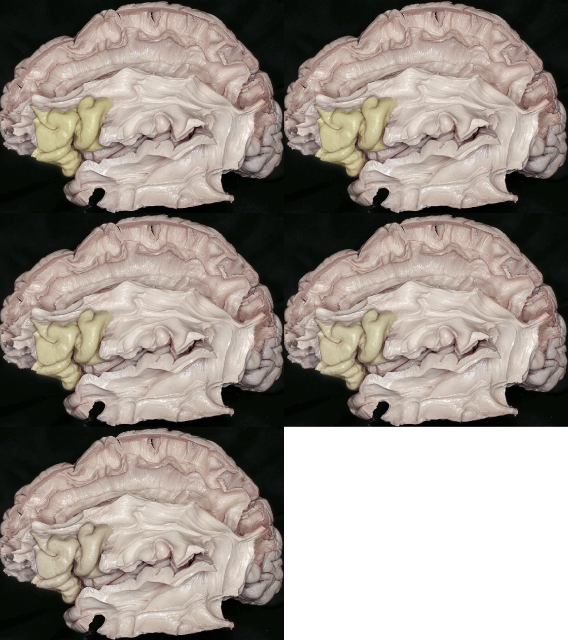
▼这是 额下回
inferior frontal gyrus
▼上纵束 被认为是一套 高级多感觉性联络系统。
The SLF is considered as a high order multisensory associative system.

▼此DTI图像显示了上纵束和弓状束的各组分。
上纵束 I(蓝绿色)为背侧通路,
上纵束 II(酒红色)为中间通路,
上纵束 III(黄色)为外侧裂上区的腹侧通路。
弓状束 分为 腹侧部(蓝色)和 背侧部(橙色)。
The compartments of the superior longitudinal fasciculus (SLF) and arcuate fasciculus (AF) can be seen on this DTI. SLF I (turquoise) is the dorsal pathway, SLF II (claret red) is the middle pathway, and SLF III (yellow) is the ventral pathway in the suprasylvian area. The ventral (blue) and dorsal (orange) segments of the arcuate fasciculus can be seen.

▼其他的长联络纤维通路有 下额枕束(下图),连接额叶和枕叶。损伤优势半球的下额枕束将引起语义性语言错乱。
Other long association fiber pathways are inferior fronto-occipital fasciculus, which is a fronto-occipital connection;
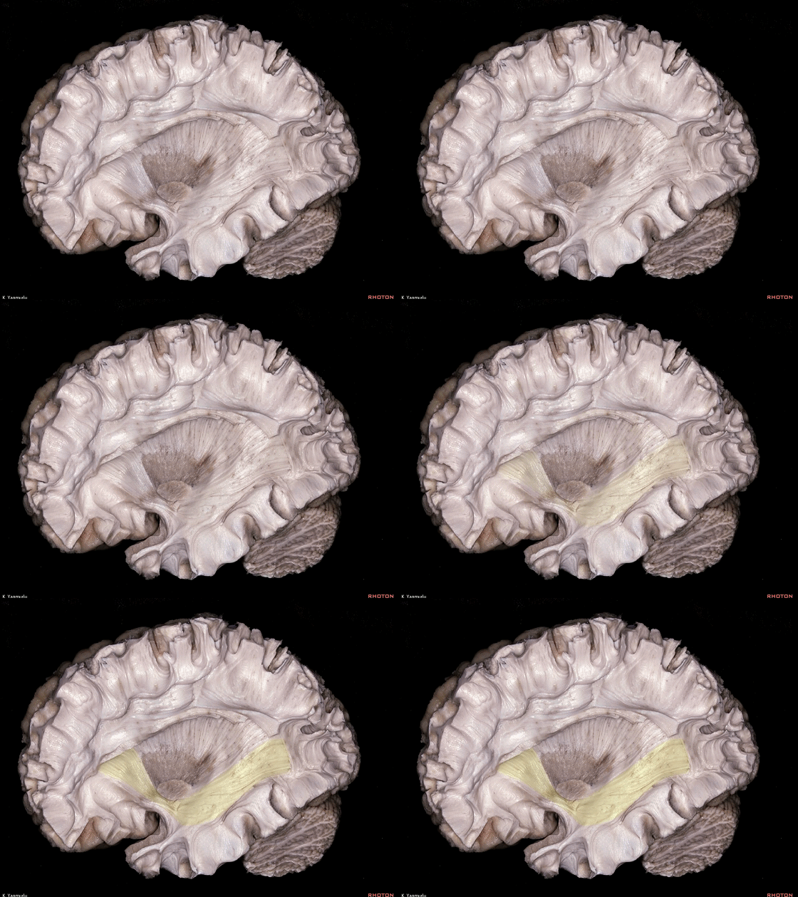
▼这是 下纵束,连接颞叶和枕叶
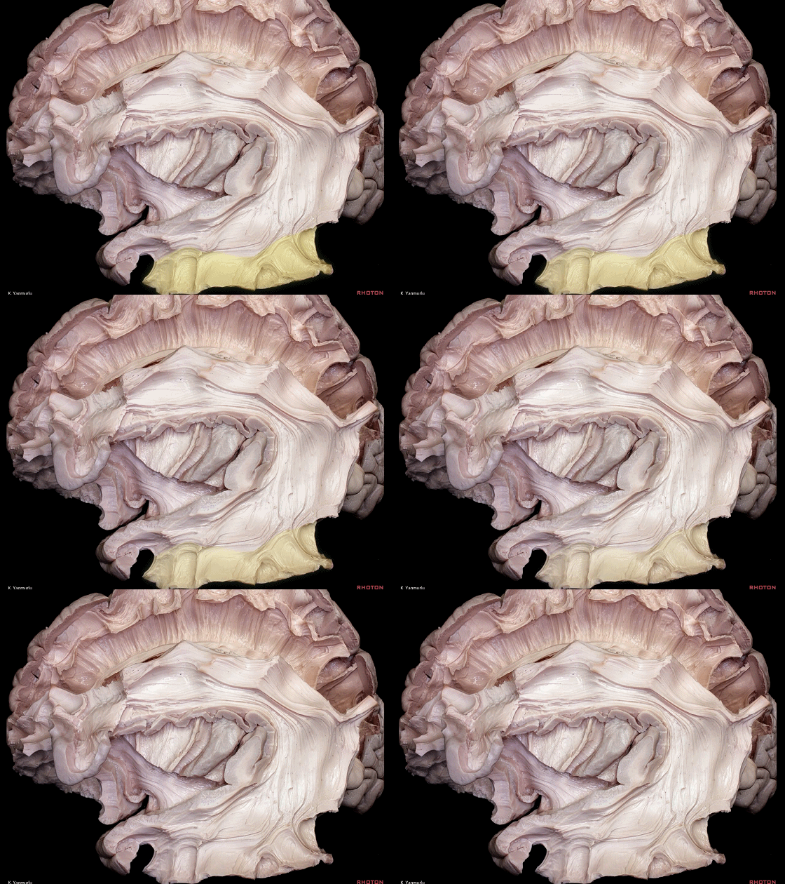
the inferior longitudinal fasciculus, which is a temporo-occipital connection;
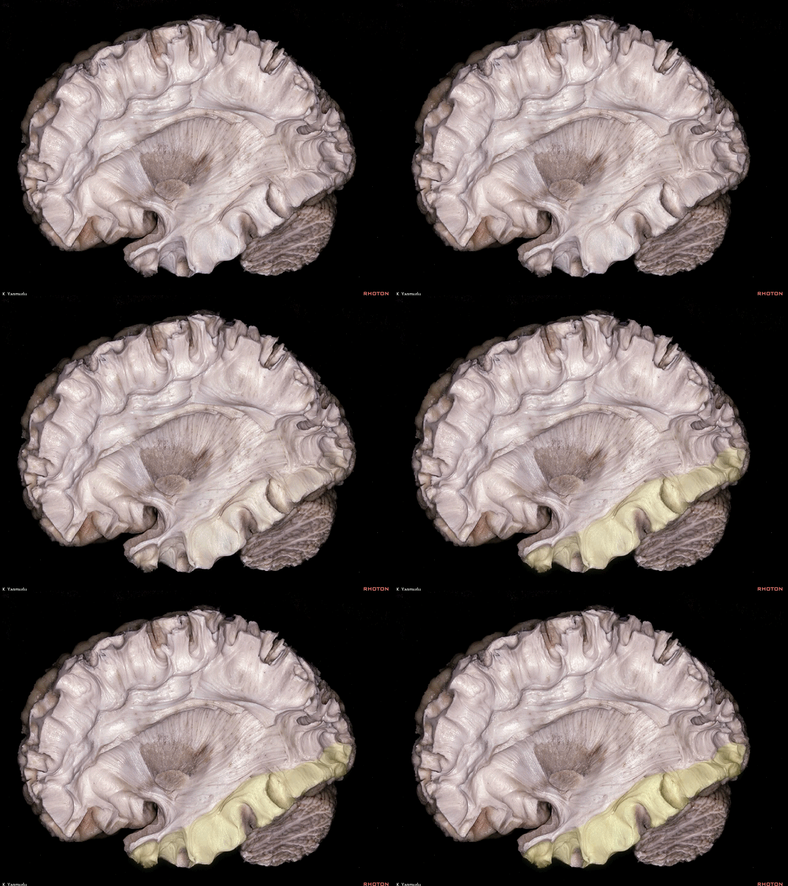
▼这是钩束,连接额叶和颞叶。
the uncinate fasciculus, which is a frontotemporal connection.
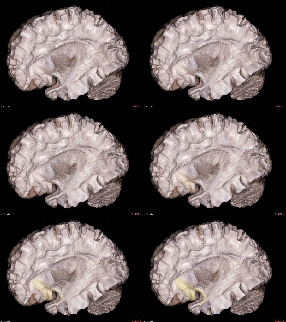
▼这是上面观。
钩束(下图)的功能在于联系情感与认知而形成 腹侧边缘通路,一些研究表明,钩束还参与了语言的语义处理、听觉记忆和声音认知。
We are looking from above. The function of the uncinate fasciculus is to link emotion and cognition as a ventral limbic pathway,Additionally, some research shows that the uncinate fasciculus is involved in semantic processing of language, auditory working memory and sound recognition.
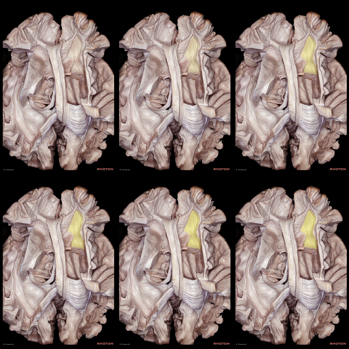
▼与钩束相对的是扣带,即 背侧边缘通路。
in contrast to the cingulum or dorsal limbic pathway.
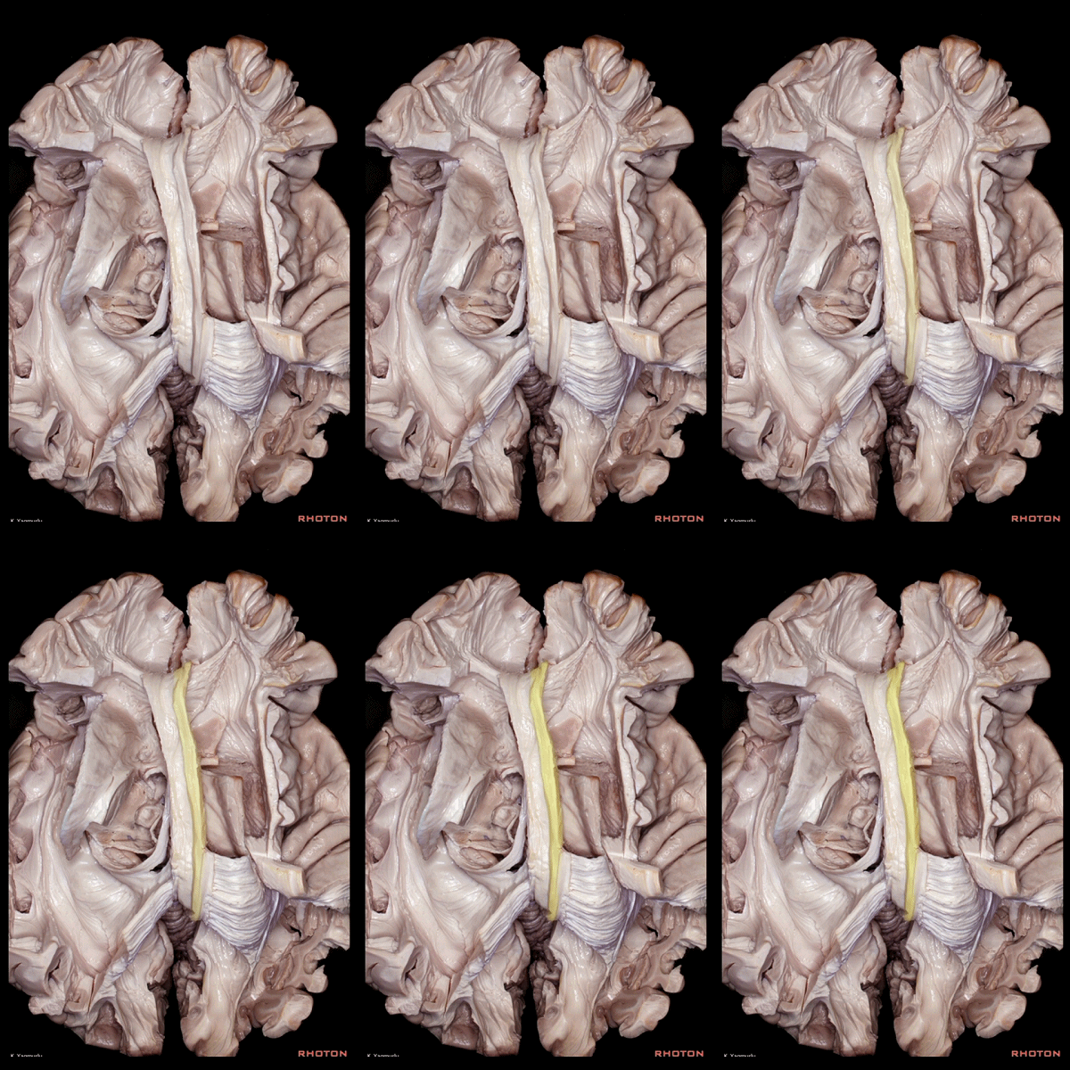
▼语言的处理过程包含一套 腹侧语义通路 和一套 背侧语音通路。
下图示 腹侧语义通路。
Language processing has a ventral semantic stream and a dorsal phonological stream
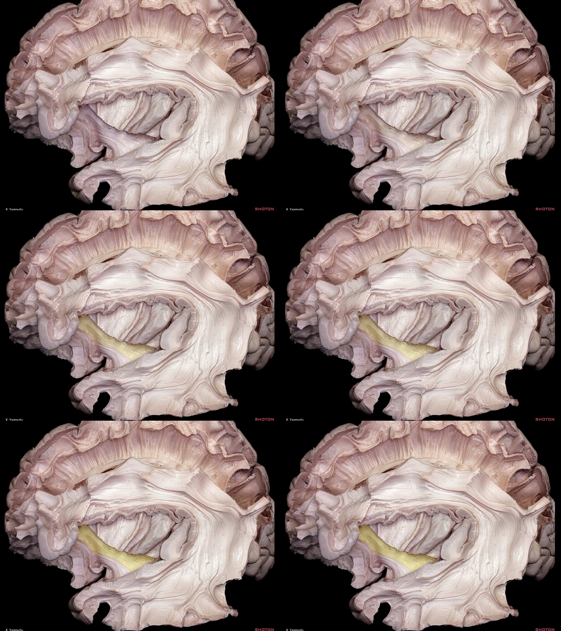
▼背侧语音通路(下图)连接Broca区和Wernicke区。
dorsal phonological stream that connect Broca's area and Wernicke's area.
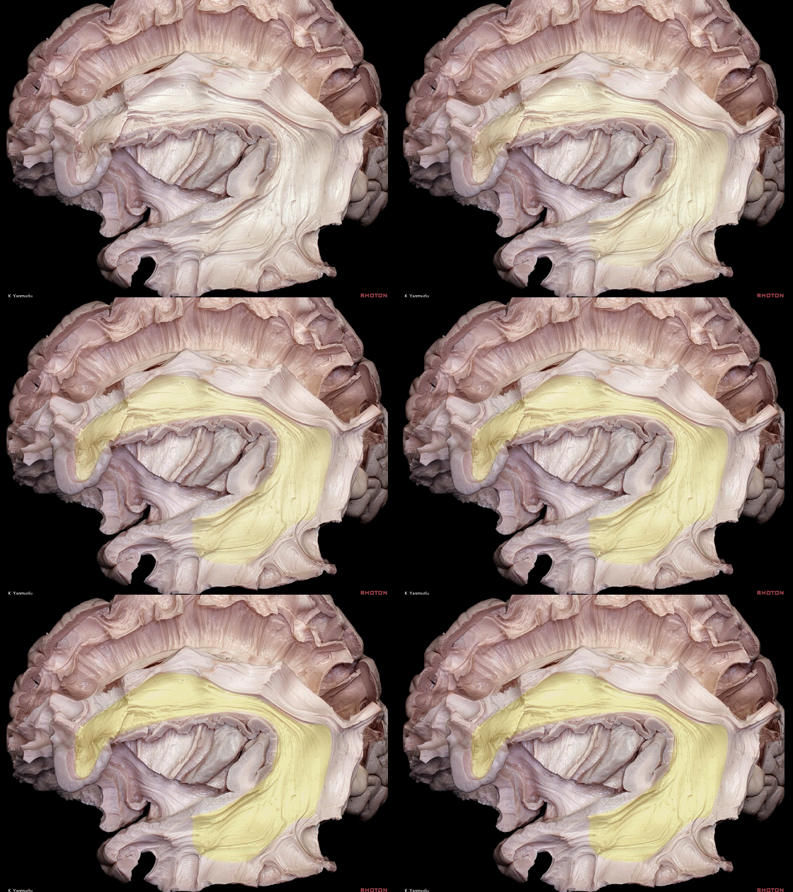
▼这是 Broca区
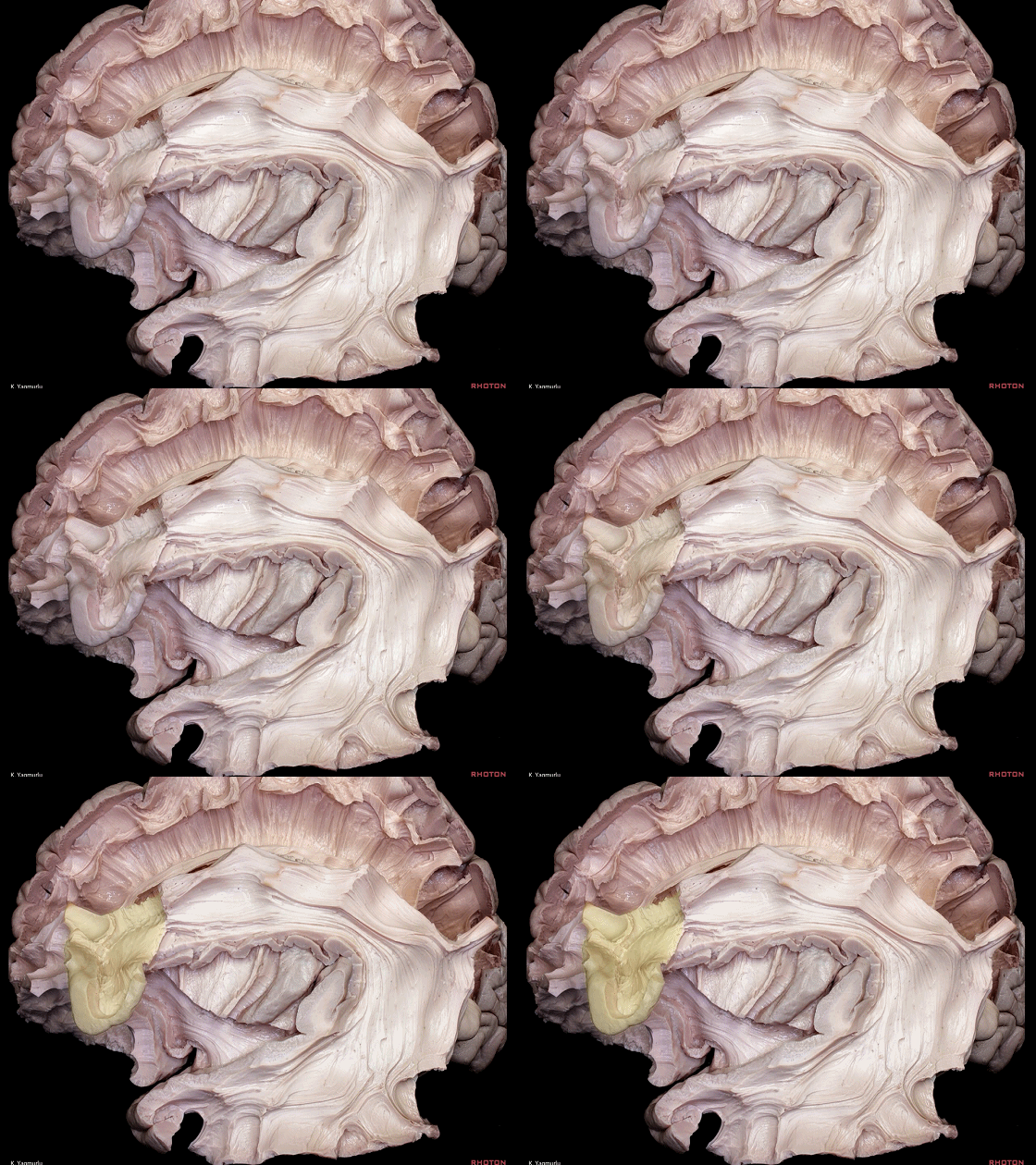
▼这是 Wernicke区
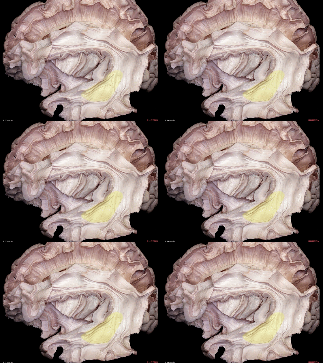
▼在背侧通路(下图)中,额顶颞区负责将发声整合成语音。其中的主要通路即为弓状束。
Within the dorsal stream, frontoparietotemporal regions are proposed as being involved in mapping auditory speech sounds to articulatory representations. The major pathway is the arcuate fasciculus.
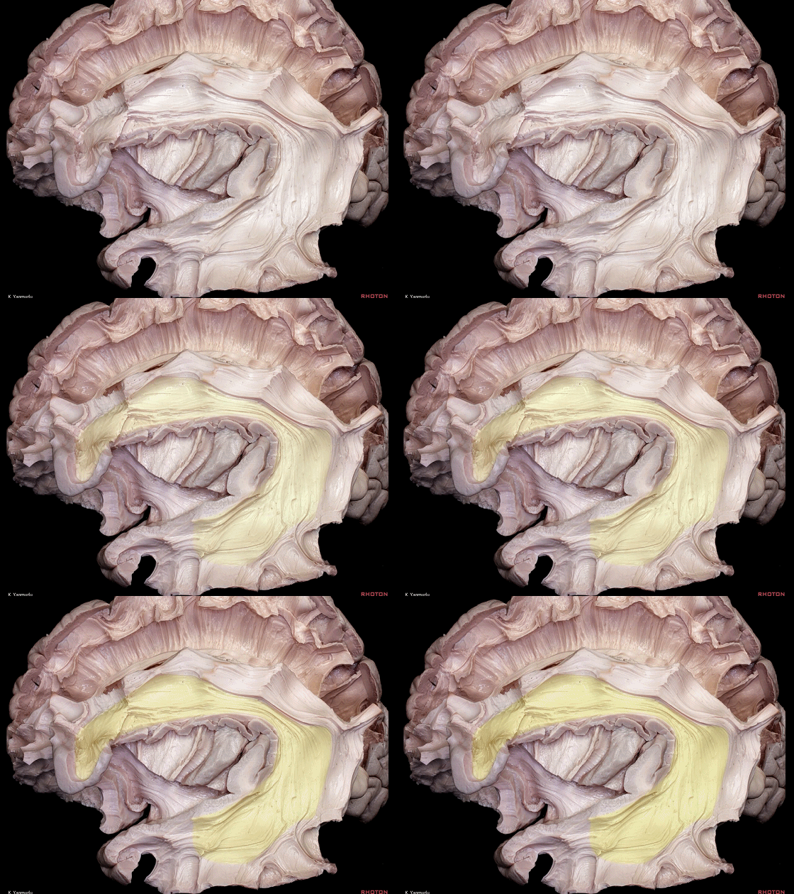
▼相对地,腹侧通路(下图)则赋予发声具体的语义。
腹侧通路中的主要通路为 下额枕束(见上)。损伤优势半球的下额枕束将引起语义性语言错乱。
In contrast, the ventral stream is proposed to be involved in mapping auditory speech sounds to meaning. The major fiber pathway in the ventral stream is the inferior fronto-occipital fasciculus.Damage to the inferior fronto-occipital fasciculus produces the semantic paraphasia in the dominant hemisphere.
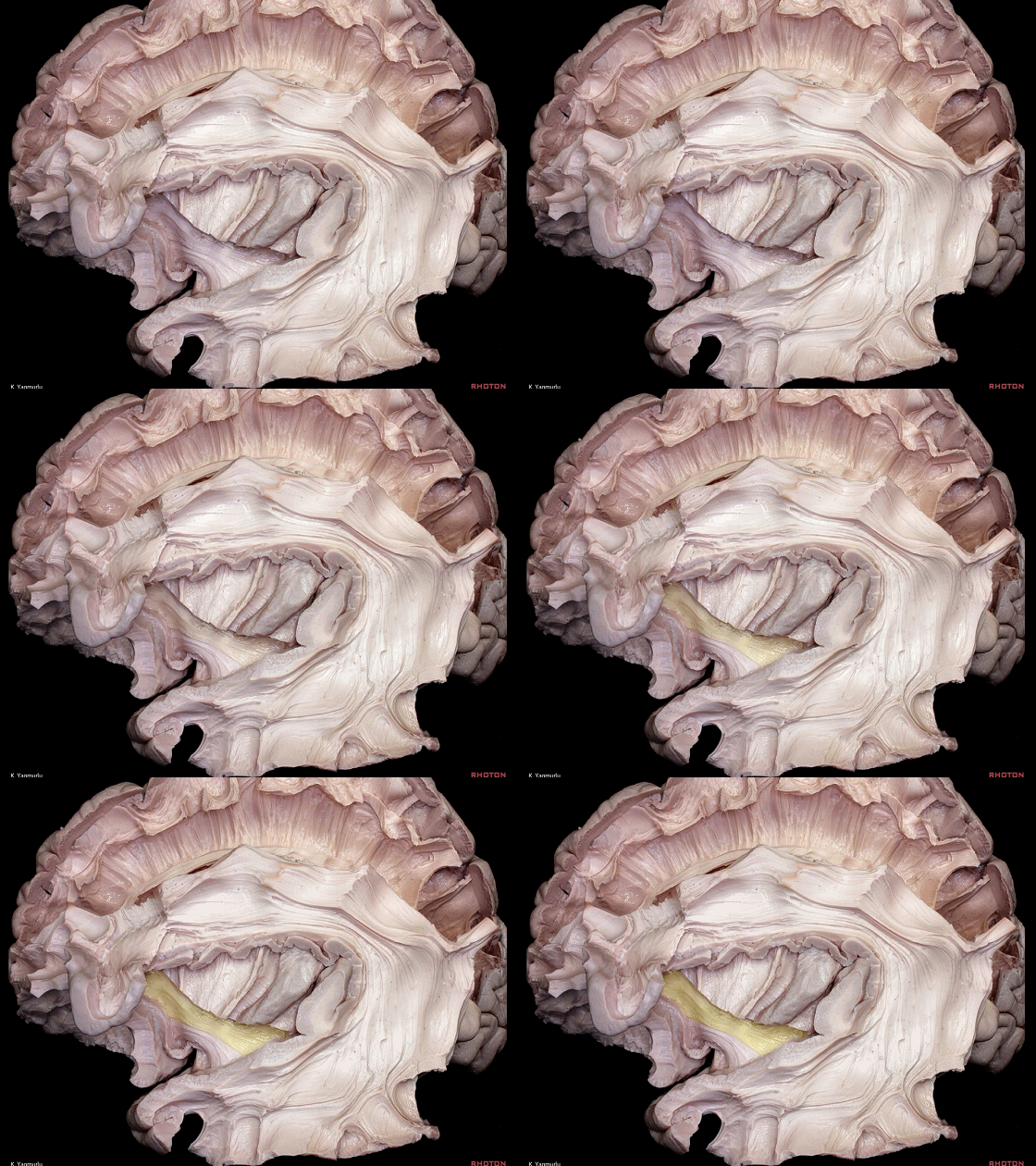
▼其他还包括外囊(下图)、钩束、下纵束、中纵束。
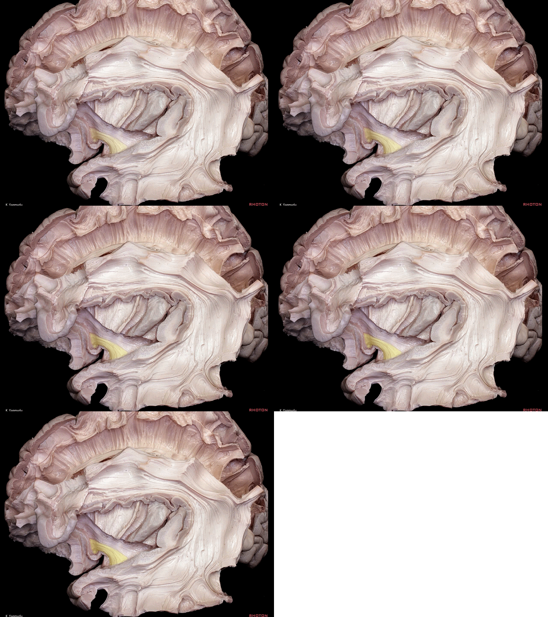
Other fiber pathways are the extreme capsule,uncinate fasciculus,inferior longitudinal fasciculus, and middle longitudinal fasciculus.
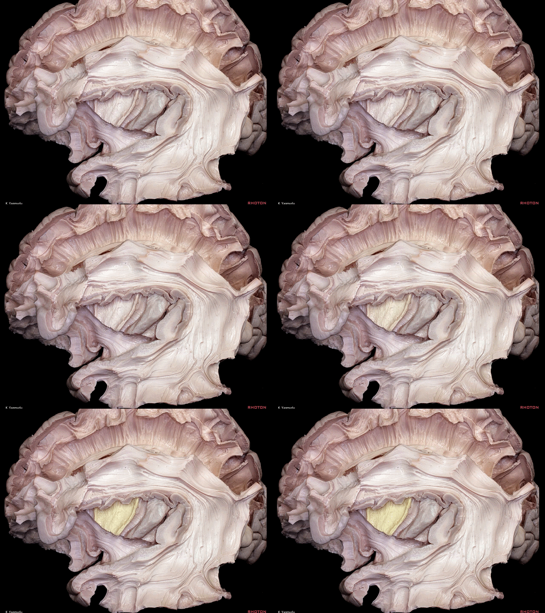
▼这是 钩束
▼这是 下纵束

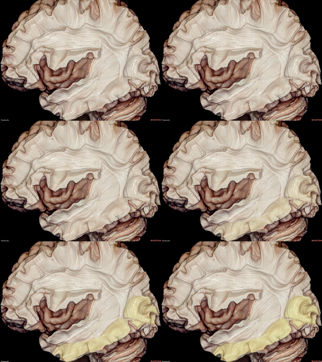
▼视觉的处理过程与语言相似,也存在两套通路。视觉信息沿此两套通路从枕叶发出。
腹侧通路,即所谓的“定性”通路,连接至颞叶,由下纵束(下图)传导,负责视物的分辨和认知。
The ventral stream, which is "what" pathway, Visual processing has two pathways like language processing. Visual information follows two main streams from the occipital lobe.travels to the temporal lobe, by means of the inferior longitudinal fasciculus, and relates to object identification and recognition.
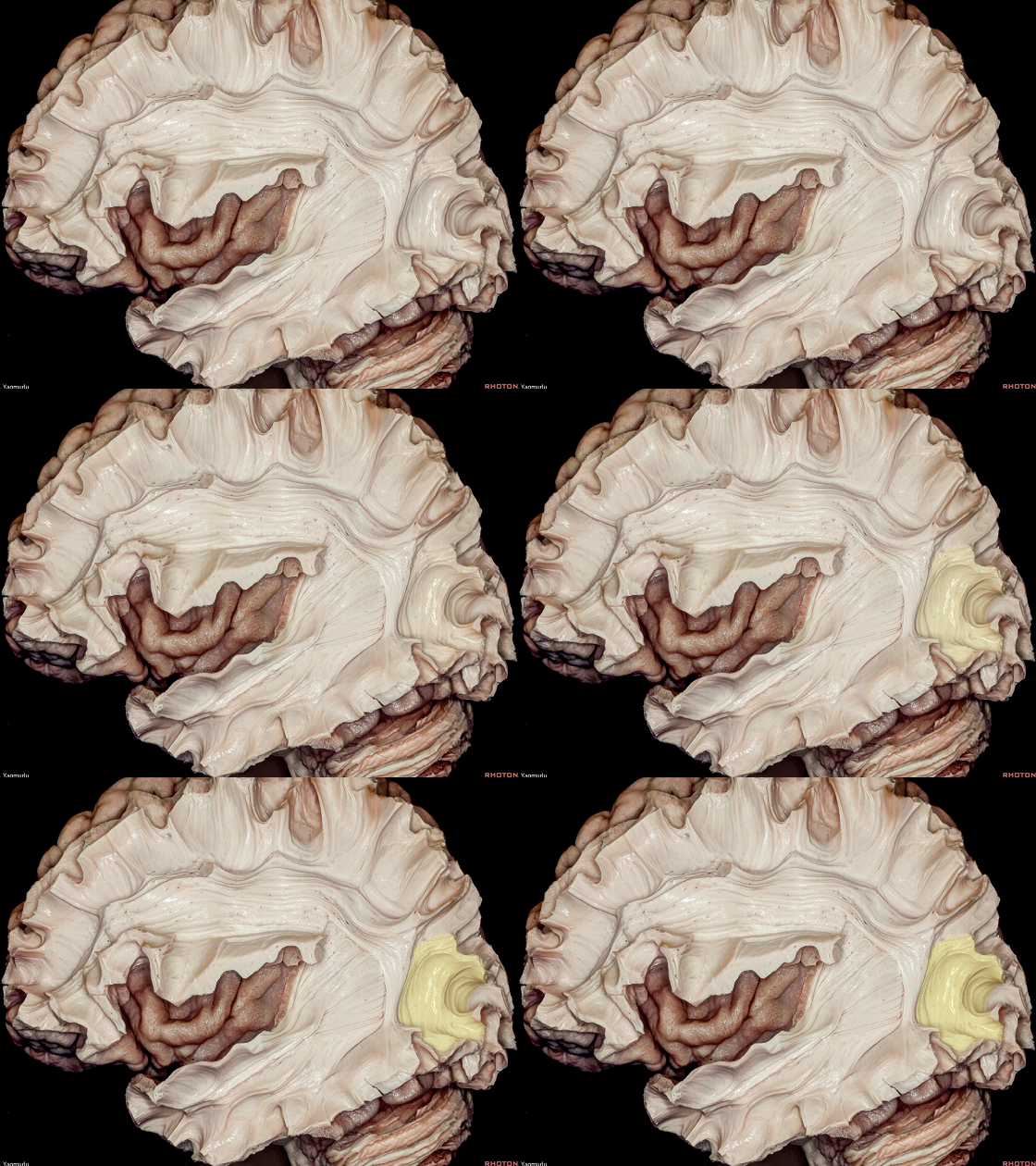
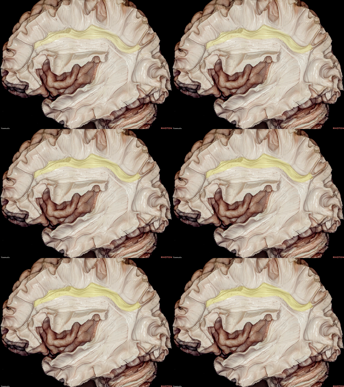
▼背侧通路,即所谓的“定位”通路,连接至顶叶,后者堪称大脑的导航中心,由枕短联络纤维(下图)和该部分上纵束 II 传导,负责视物的空间定位。
The dorsal stream, which is "where" pathway, travels to the parietal lobe, which is the navigation center of the cerebrum,by means of the occipital short association fibers and the part of the SLF II, and relates to processing location of object in space.
▼下图示上纵束 II

▼下面介绍联合纤维
胼胝体(下图)为主要的半球间联合纤维通路,连接绝大部分新皮层区域。
The corpus callosum is the major interhemispheric commissural pathway that connects most neocortical areas.
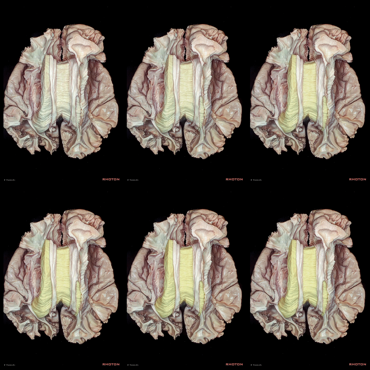
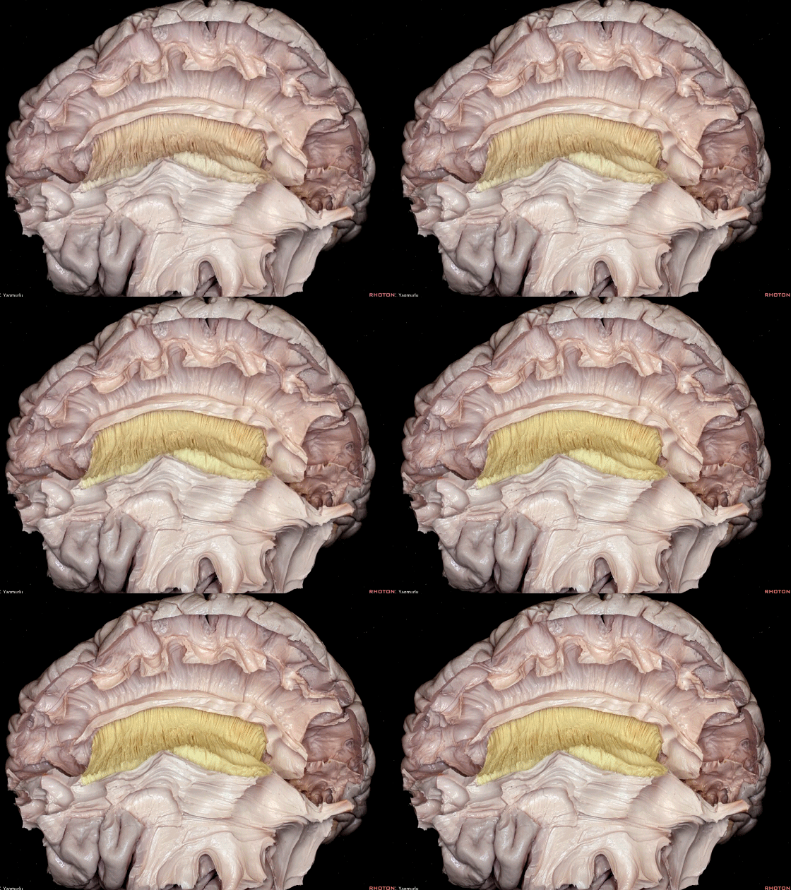
▼胼胝体纤维(下图)起自一侧半球的皮层区域汇聚于侧脑室上方,随后进入胼胝体并到达对侧半球。其在扣带下方跨过中线而构成侧脑室顶壁。
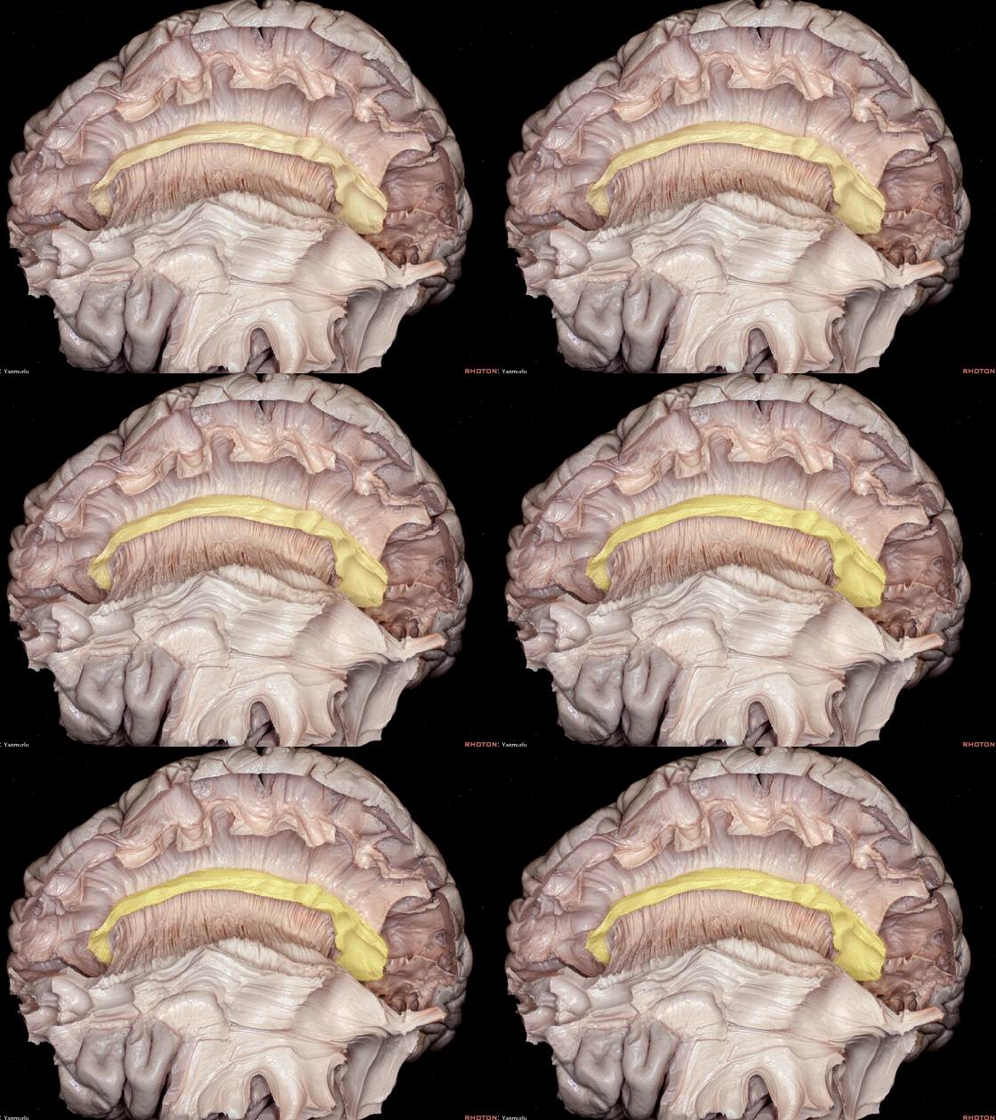
The callosal fibers emanating from cortical areas in one hemisphere gather above the lateral ventricle and enter the corpus callosum to reach the other hemisphere.They cross the midline by passing below the cingulum to form the roof of the lateral ventricle.
▼这是 扣带回
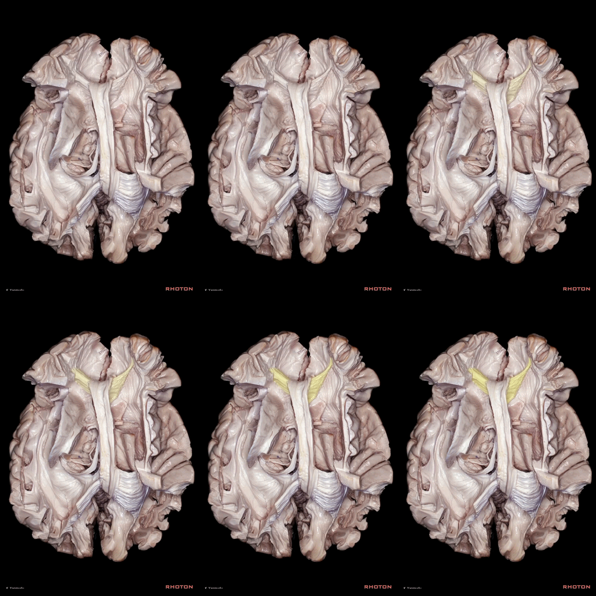
▼胼胝体膝部发出的纤维斜行转向前方,以连接双侧额区,即形成所谓的 小钳(下图)。
The callosal fibers arising from the genu of the corpus callosum turn in an anterior oblique direction, to interconnect the frontal regions,where they are called the forceps minor.
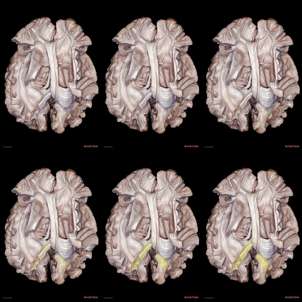
▼胼胝体压部发出的纤维斜行转向后方,以连接双侧顶枕区,即形成所谓的 大钳(下图)。
At the level of the splenium, fibers turn in a posterior oblique direction, to interconnect the parieto-occipital regions, where they are called the forceps major.
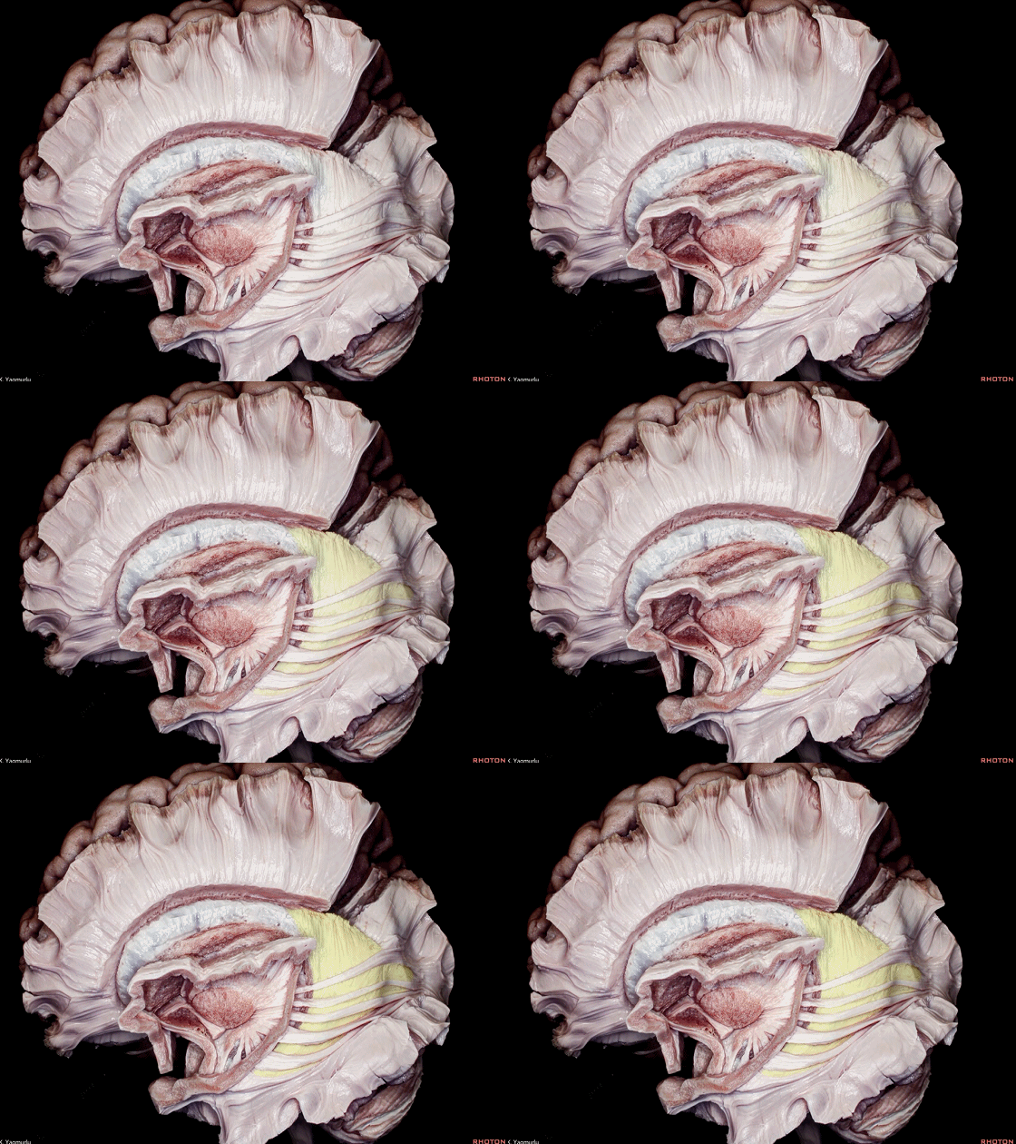
▼发自胼胝体压部的纤维,有一束称为 胼胝体毯(下图)
The callosal fibers arising from the splenium, named the tapetal fibers,
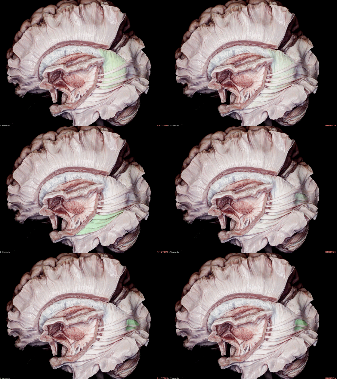
▼胼胝体毯 向下展开而构成侧脑室 房部、颞角、枕角(下图)的顶壁和外侧壁。
tapetal fibers sweep downward to form the roof and lateral wall of the atrium, temporal and occipital horns of the lateral ventricle.
▼其他联合纤维包括 前连合 和 后连合 (下图),均跨过中线。
Other commissural pathways are the anterior and posterior commissures, which traverse the midline.
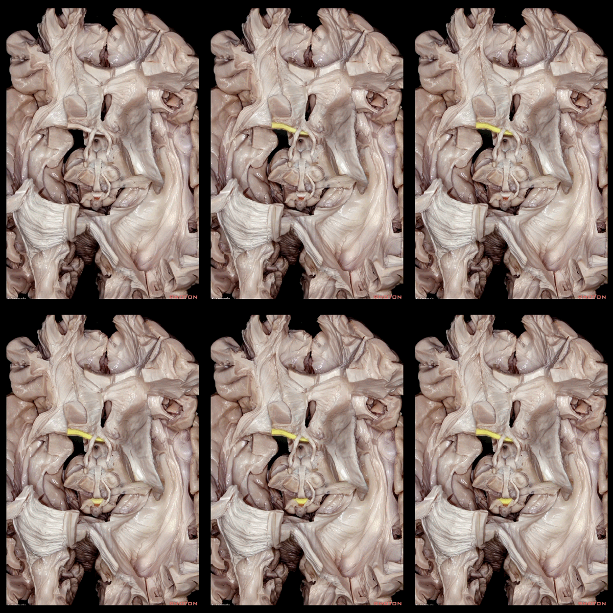
▼前连合(下图)形似自行车的车把,位于穹窿柱(下图)的前方。
前连合参与视觉处理,并协同于胼胝体压部。
前连合可在先天性胼胝体缺如的患者中代偿半球间视觉信息的整合。
The anterior comissure resembles bicycle handlebars located immediately in front of the columns of the fornix,and plays a role in visual processing together with the splenium of the corpus callosum.The anterior commissure may compensate for congenitally absent corpus callosum by integrating interhemispheric visual information.
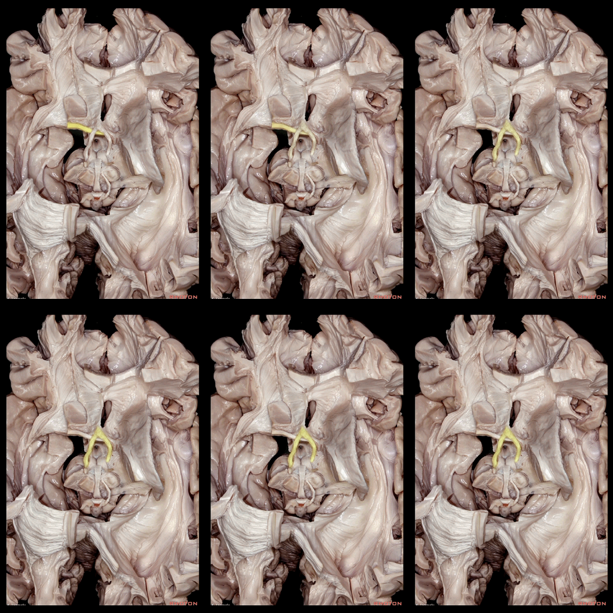
▼这是 胼胝体压部
the splenium of the corpus callosum
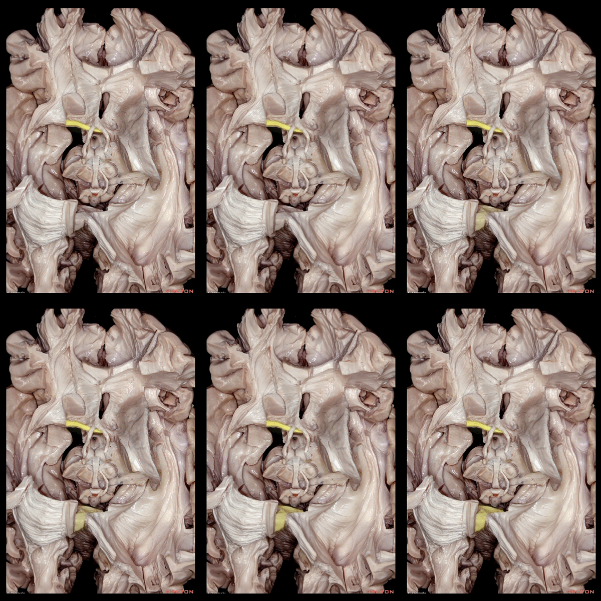
▼这是 乳头体
The mamillary bodies
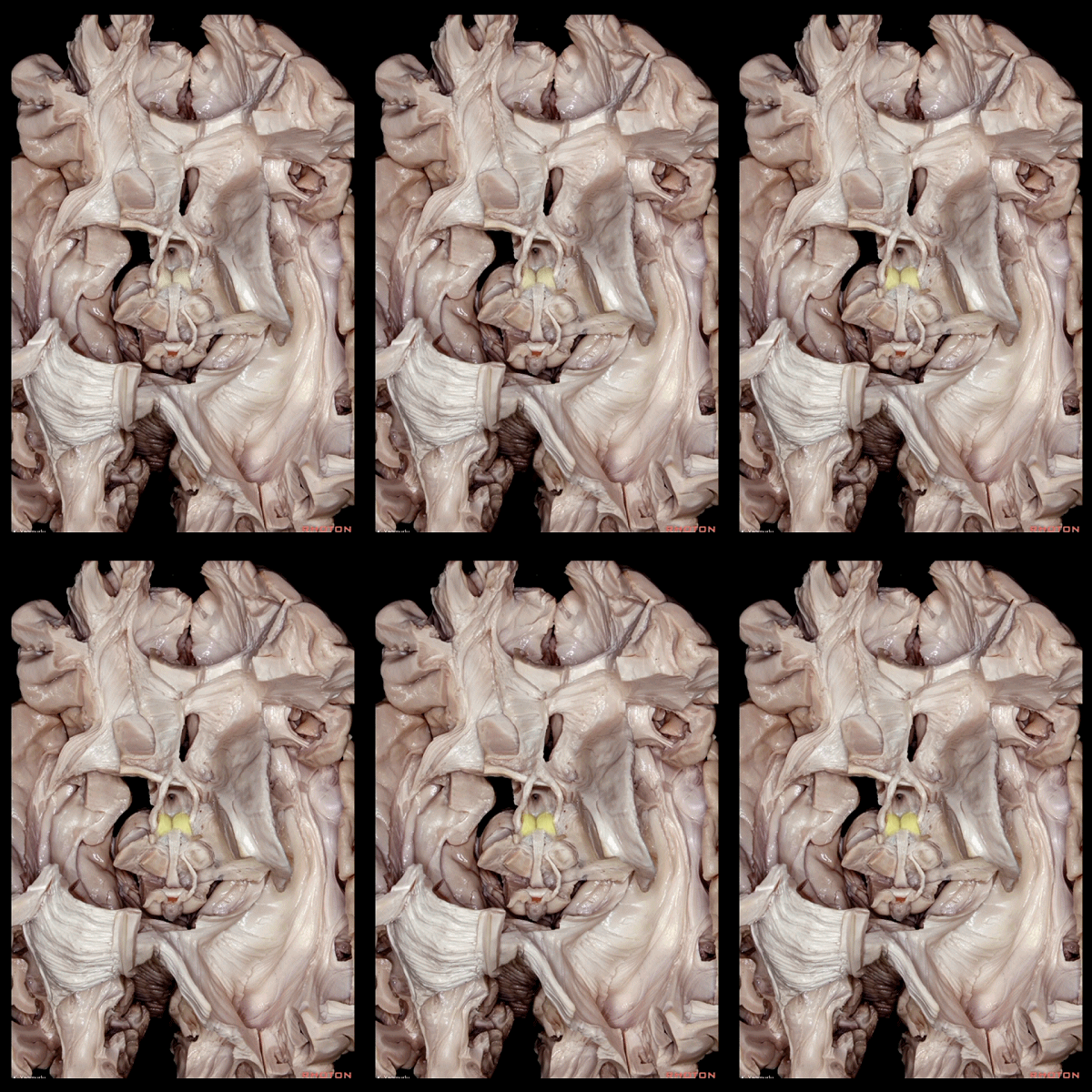
▼这是 第三脑室底壁
the floor of the third ventricle
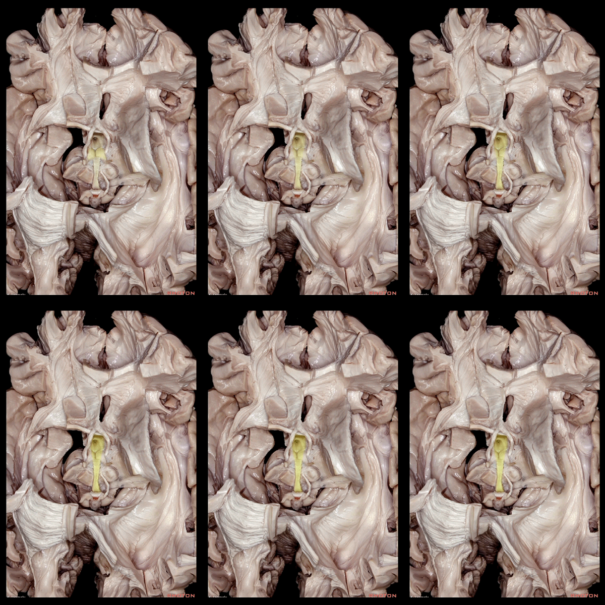
▼后连合(下图)位于中脑导水管上端开口的背侧。
The posterior commissure is located on the dorsal aspect of the upper end of the cerebral aqueduct.
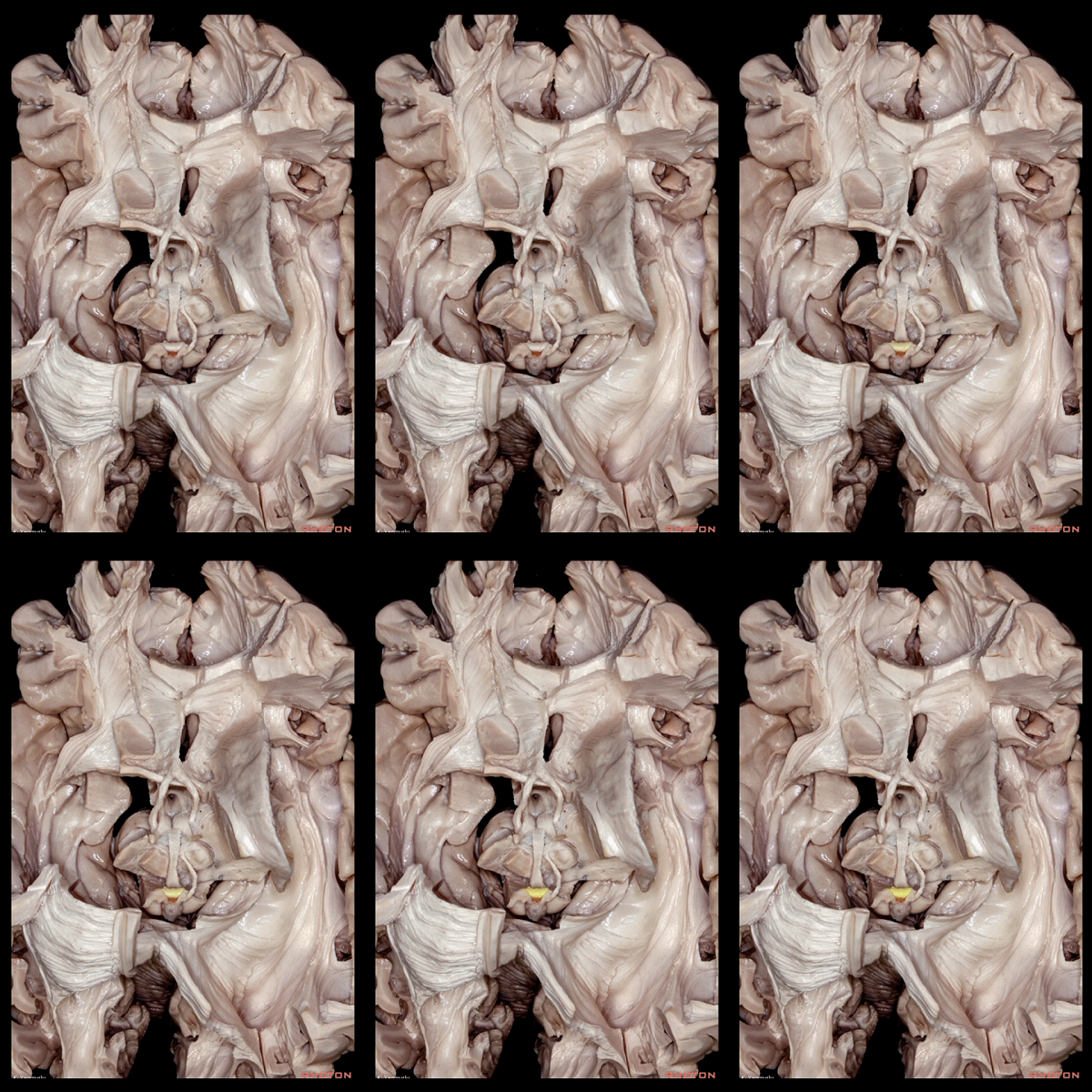
▼这是 中脑导水管
cerebral aqueduct
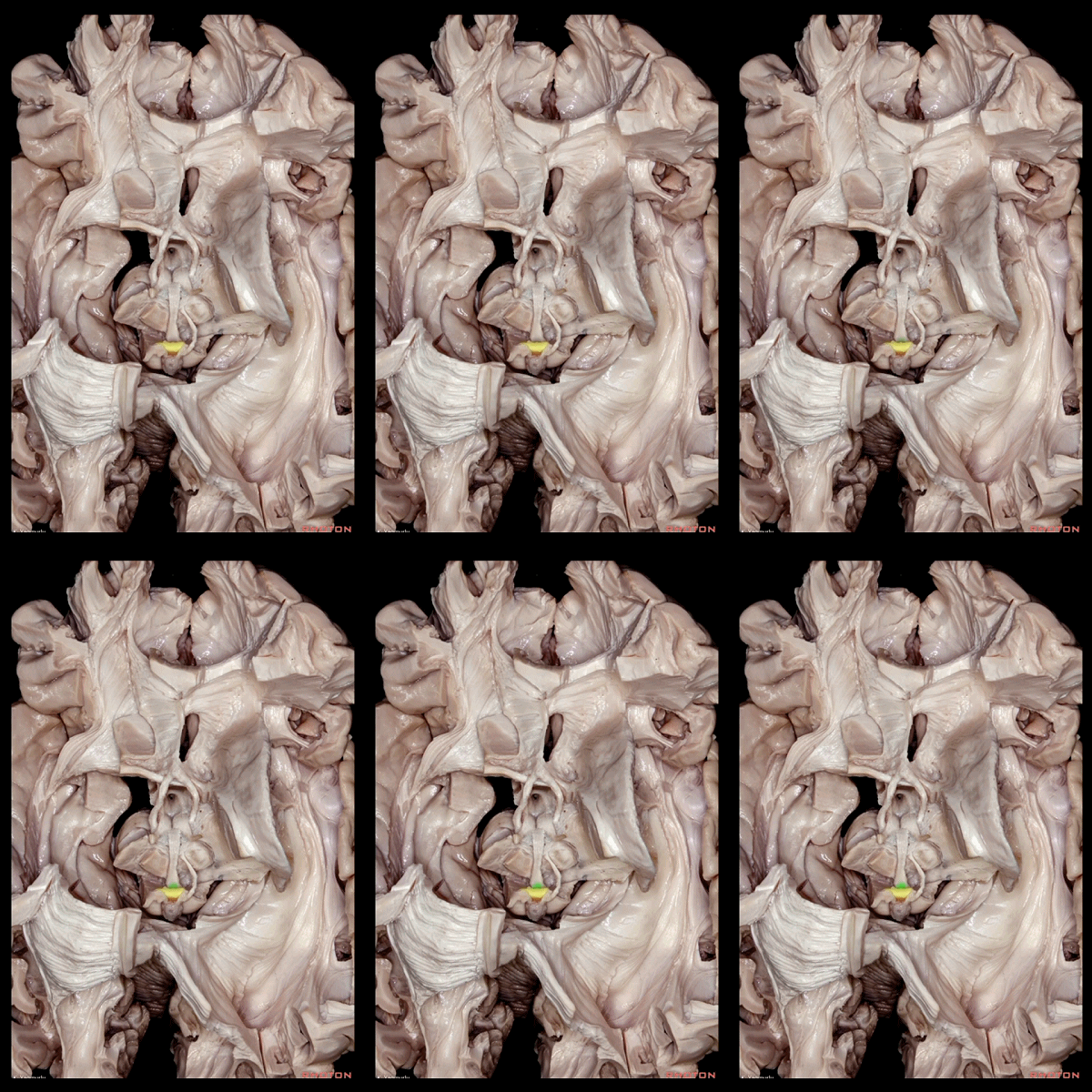
▼从大脑内侧面来看前连合和后连合
The anterior and posterior comissures can be seen in this medial view.
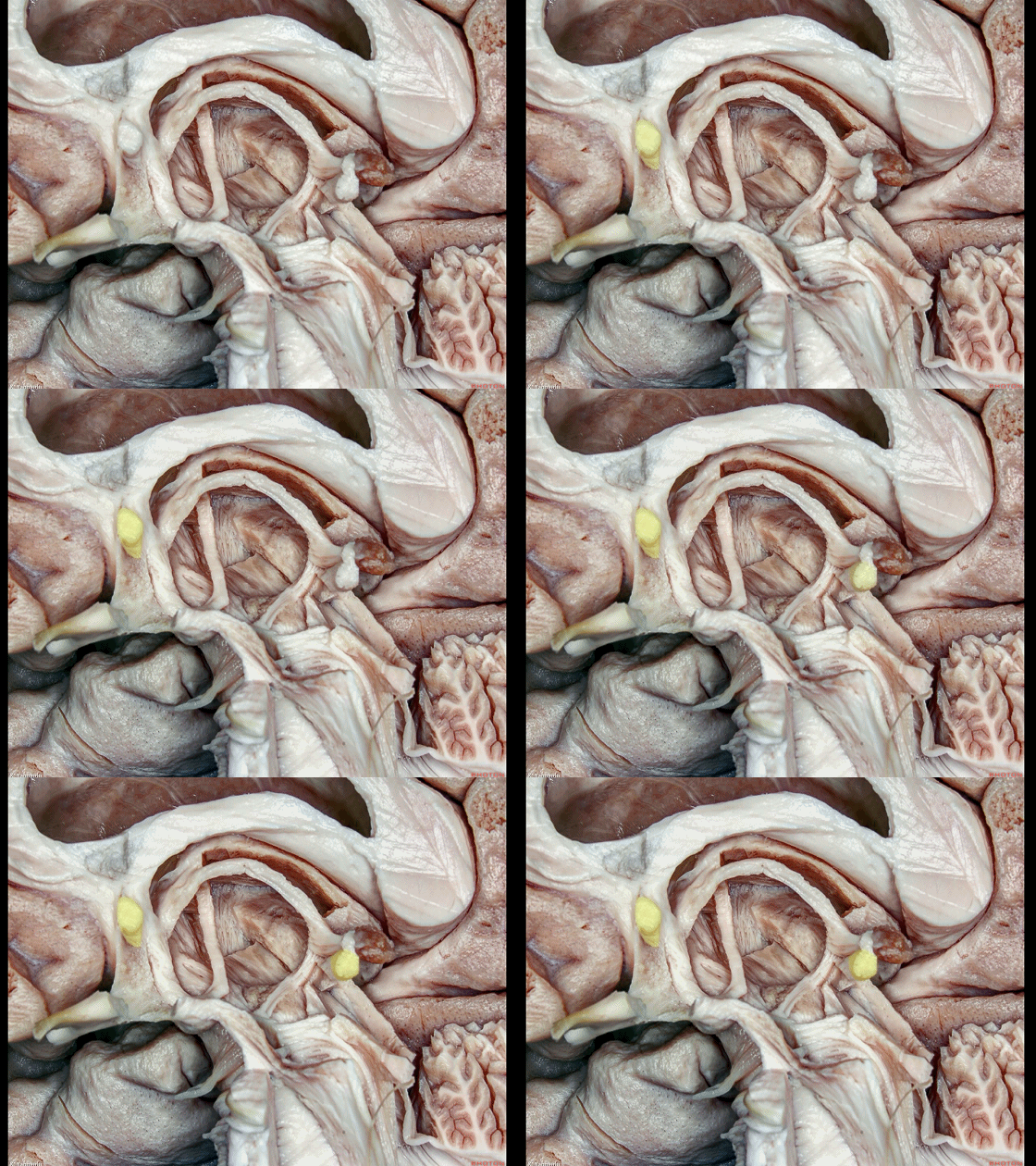
▼后连合(下图)起自其神经核,参与瞳孔对光反射。
The posterior commissure originates from its nucleus,The posterior commissure is involved in pupillary light reflex.
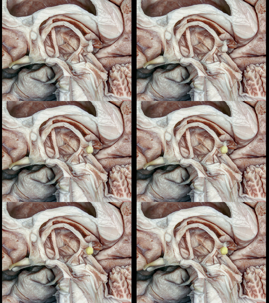
▼后连合核团位于动眼神经核(下图)前方。
located in front of the oculomotor nucleus.
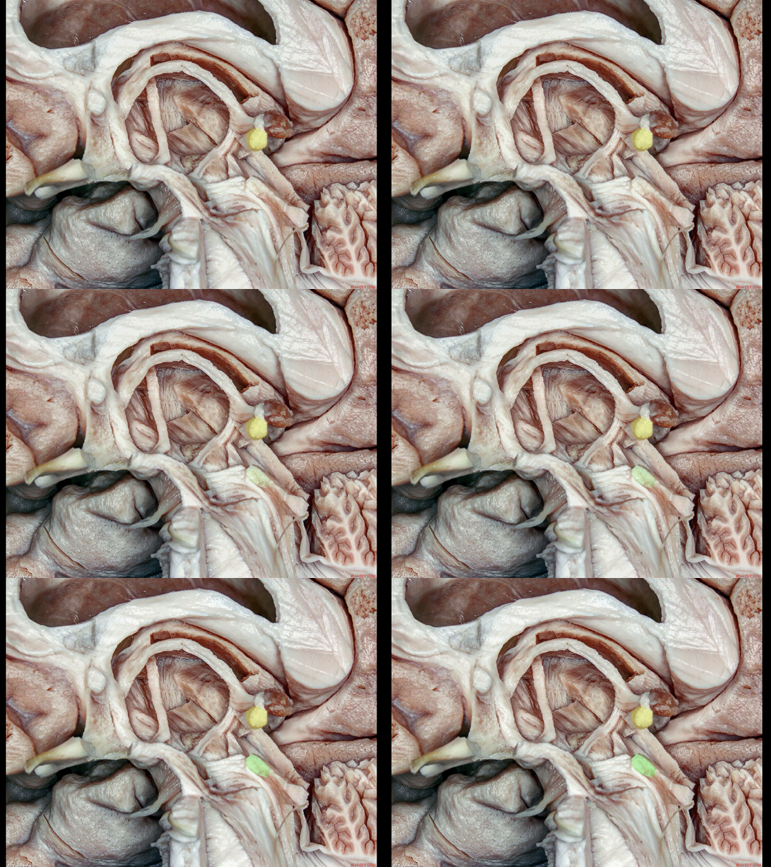
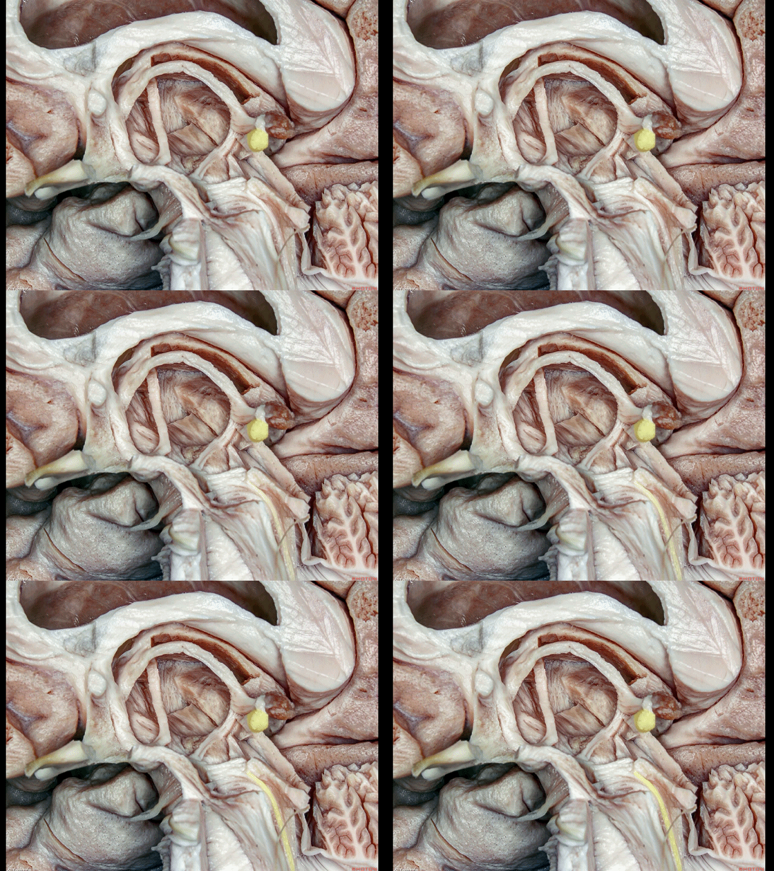
▼后连合向下续于内侧纵束(下图)。
The posterior commissure continues downward into the medial longitudinal fasciculus.
▼这是 前连合
anterior commissure
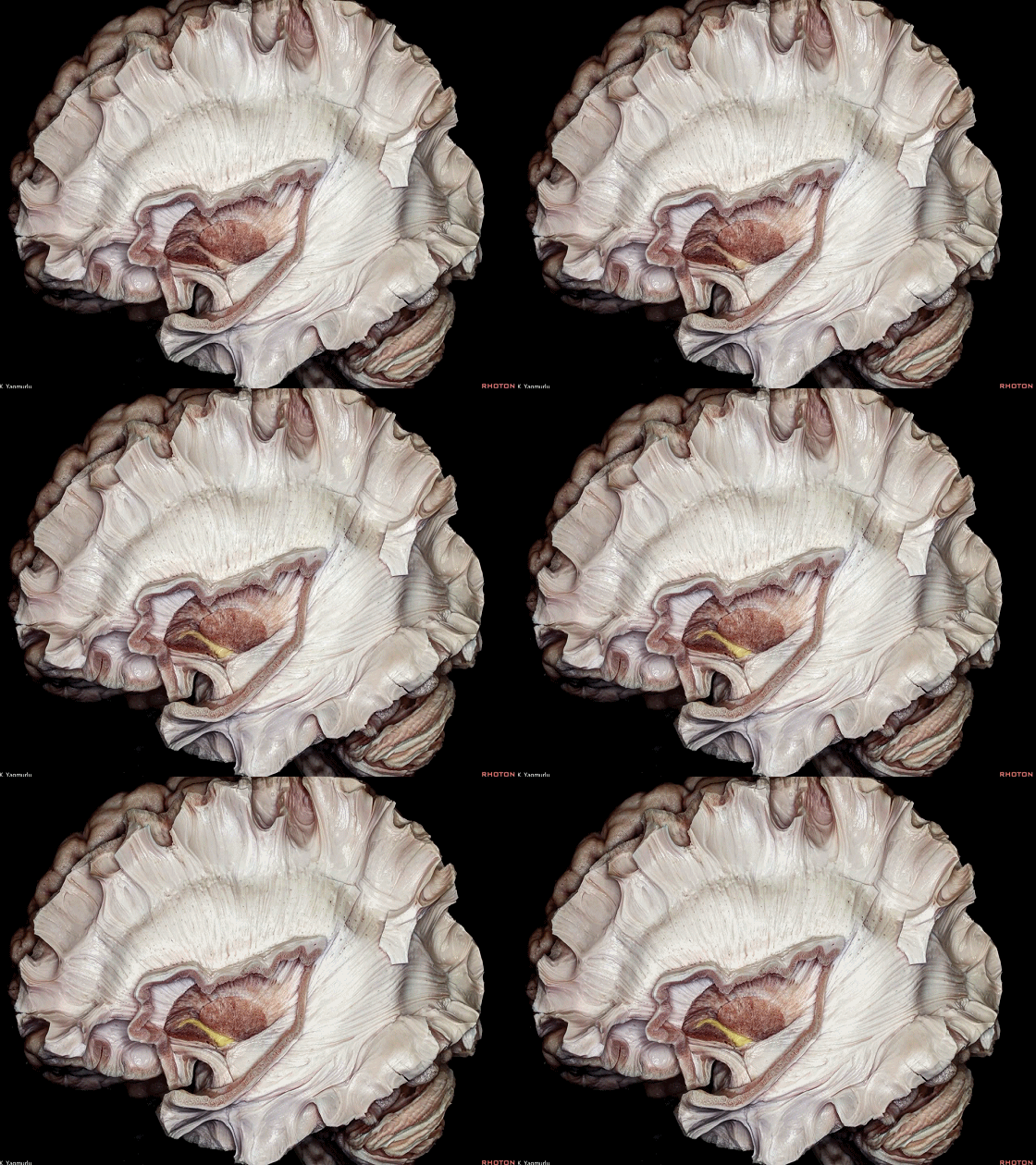
▼前连合越过中线,毗邻 壳核(下图)基底部
The anterior commissure crosses the midline at the base of the putamen,
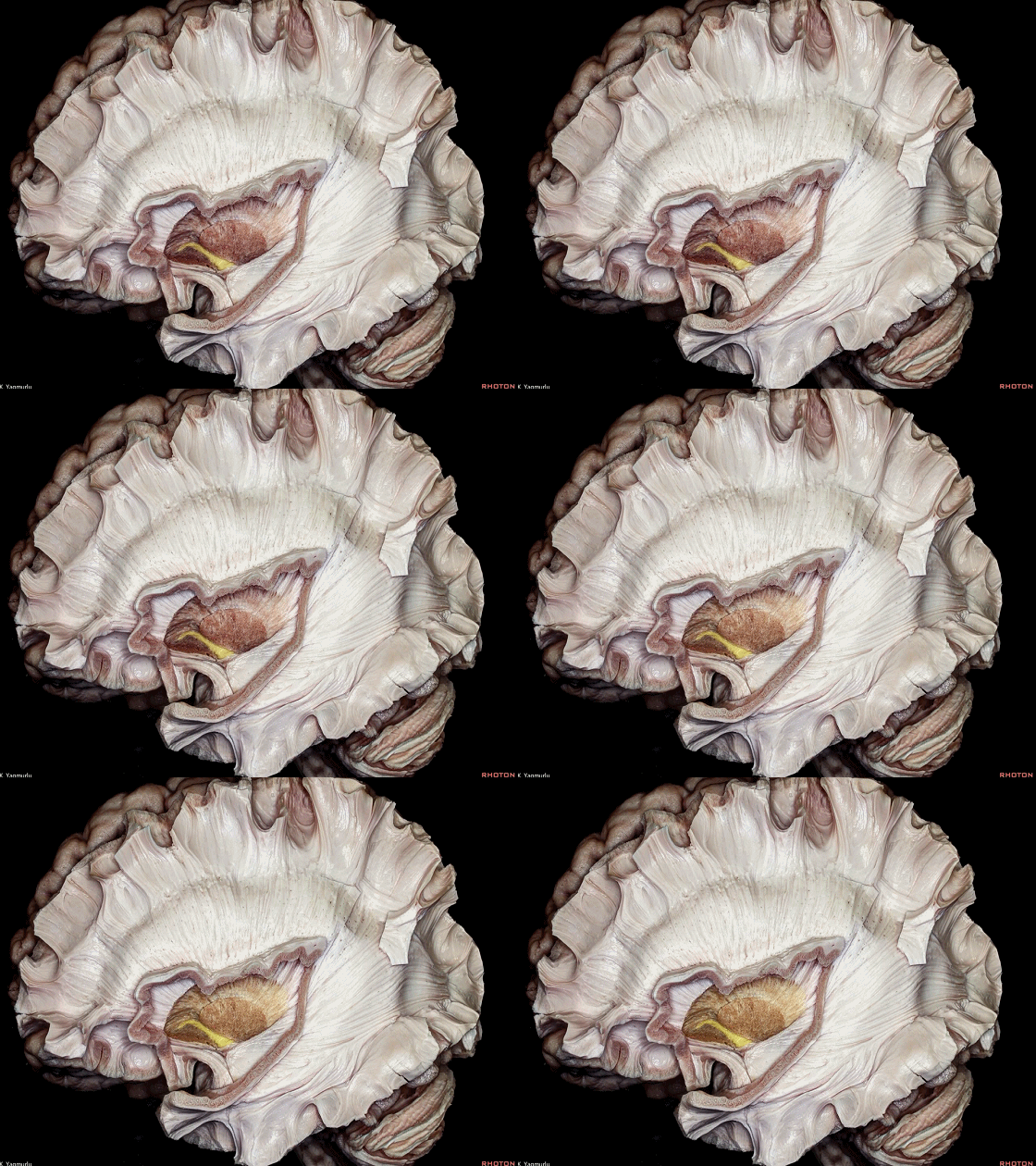
▼前连合连接双侧眶额叶、枕叶、以及颞叶,尤其是杏仁核(下图)。
to connect the orbitofrontal, occipital, and temporal lobes, especially amygdala.
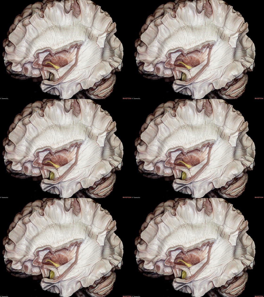
▼前连合包含一前脚行向前方,一后脚伸向外侧。
It has an anterior crus that extends forward, and a posterior crus that extends laterally.
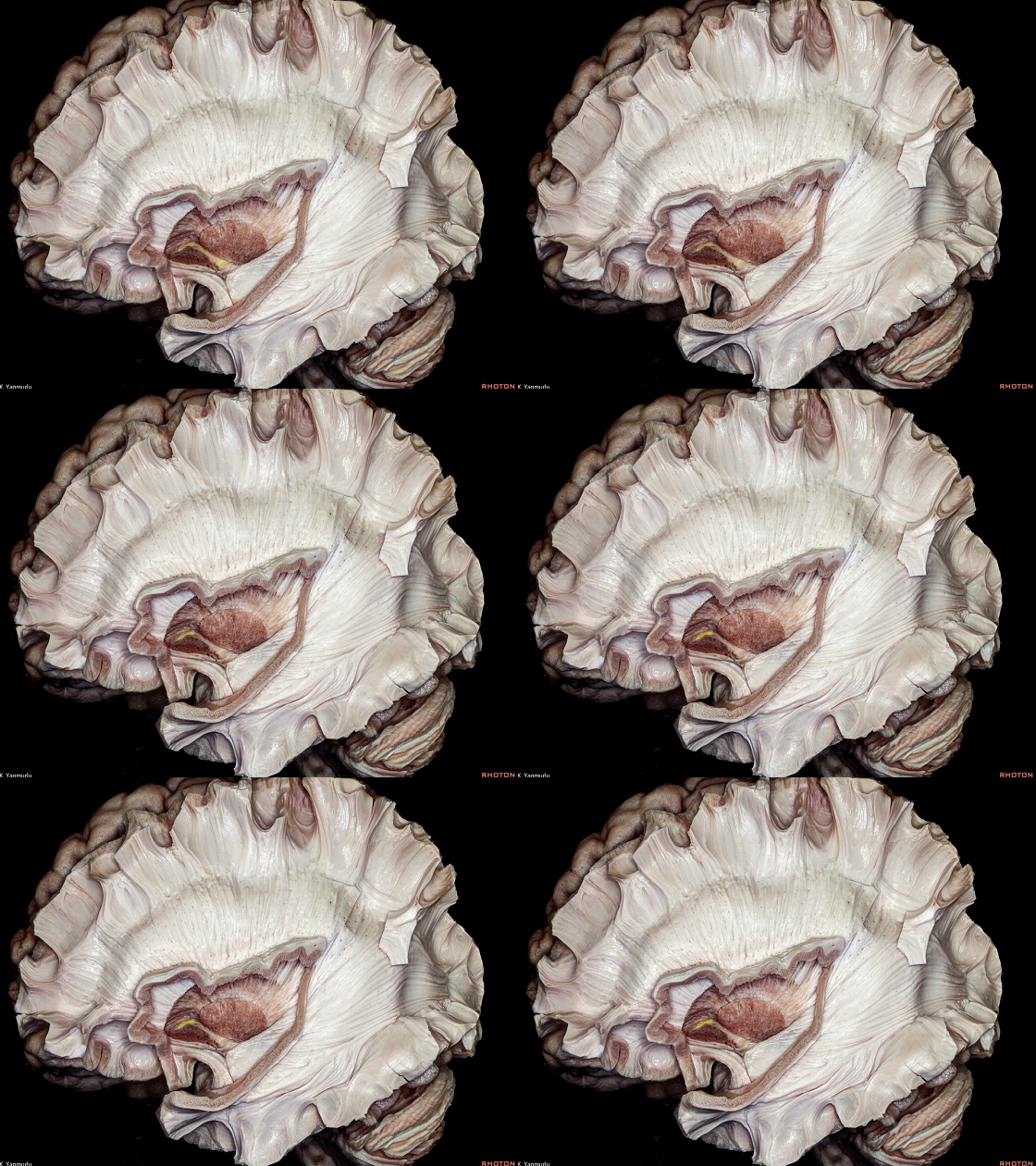
▼前连合后脚进一步分别向颞叶和枕叶延伸。
The posterior crus is divided into temporal and occipital extensions.
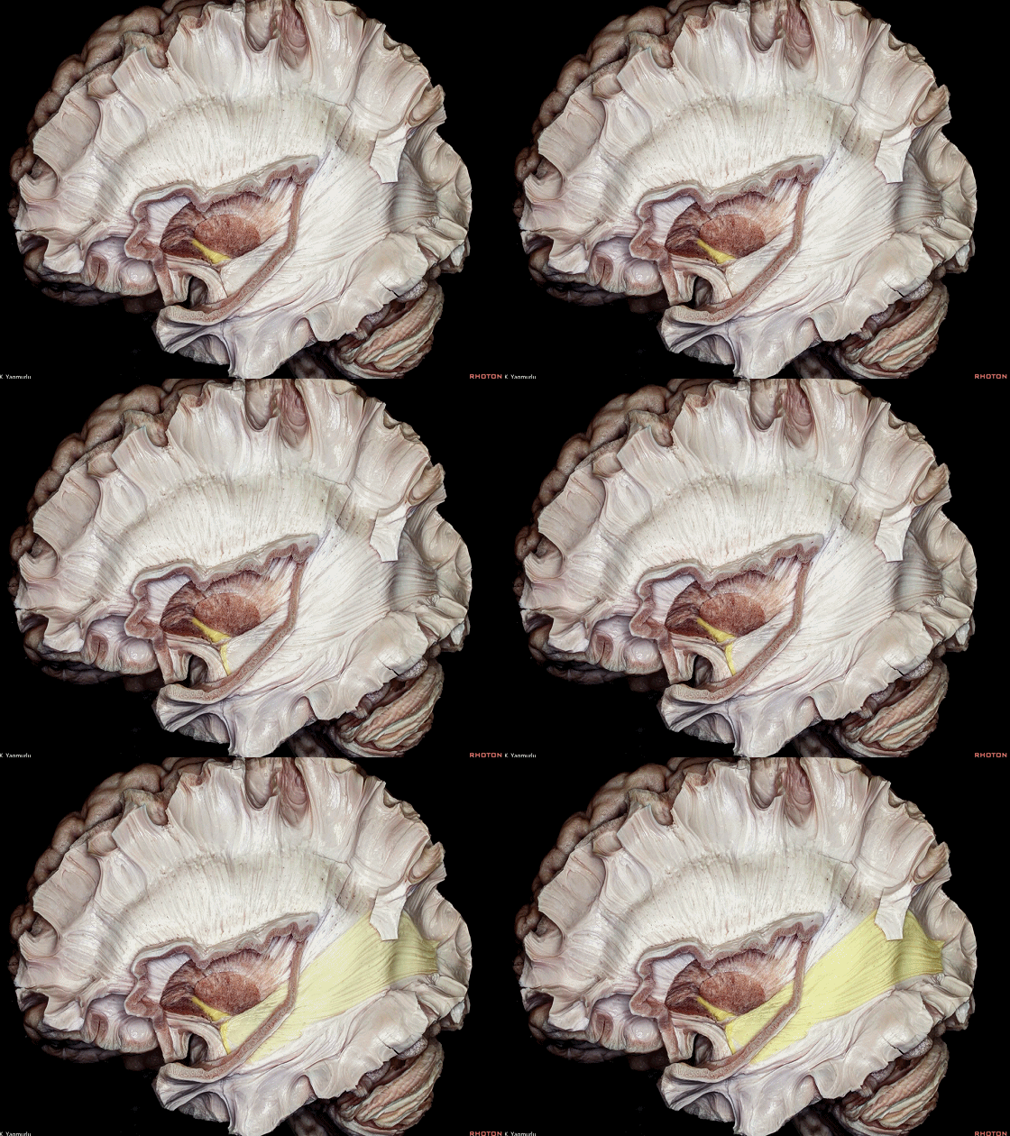
▼临床上,有观点认为痫性活动可经由前连合传递至对侧的颞叶内侧部,因此,用于治疗难治性癫痫的大脑半球切除术,包含了前连合的切除。
From clinical perspective, it was suggested that seizure activity can propagate to the contralateral medial temporal lobe by way of the anterior commissure,so its cutting is part of the hemispherotomy procedure for control of intractable seizures.
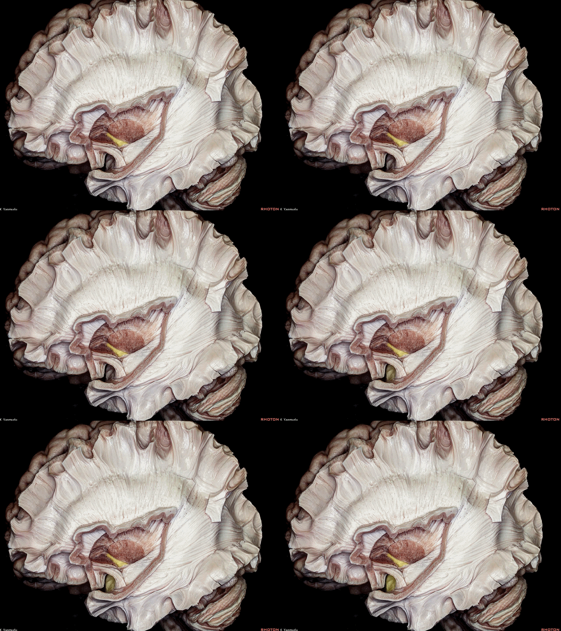

▼讨论完联络纤维和联合纤维,我们来看 投射纤维。
额顶叶投射纤维,即所谓的 放射冠(下图),呈垂直方向走行。
After looking at the association and commissural fiber pathways, let's move on to the projection fiber pathways.The frontoparietal projection fibers, which have a vertical direction,can be called as the corona radiata,
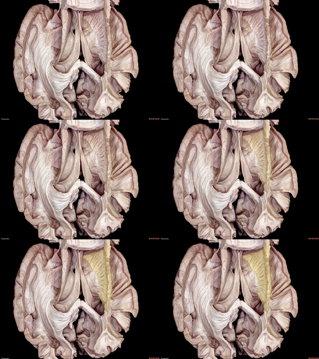
▼枕叶投射纤维(下图),即所谓的矢状层,呈水平方向走行。
and the occipital projection fibers, which have a horizontal direction, can be called as the sagittal stratum.
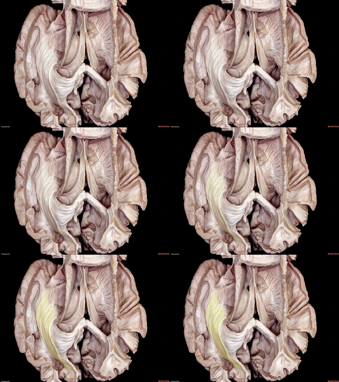
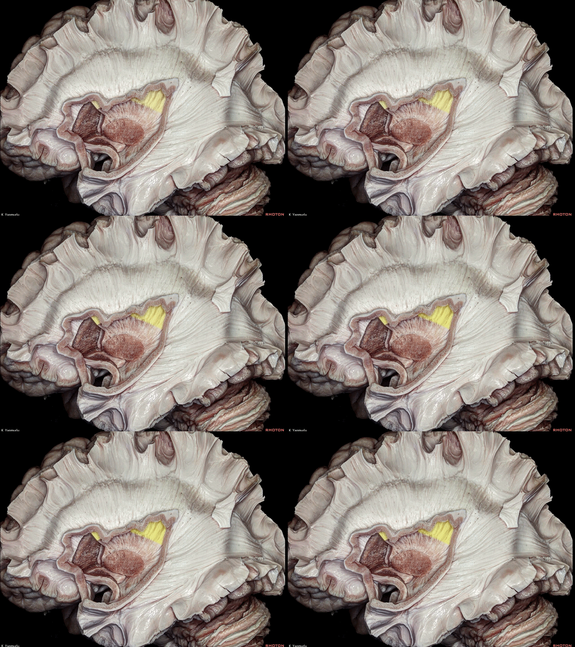
▼放射冠(下图)的纤维在壳核以上,包含外囊和内囊。
The corona radiata is formed by the external and internal capsules above the upper level of the putamen.
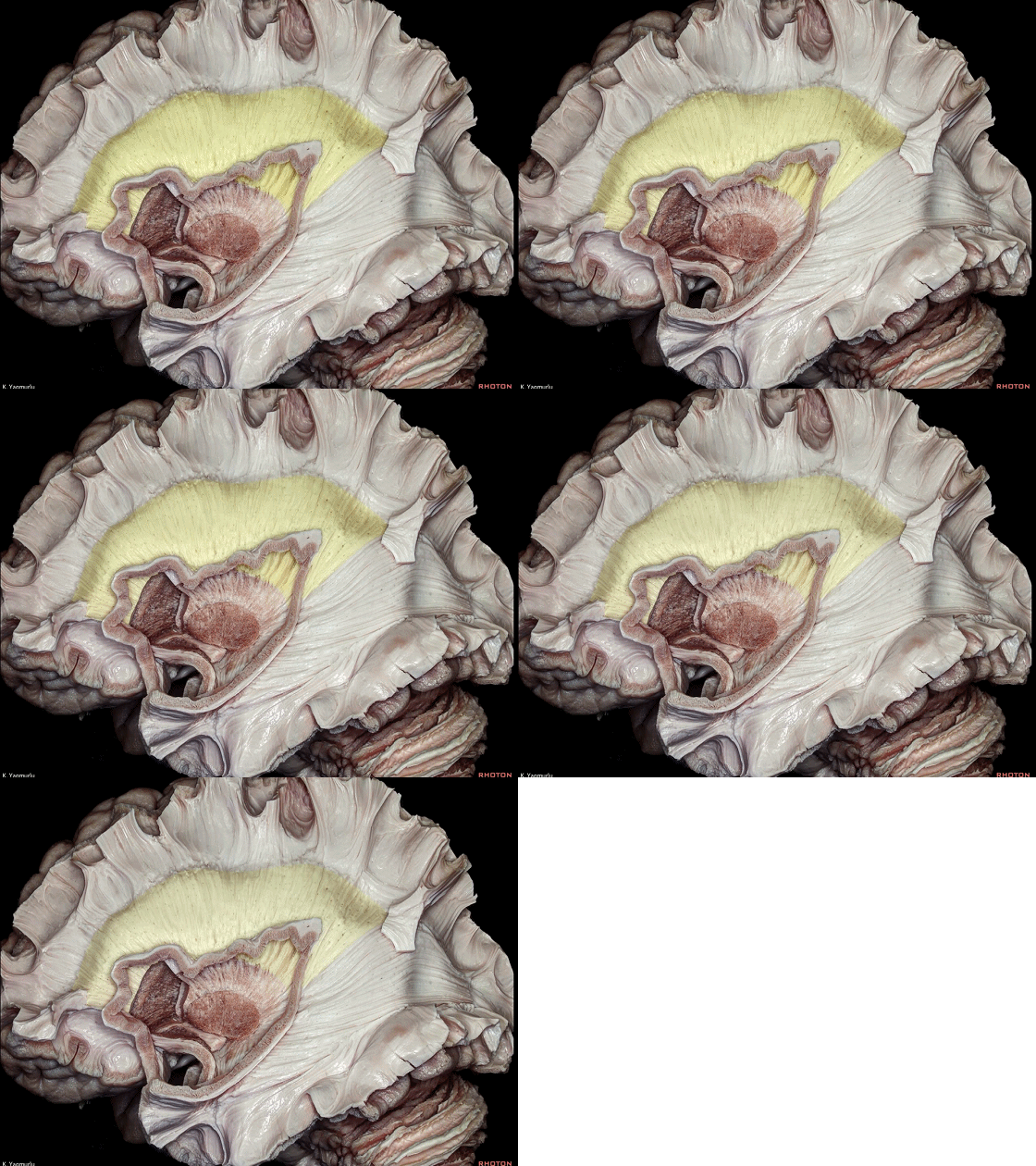
▼这是 外囊
external capsule
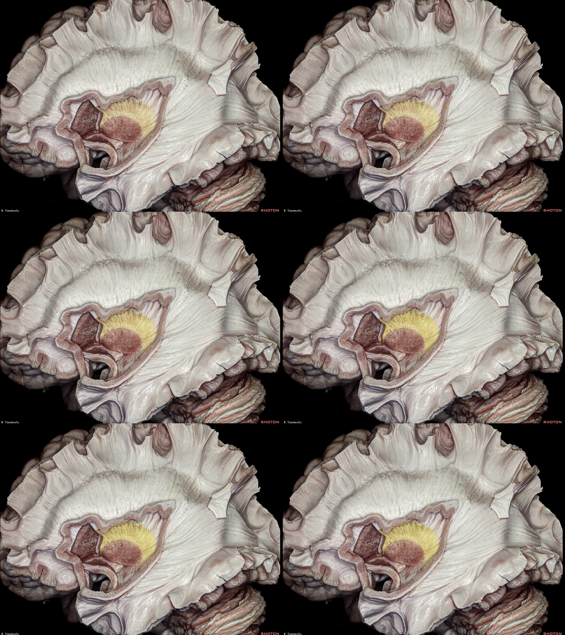
▼这是 内囊
internal capsule
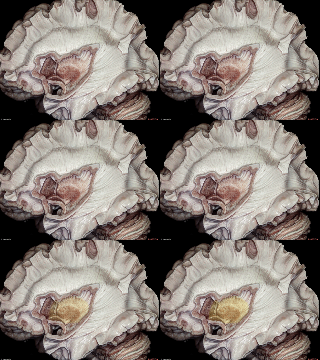
▼这是 壳核
▼半卵圆中心(下图)是位于胼胝体层面上方的结构
The centrum semiovale is located above the level of corpus callosum,
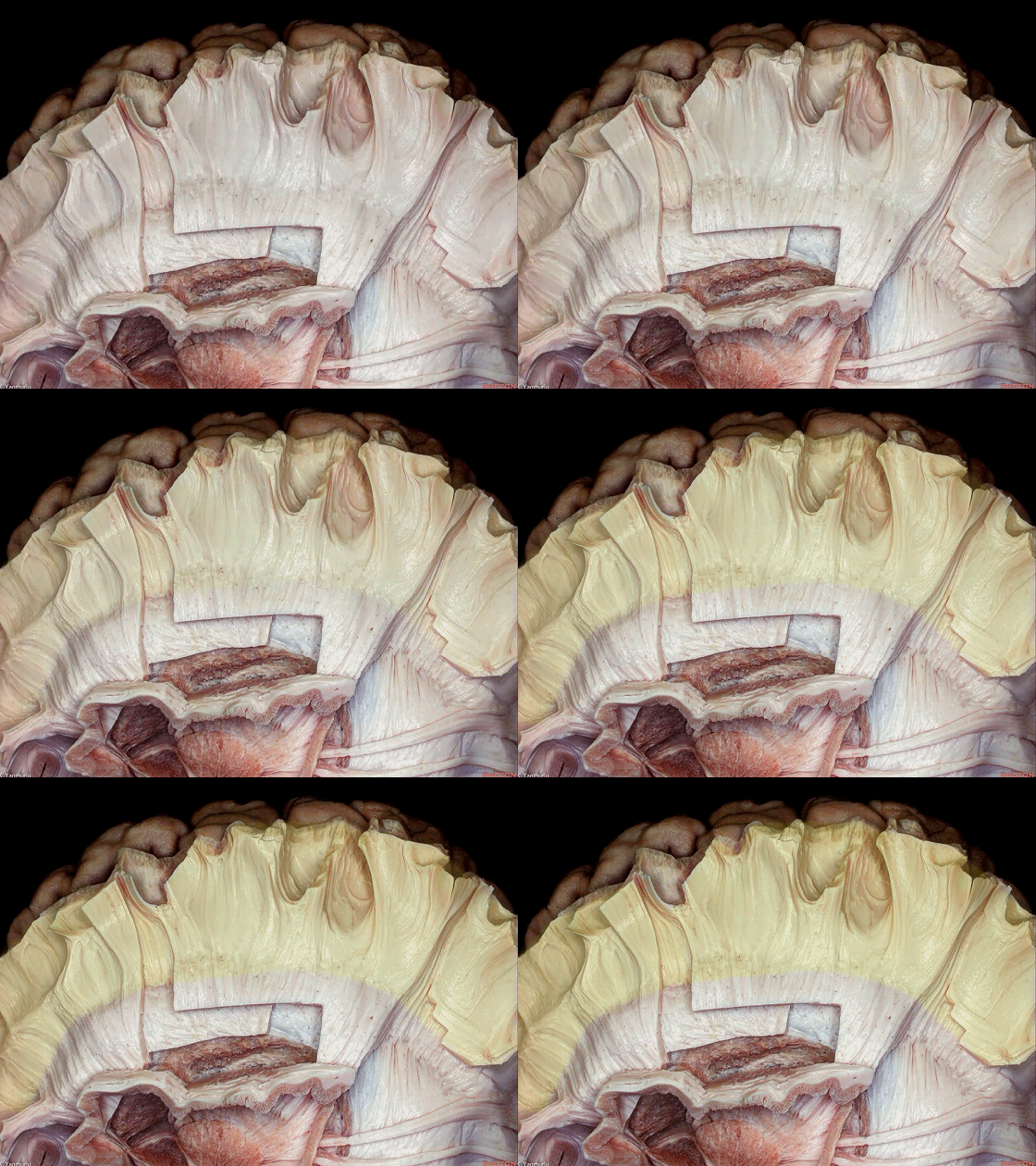
▼半卵圆中心 包含 上纵束(联络纤维 下图)、放射冠(投射纤维)、胼胝体(联合纤维)。
The centrum semiovale consists of the superior longitudinal fasciculus association fibers, corona radiata projection fibers and callosal comissural fibers.
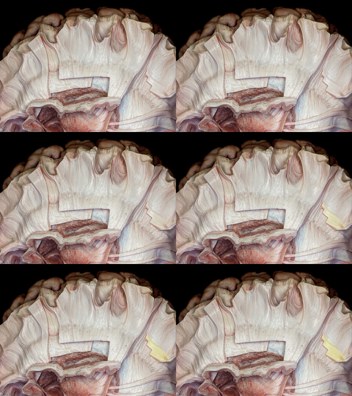
▼下图示 放射冠(投射纤维)
▼下图示 胼胝体(联合纤维)
以上是对三种大脑纤维传导通路的系统性介绍。
We have seen all three types of fiber pathways of the cerebrum.
![]()
▼接下来将从外向内逐层解剖上述纤维传导通路,以及核心区灰质和边缘系统。
Now, we are going to look at all these fiber pathways from the lateral to medial direction in a stepwise manner, as well as the central core and limbic system.

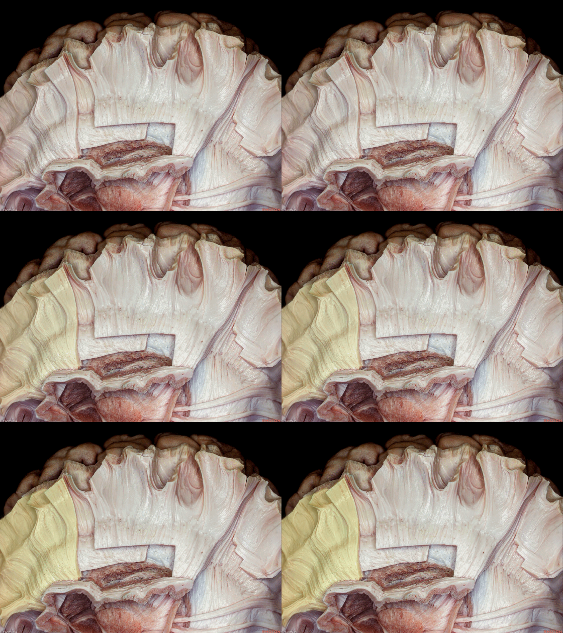
▼去除皮层灰质即显露 短联络纤维(下图),其连接相邻脑回。
The removal of the grey matter exposes the short association fibers, which interconnect neighboring gyri.
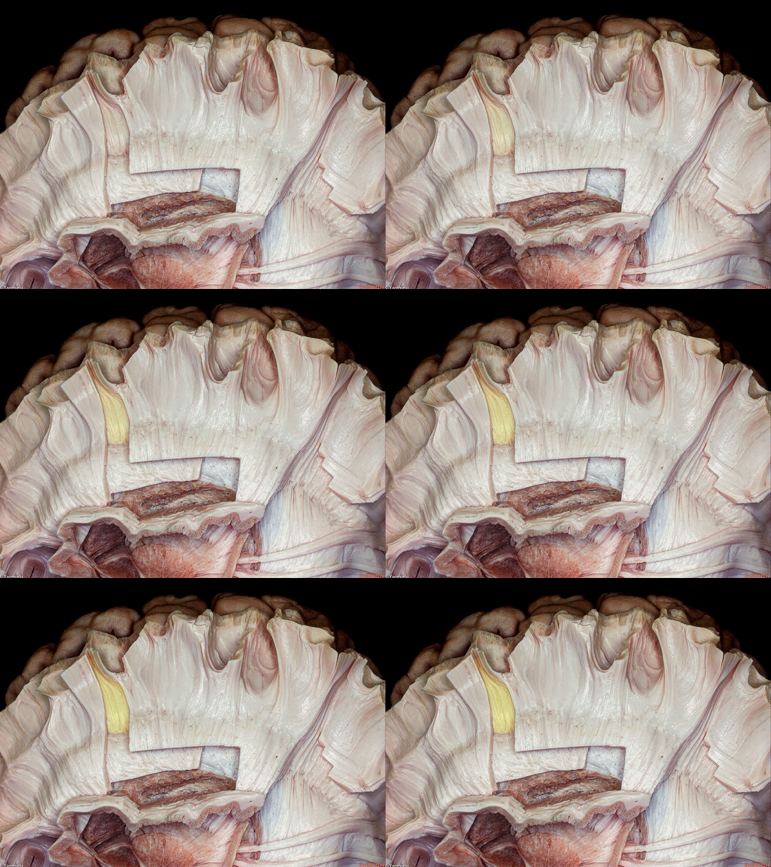
▼去除短联络纤维即显露深部的 长联络纤维(下图)。
Removal of the short association fibers exposes the long association fibers at a deeper position.
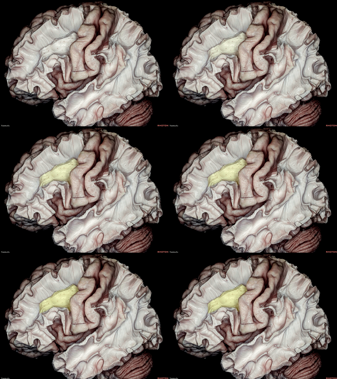
▼最为表浅的长联络纤维是上纵束(I、II),弓状束
上纵束(I、II 下图)连接额叶和顶叶。
The most superficial long association fibers are the superior longitudinal fasciculus (SLF II, SLF III), which connects the frontal and parietal lobes;and the arcuate fasciculus.
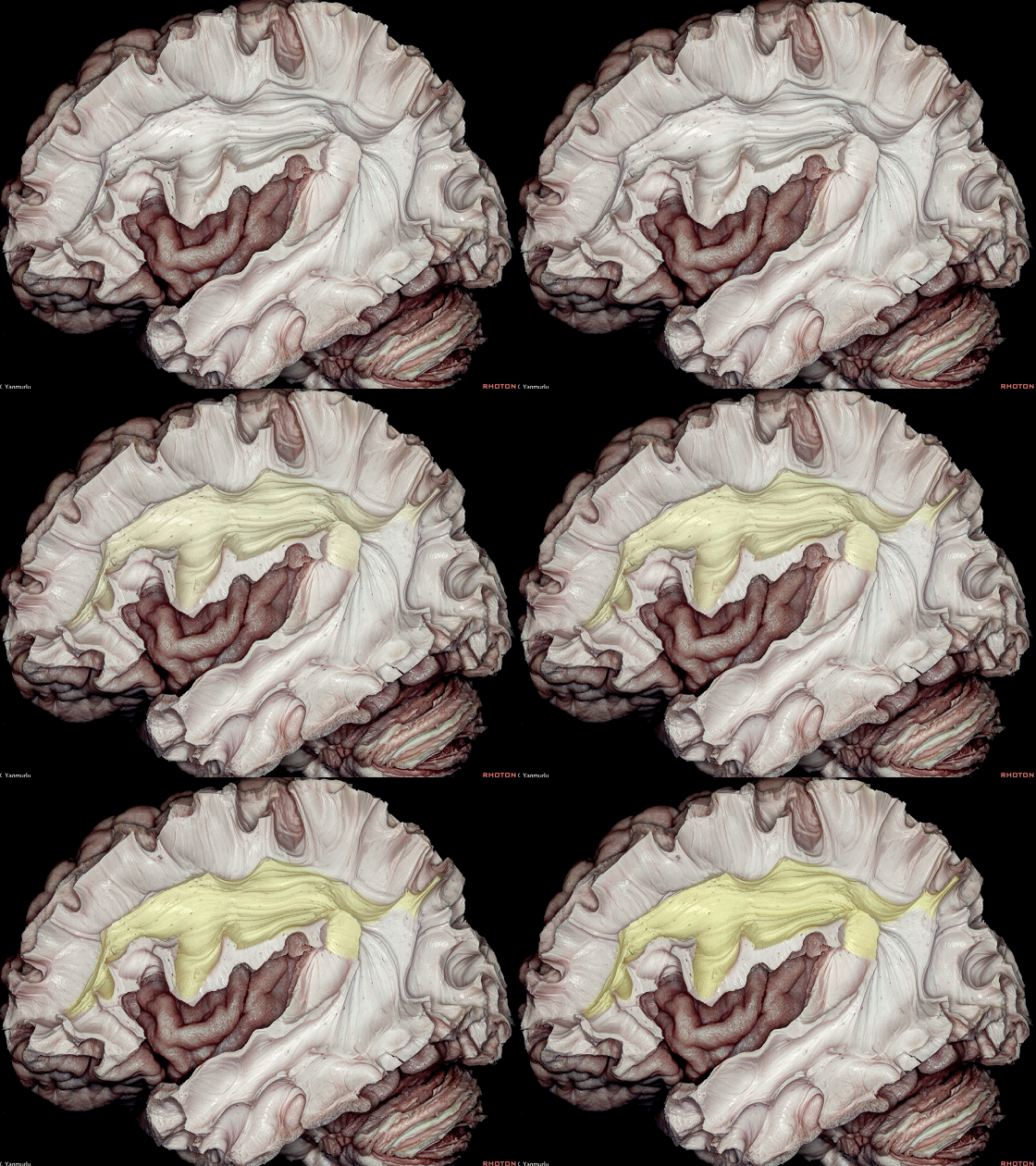
▼弓状束(下图)连接 额叶和颞叶。
the arcuate fasciculus, which connects the frontal and temporal lobes.
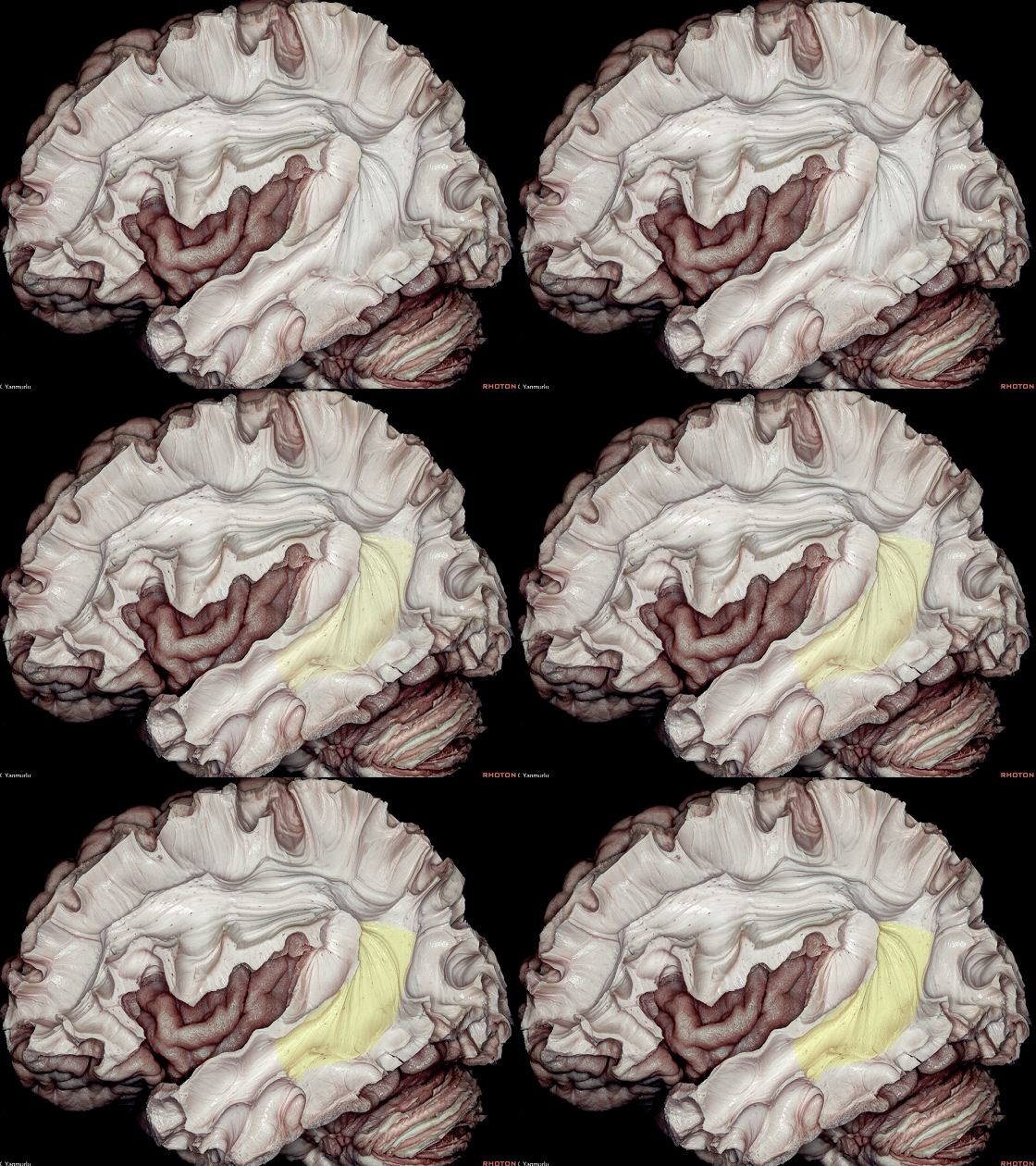
▼去除弓状束即显露 中纵束 和 下纵束。
中纵束(下图)位于颞上回内
The removal of the arcuate fasciculus exposes the middle longitudinal fasciculus and inferior longitudinal fasciculus.The middle longitudinal fasciculus lies within the superior temporal gyrus
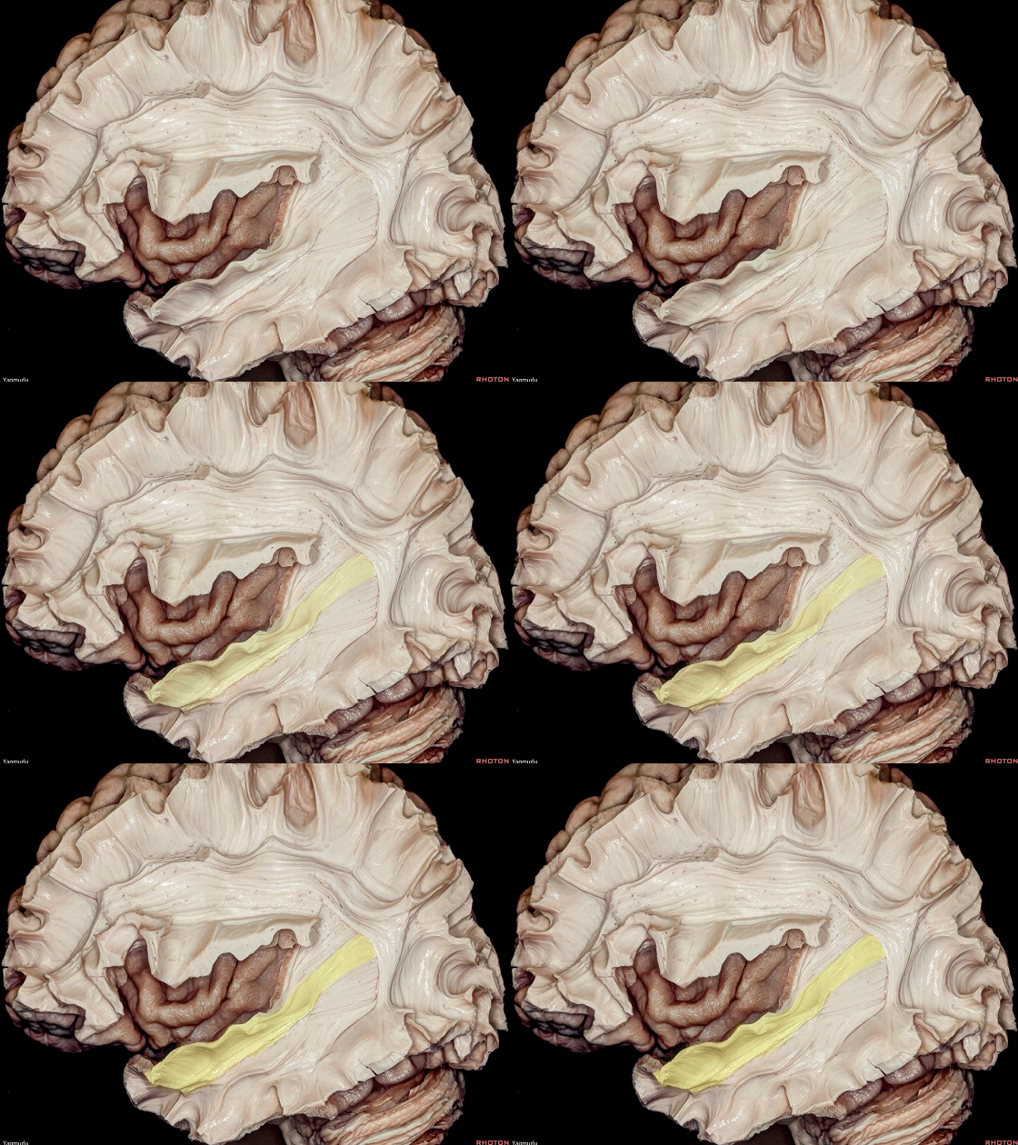
▼中纵束连接 颞极 和 顶叶下部,尤其是角回(下图)。
中纵束参与声音的空间定位。
The middle longitudinal fasciculus connects the temporal pole to the inferior parietal lobe, especially the angular gyrus.The middle longitudinal fasciculus is involved in location of sound in space.
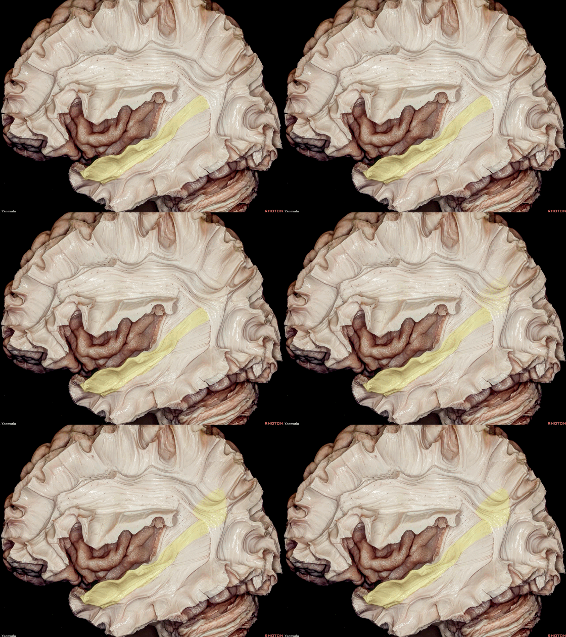
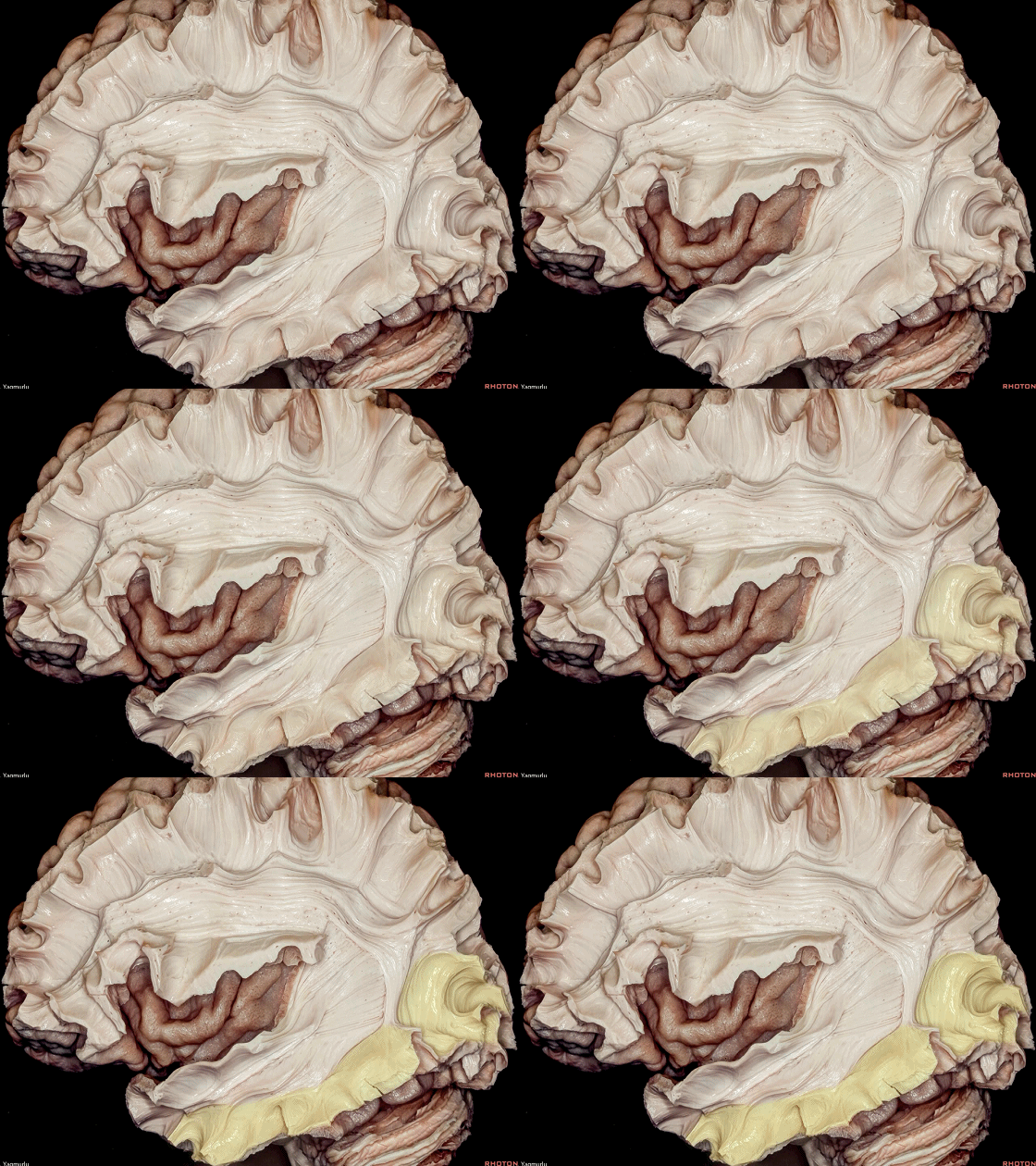
▼下纵束(下图) 位于颞下回内。
下纵束连接颞极与枕叶背外侧,而未连接视觉皮层。
the inferior longitudinal fasciculus lies within the inferior temporal gyrus.The inferior longitudinal fasciculus connects the temporal pole to the dorsolateral occipital lobe,without reaching the visual cortex.
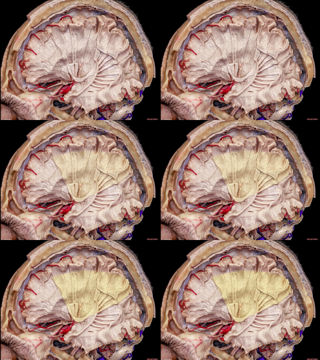
▼该标本中,额顶部岛盖已被去除而显露出 岛叶(下图)。
In this dissection, the frontoparietal operculum has been removed to expose the insula.
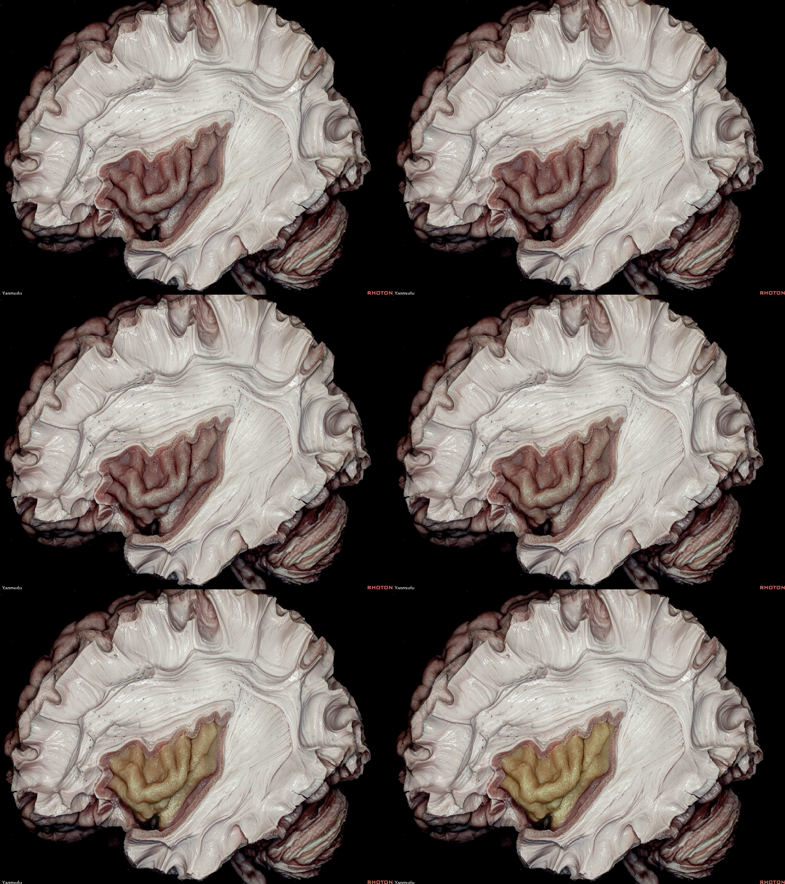
▼这是 岛中央沟
The central insular sulcus
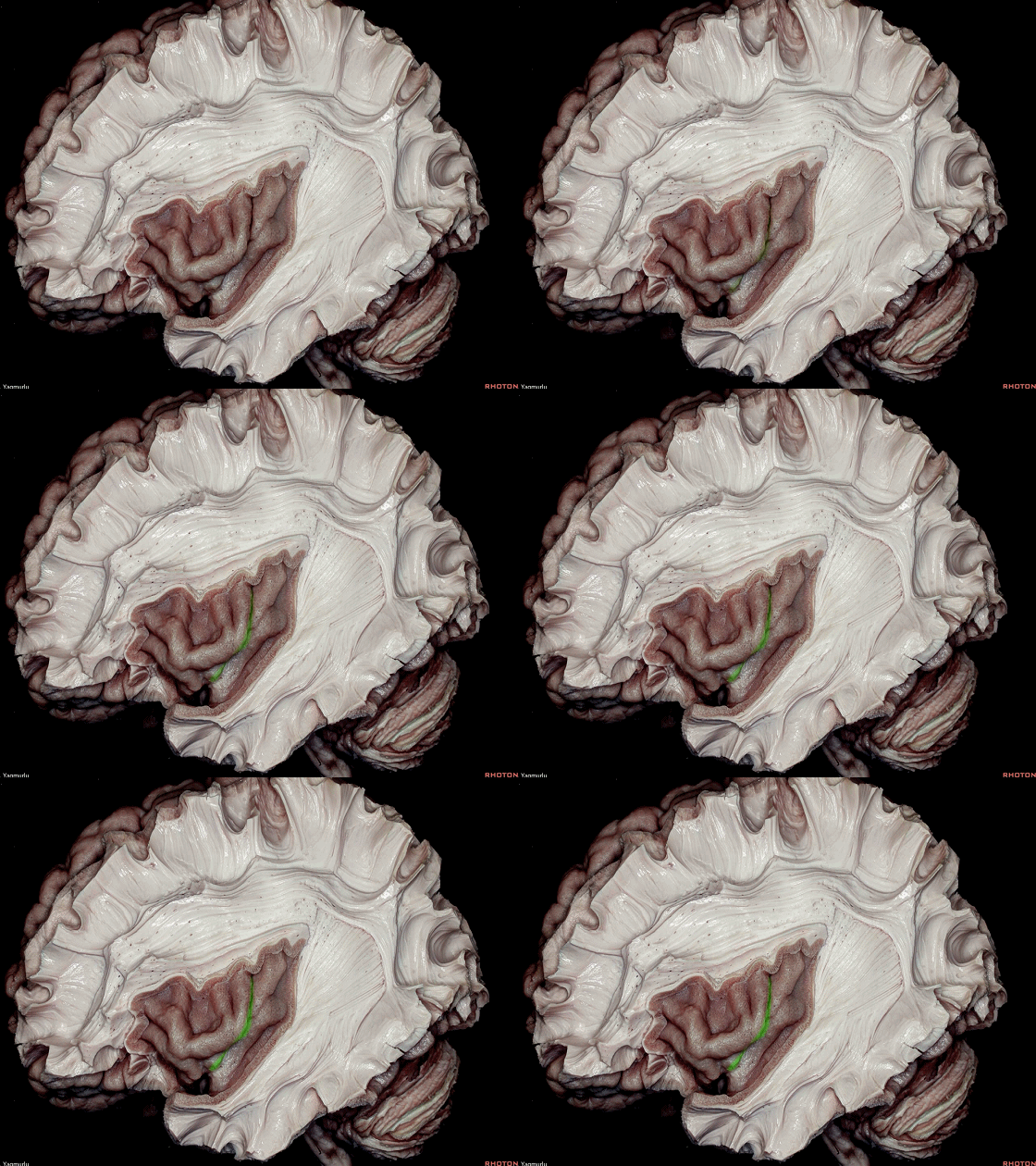
▼岛中央沟的走行 几乎平行于 半球凸面的中央沟(下图)
The central insular sulcus courses almost parallel with the central sulcus on the convexity
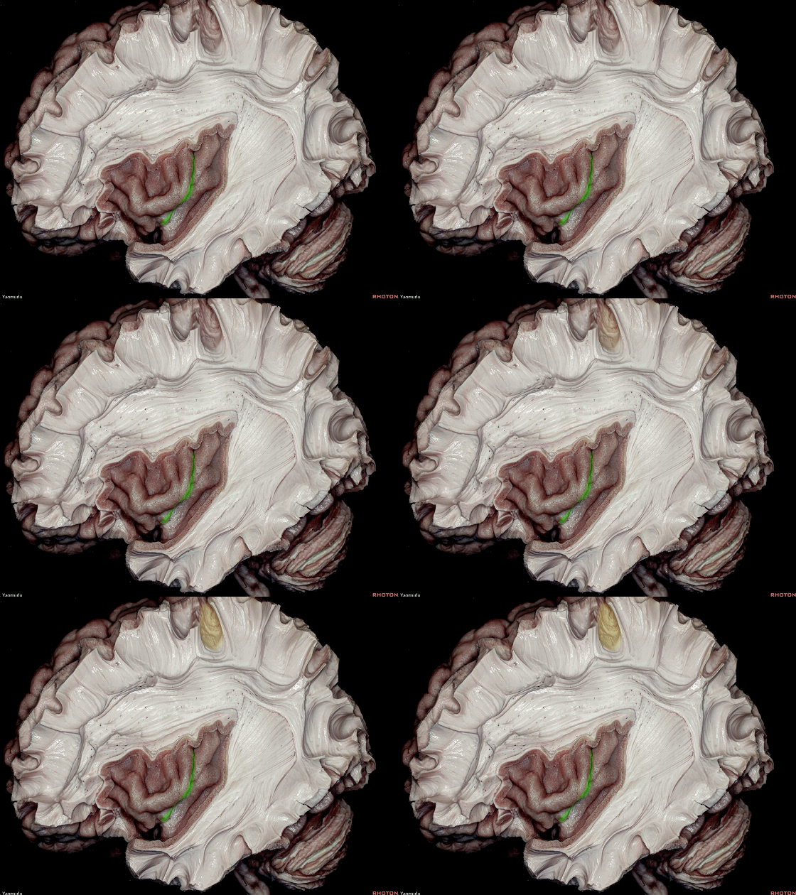
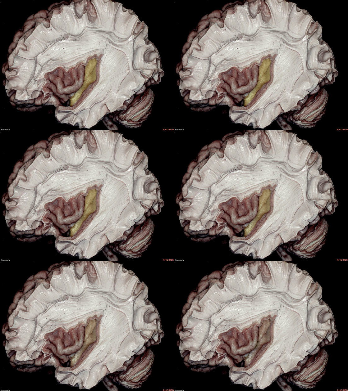
▼岛中央沟 将岛叶分成 岛短回 和 岛长回。
The central insular sulcus divides the insular cortex into short and long insular gyri.
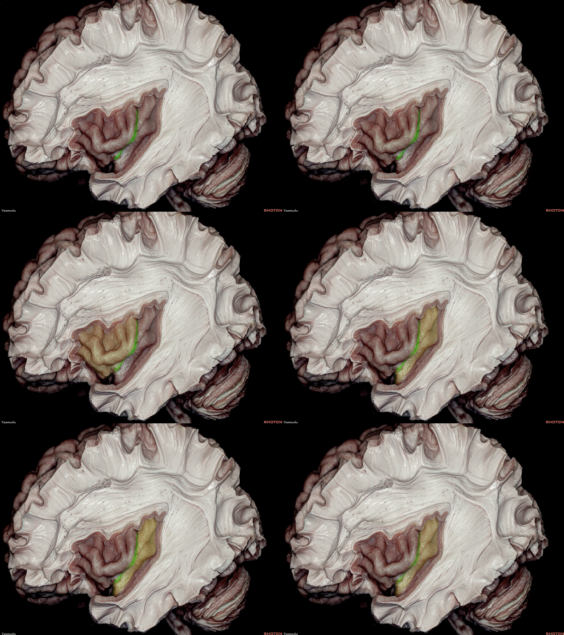
▼岛短回(下图)位于岛中央沟前方。
The short insular gyri are located anterior to the central insular sulcus
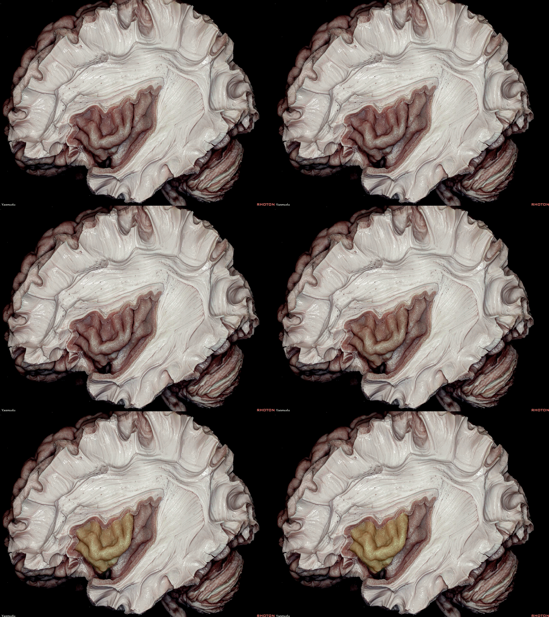
▼岛长回(下图)位于岛中央沟后方。
the long insular gyri are located posterior to the central insular sulcus.
▼岛叶被界沟环绕,分别是 上界沟、下界沟、前界沟。
The insula is encircled by limiting sulci, which are the superior,inferior and anterior limiting sulci.
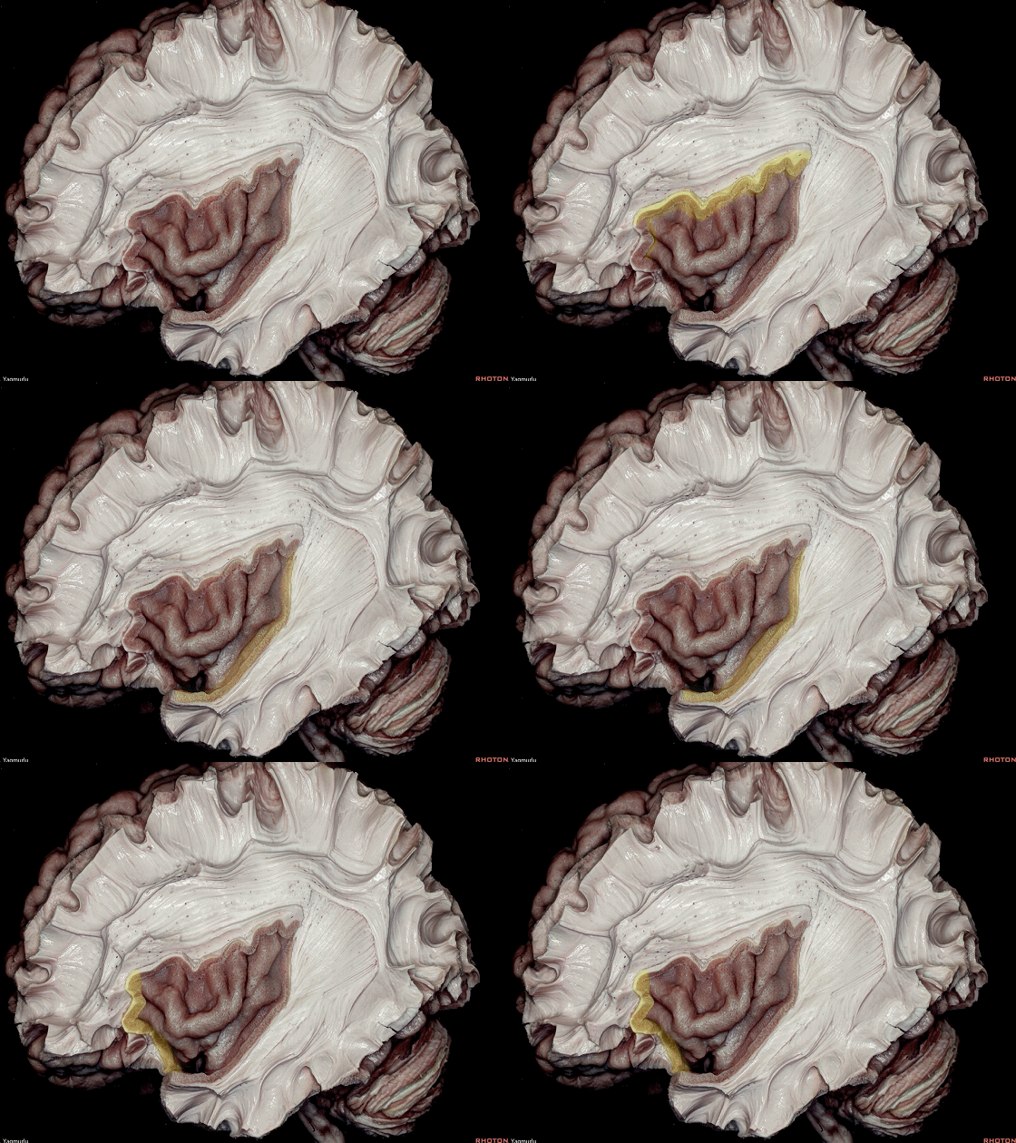
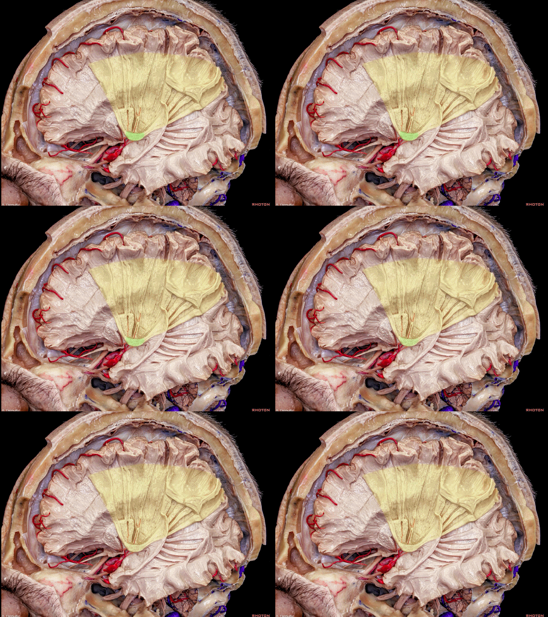
▼前界沟 和 上界沟 的交点称为 岛前点 (下图绿色部分)
The junction of the anterior and superior limiting sulci is called the anterior insular point
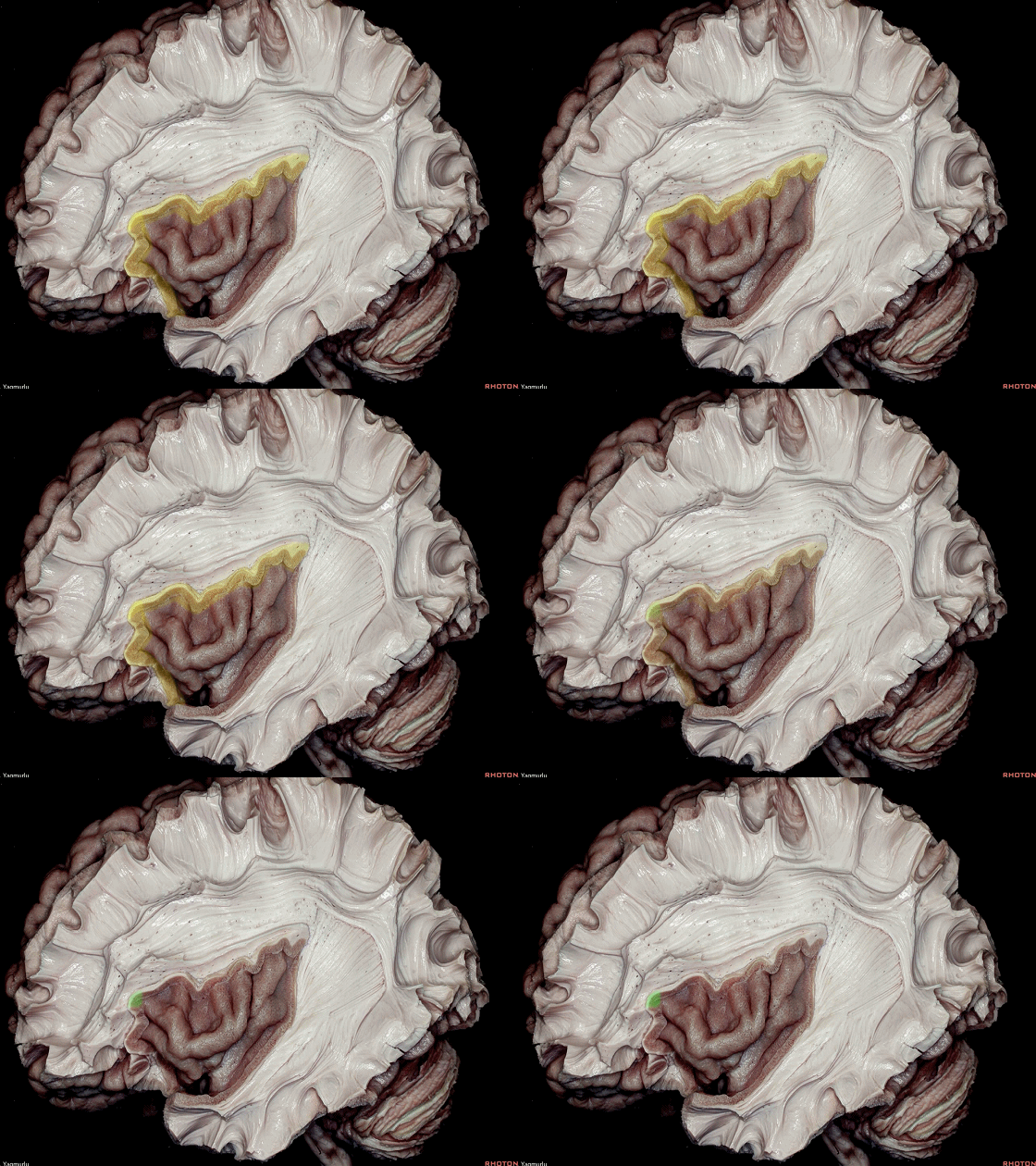
▼上界沟和下界沟的交点称为 岛后点(下图绿色部分)。
上述岛点和界沟是岛叶手术中的重要标志
the junction of the superior and inferior limiting sulci is called the posterior insular point.These points and limiting sulci are important during insular surgery.
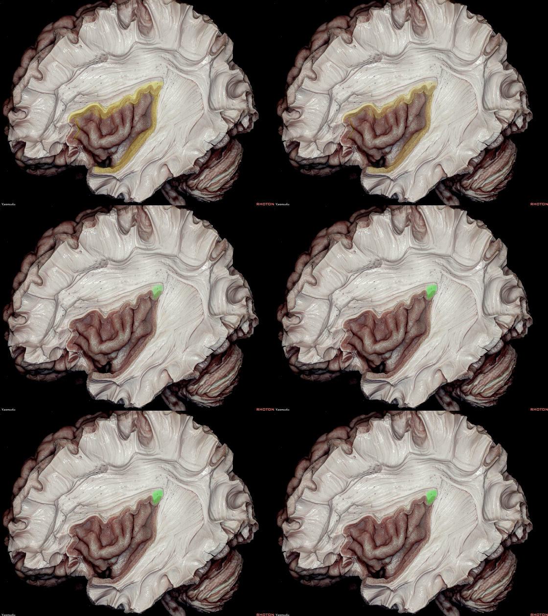

▼在轴位上,我们来看核心区
An axial plane, we are looking at the central core area
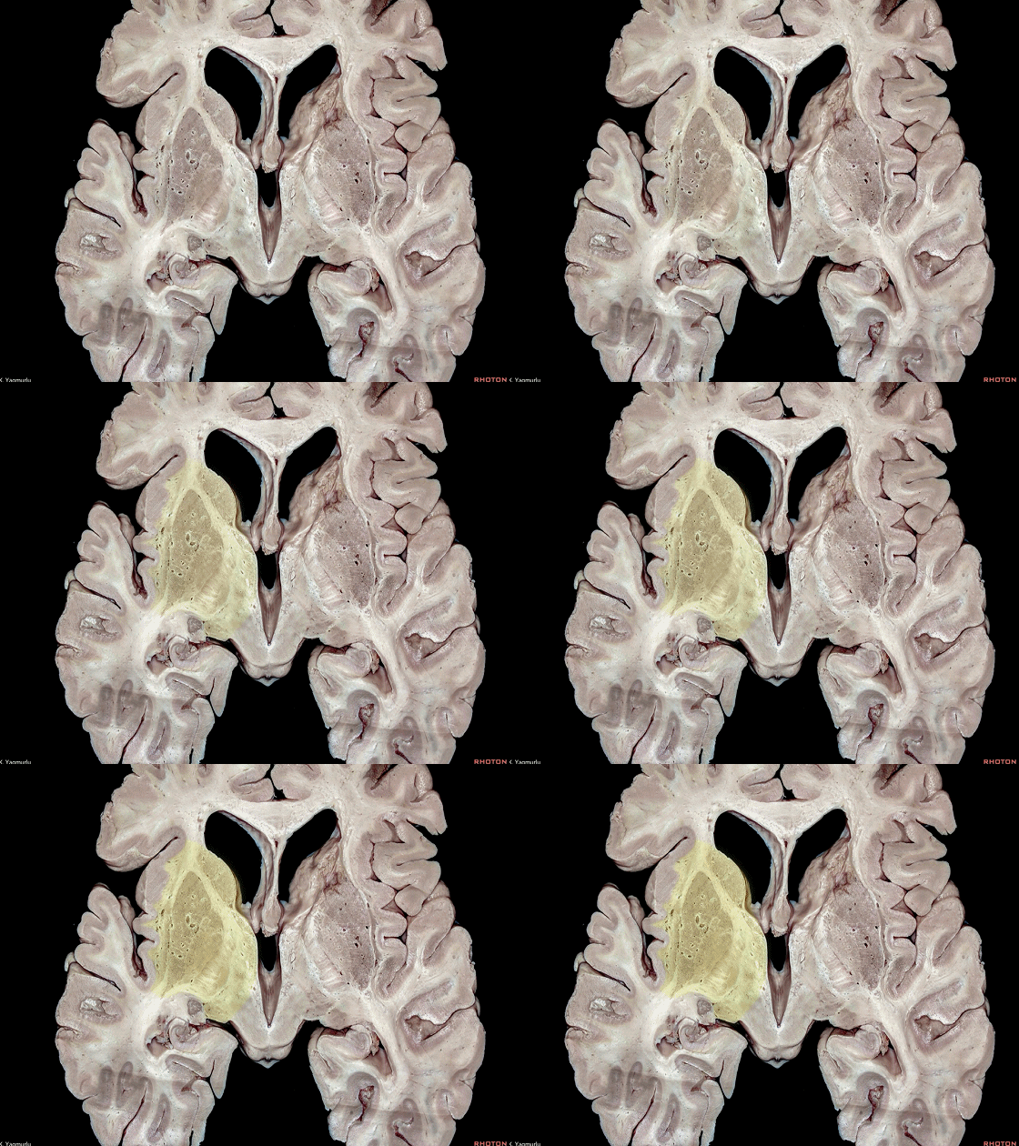
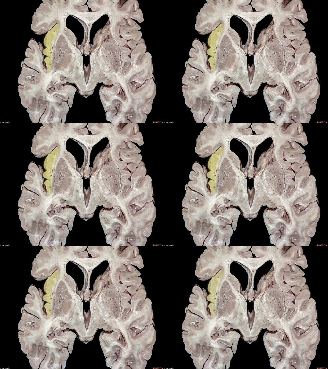
▼核心区外侧是 岛叶皮层(下图)
located between the insular cortex laterally
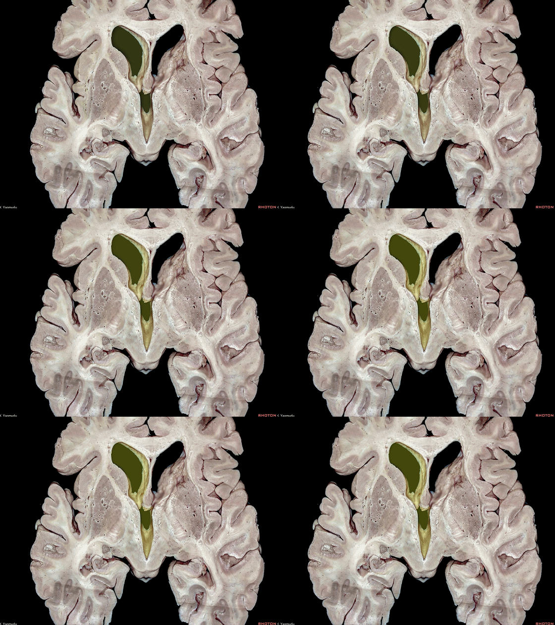
▼核心区内侧是 脑室系统(下图)
and the ventricles medially.
▼核心区包括最外囊、屏状核、外囊、壳核、苍白球、尾状核、内囊、丘脑。 按从外到内分布。
下图示 最外囊
The central core includes the extreme capsule, claustrum, external capsule, putamen, globus pallidus, caudate nucleus,internal capsule and thalamus.are positioned in the central core, from the lateral to medial.
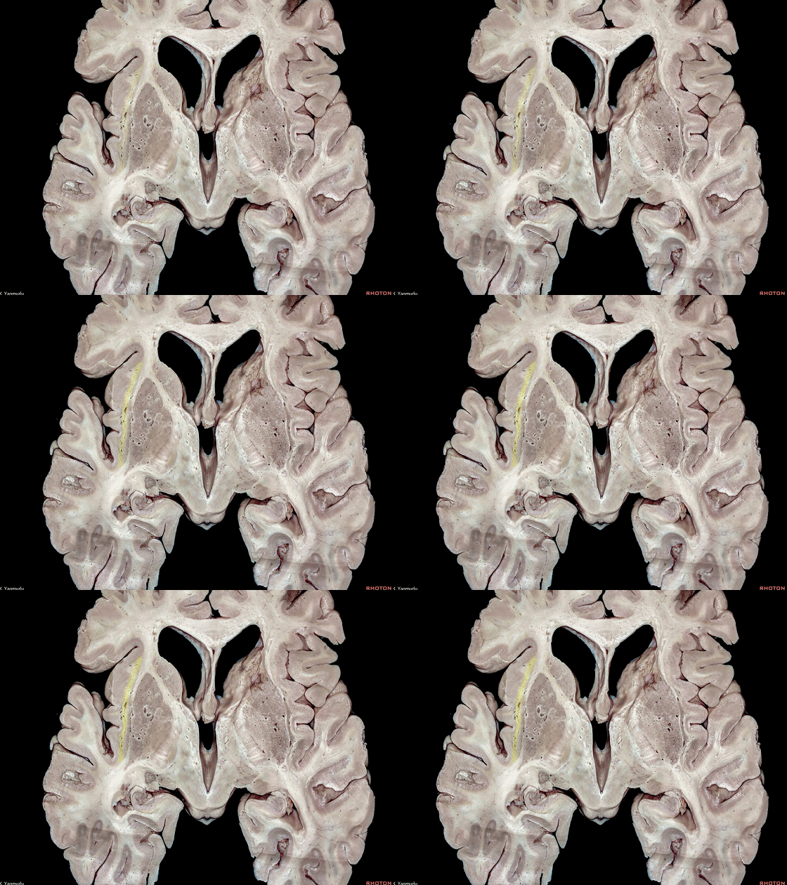
▼这是 屏状核。
claustrum.
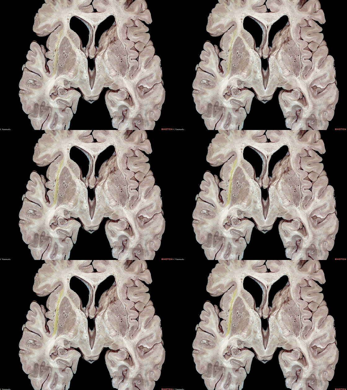
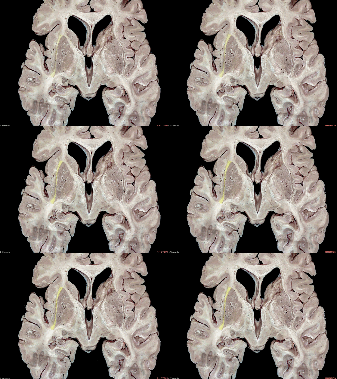
▼这是 外囊
external capsule
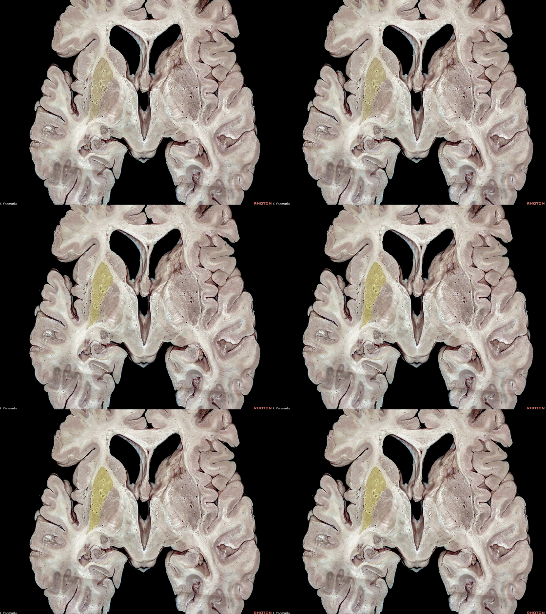
▼这是 壳核。
putamen.
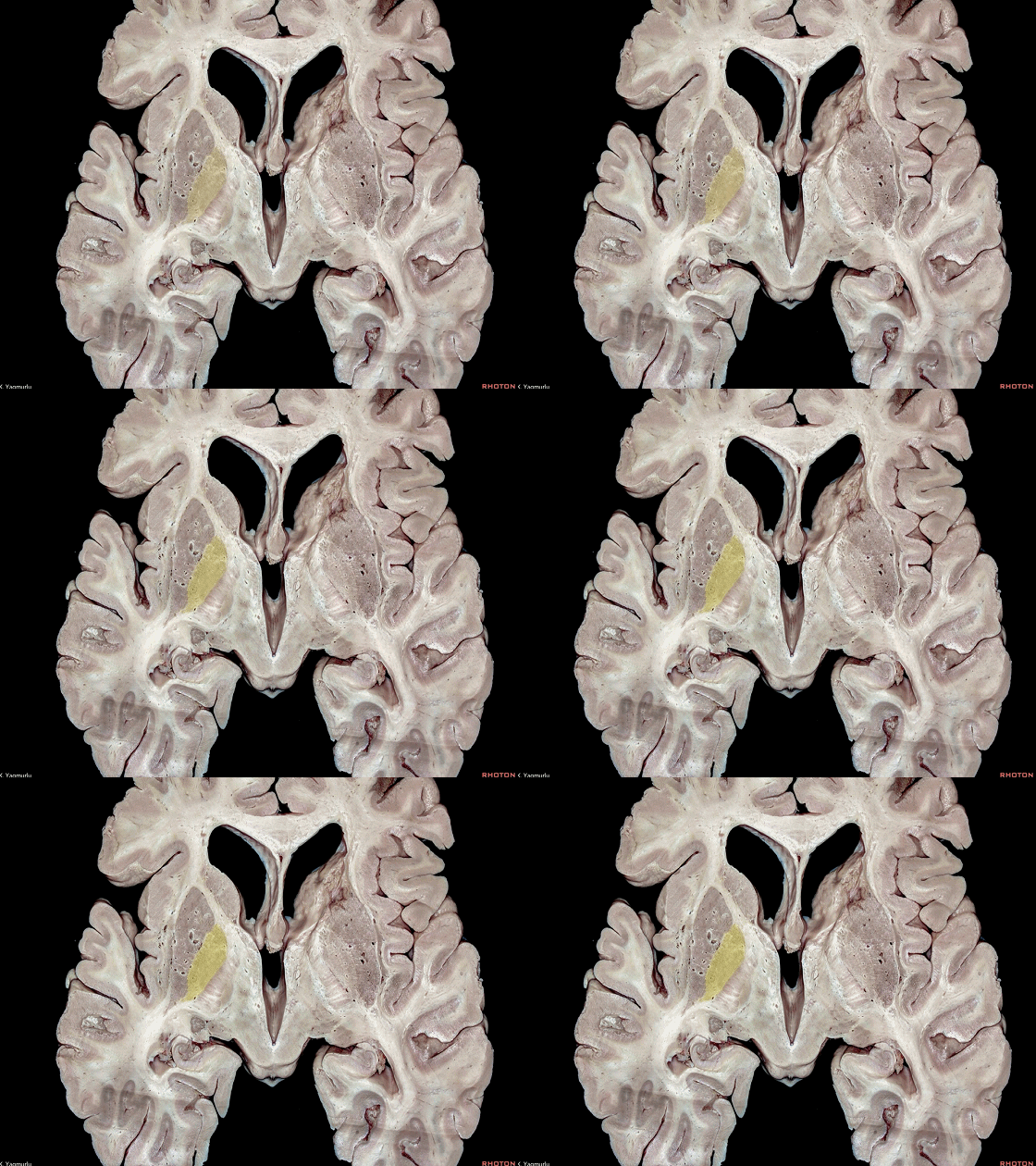
▼这是 苍白球。
globus pallidus.
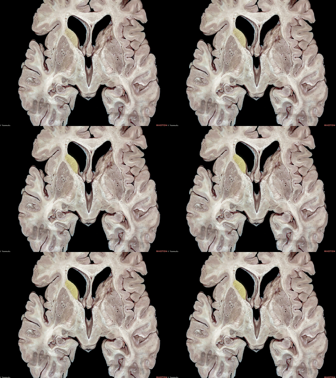
▼这是 尾状核。
caudate nucleus.
▼这是 内囊
internal capsule
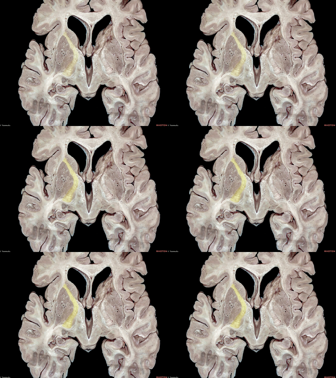
▼这是 丘脑。
thalamus.
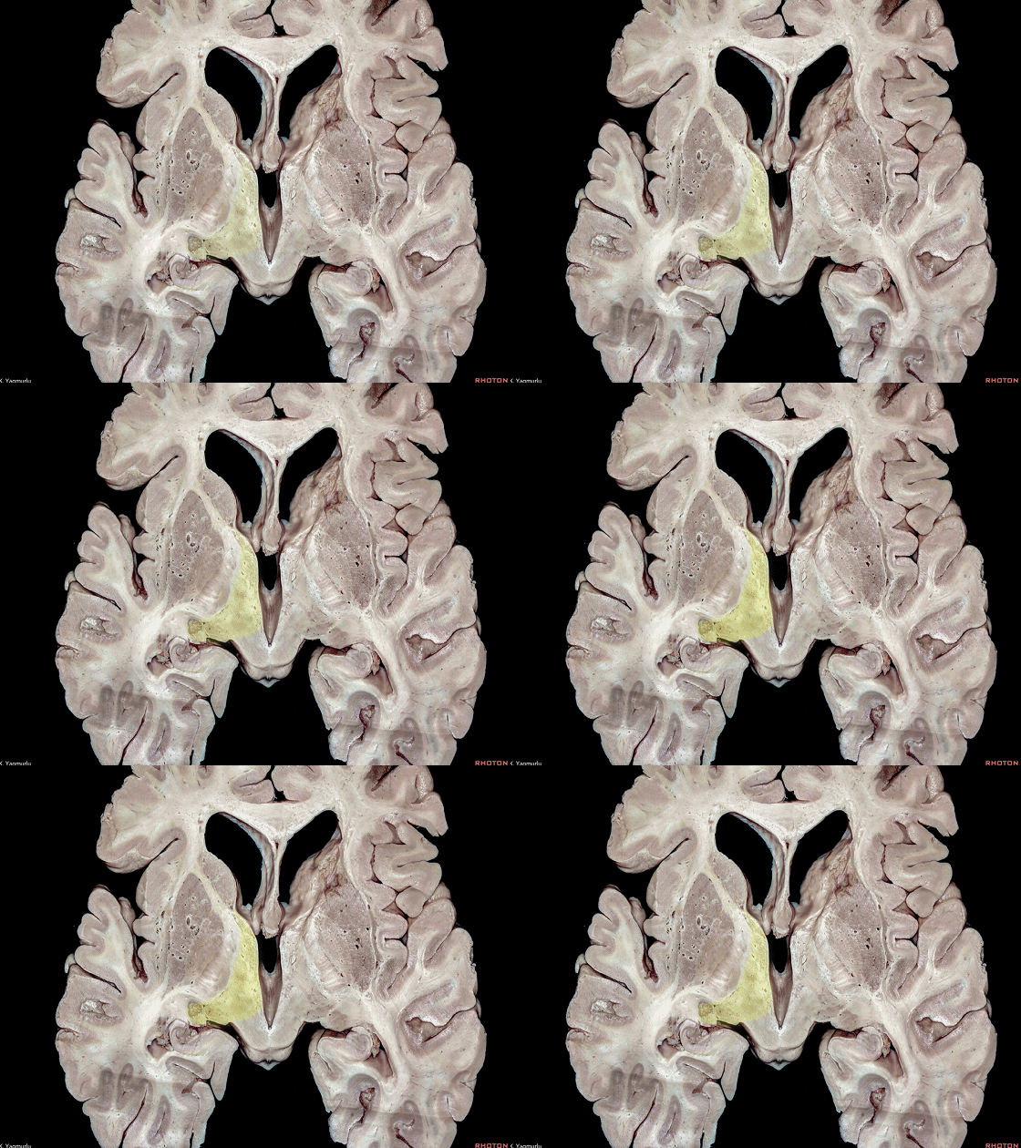
▼从侧面来看,这是 岛叶皮层。
The insular cortex.
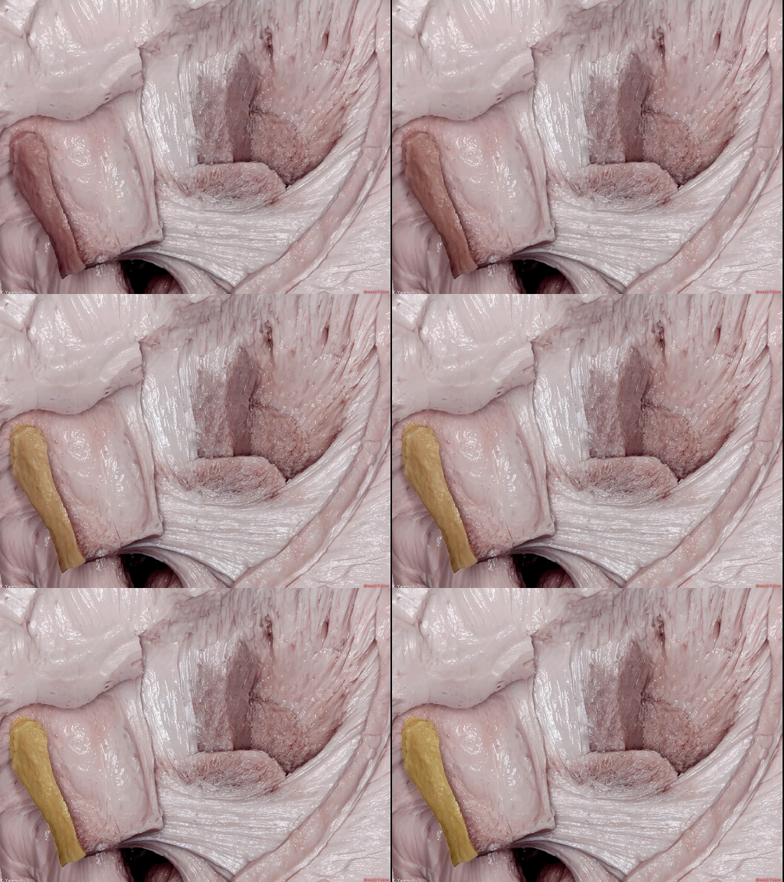
▼这是 最外囊。
extreme capsule.
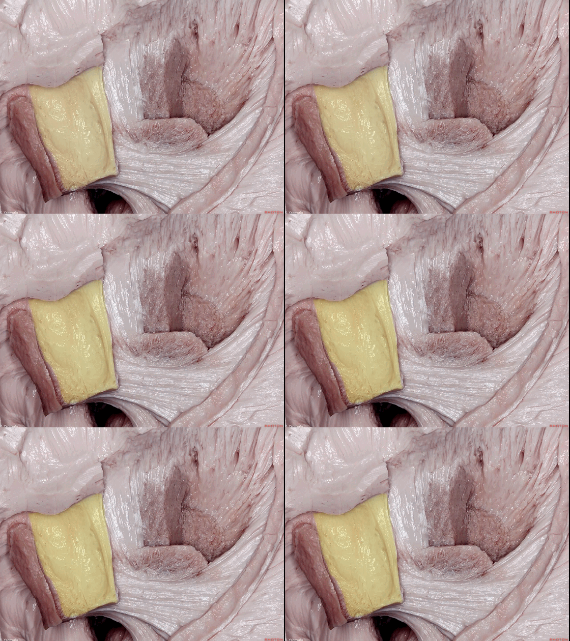
▼这是 屏状核。
claustrum.
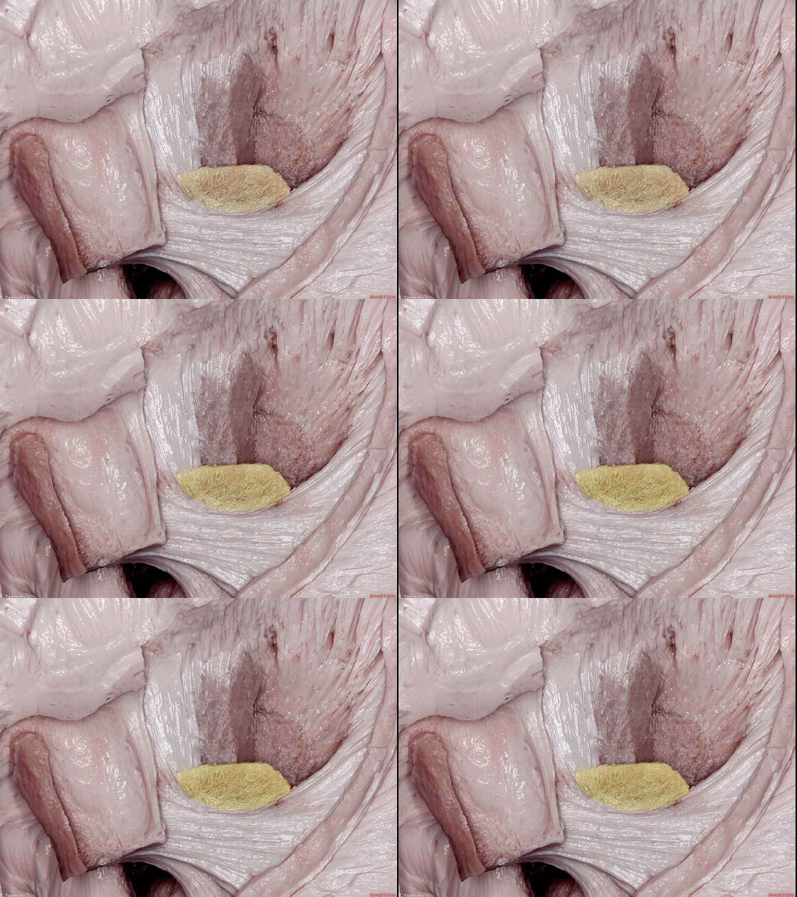
▼这是 外囊。
external capsule.
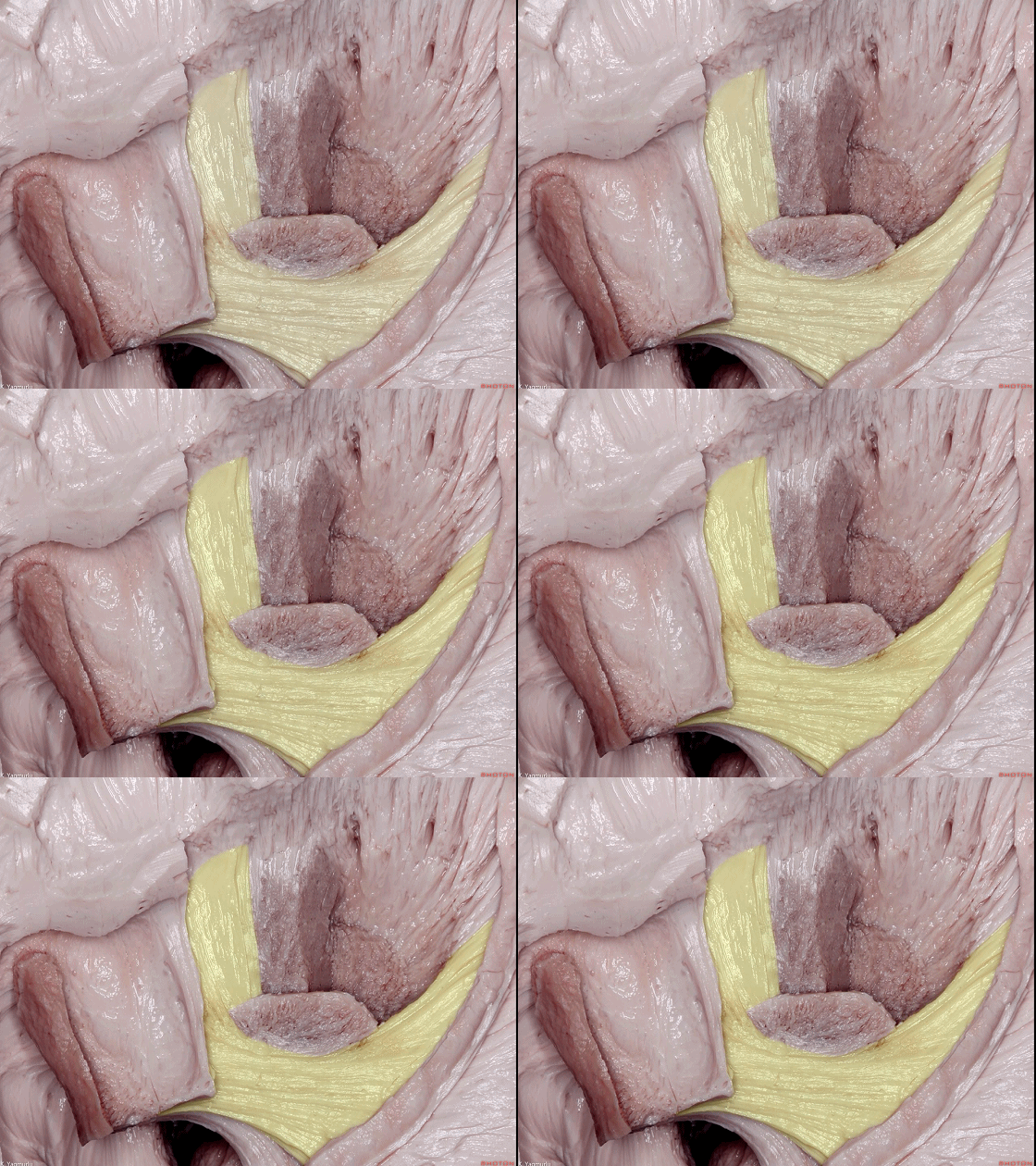
▼这是 壳核。
putamen.
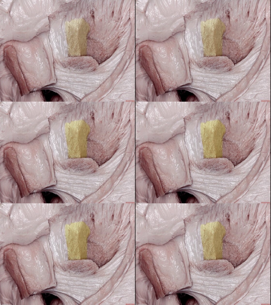
▼这是 苍白球。
globus pallidus.
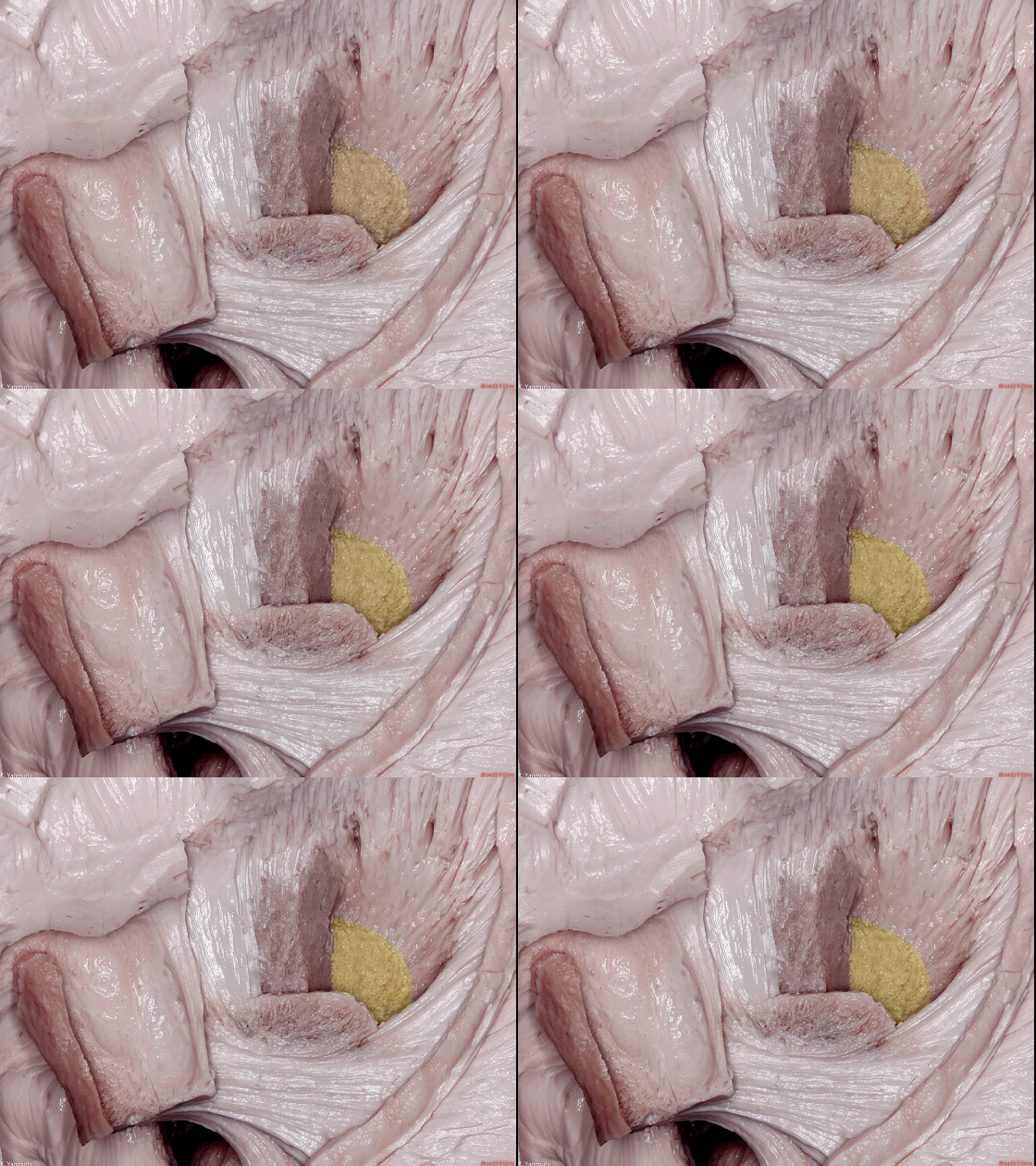
▼这是 内囊。
internal capsule.
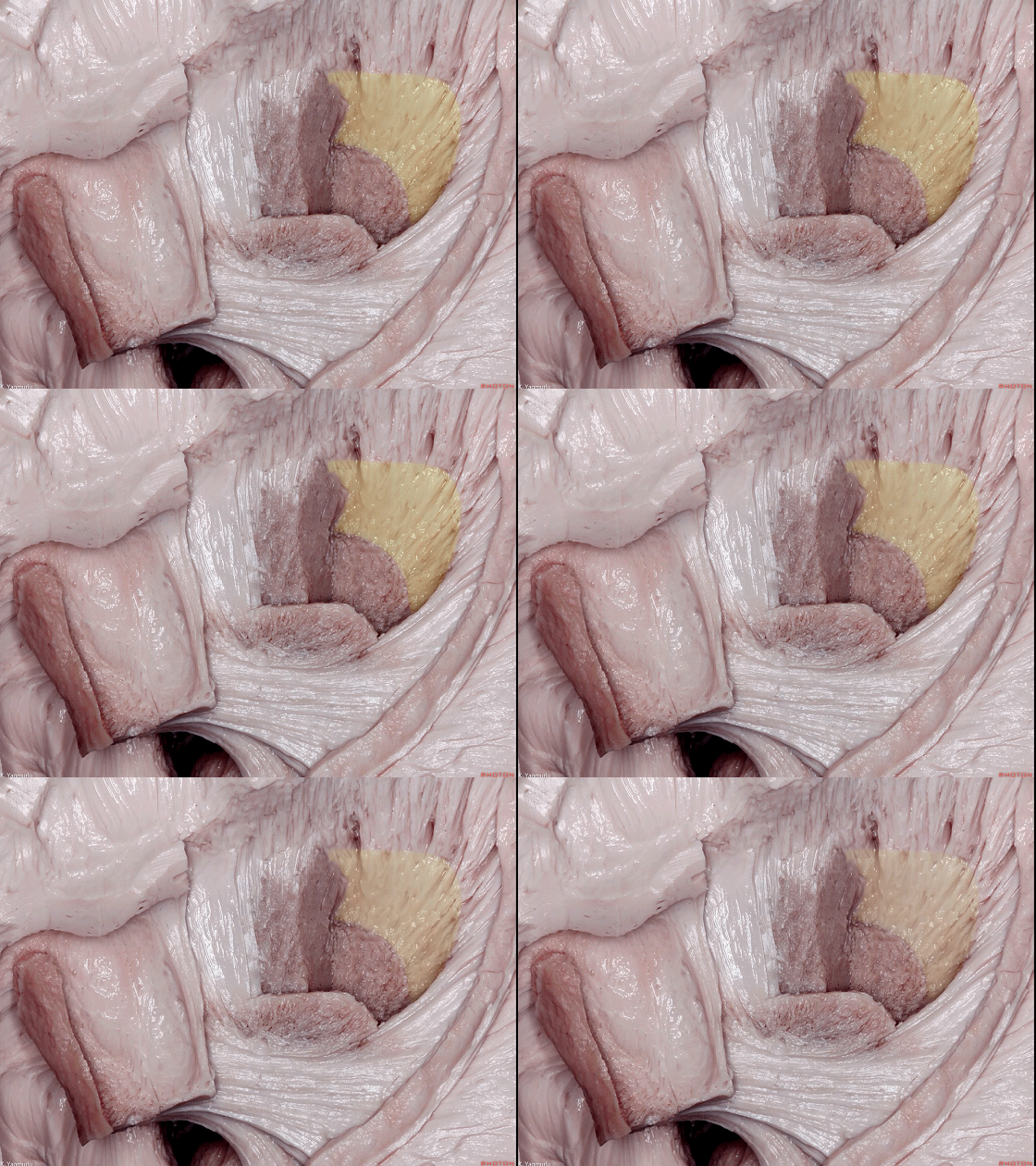
▼去除岛叶皮层,即可见 最外囊(下图),由短联络纤维构成,连接岛回与岛盖。
最外囊的功能尚未明了,但目前认为是语义性语言通路的一部分。
After removal of the insular cortex, we can see the extreme capsule, which is composed of short association fibers providing the connections among the insular gyri and the operculi. The function of the extreme capsule is not clear, but it has been suggested as a part of the semantic language stream.
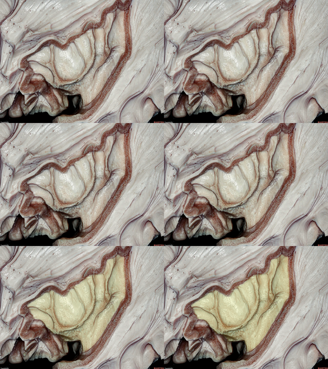
▼继续深入,外囊 和 屏状核 即显露于最外囊的深面。
下图示 外囊。
In further dissection, the external capsule and claustrum were exposed by removing of the extreme capsule.
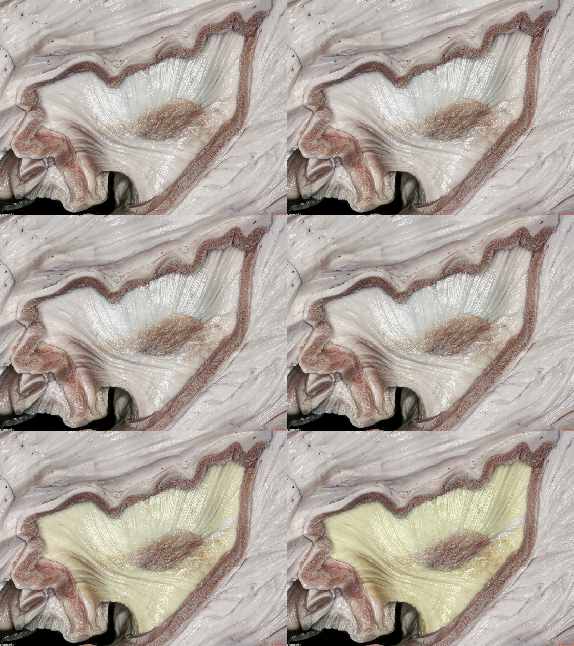
▼下图示 屏状核。
claustrum .
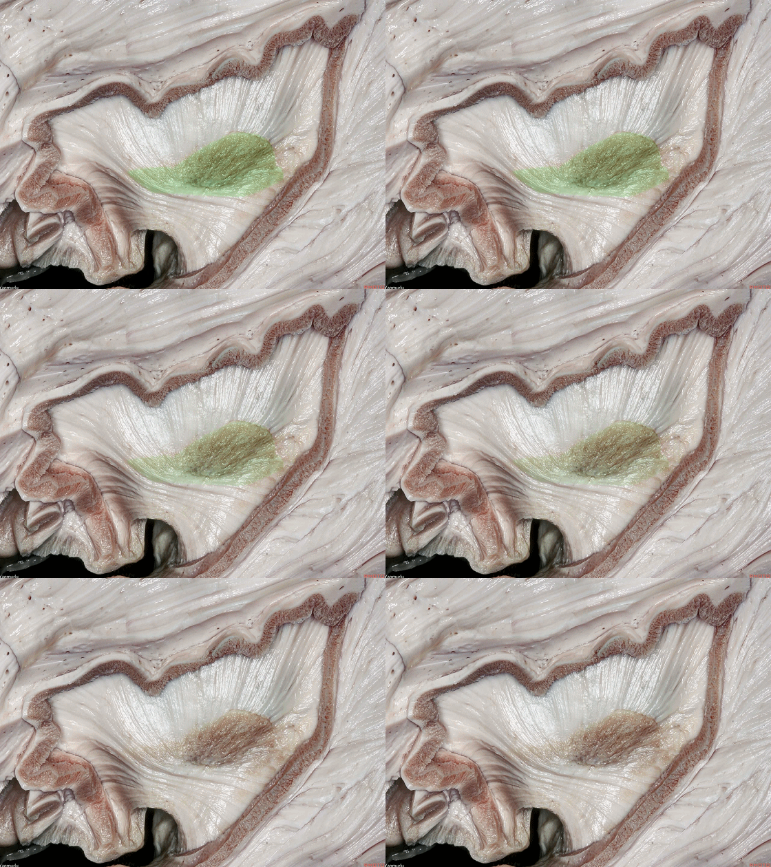
▼屏状核与外囊可进一步分为 背侧部 和 腹侧部 。
The claustrum and external capsule can be divided into dorsal and ventral parts.
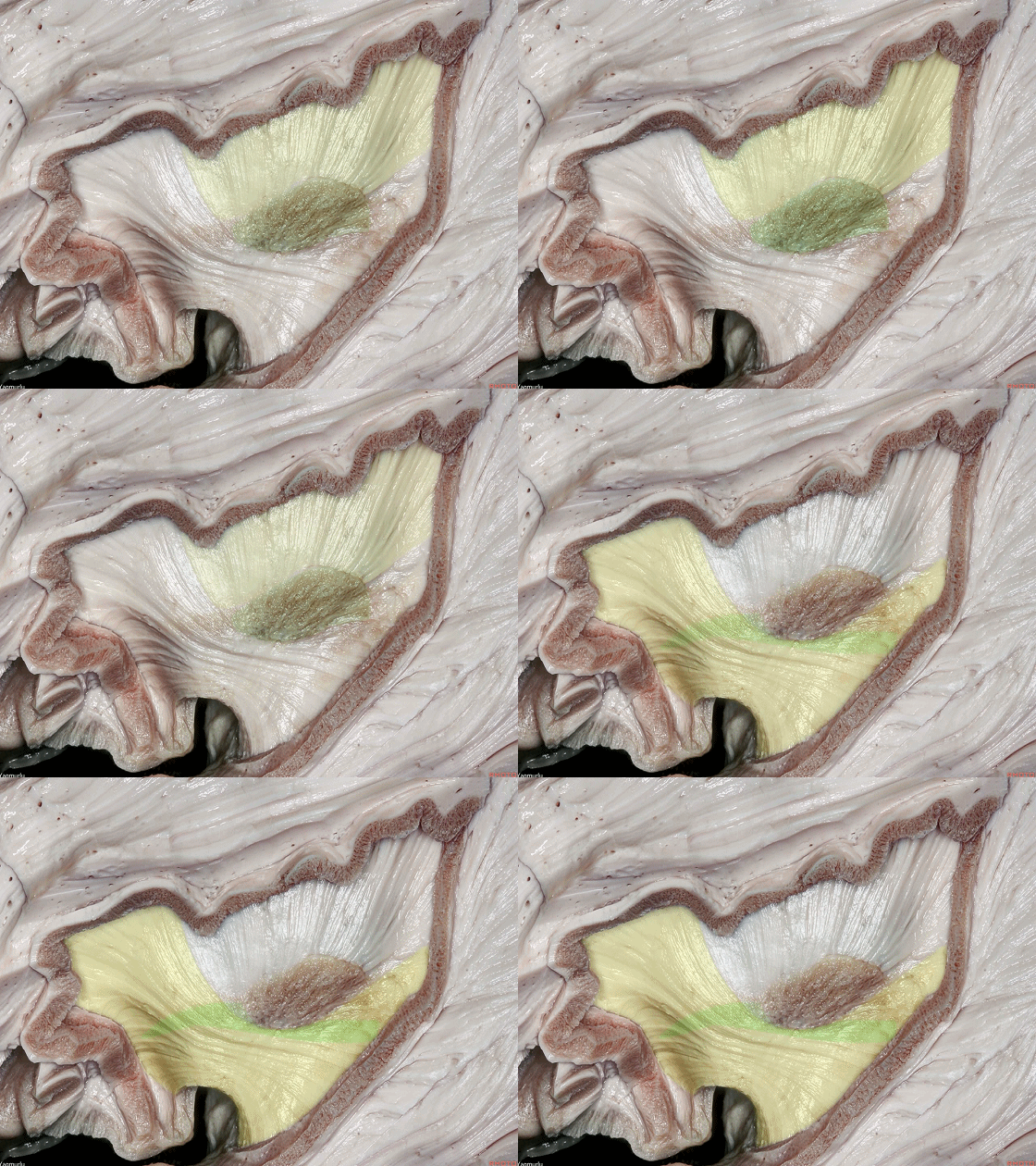
▼这是 背侧屏状核。
The dorsal claustrum is here.
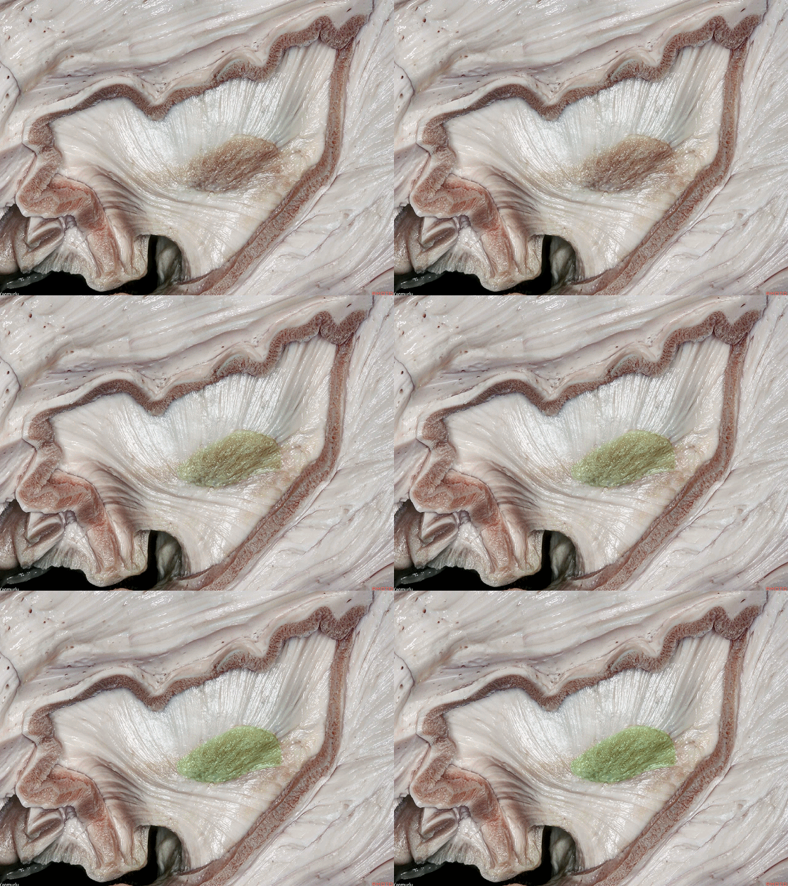
▼这是 背侧外囊,其由屏状皮质纤维构成。
and the dorsal external capsule,which is formed by claustrocortical fibers, is here.

▼这是 腹侧屏状核。
The ventral claustrum.
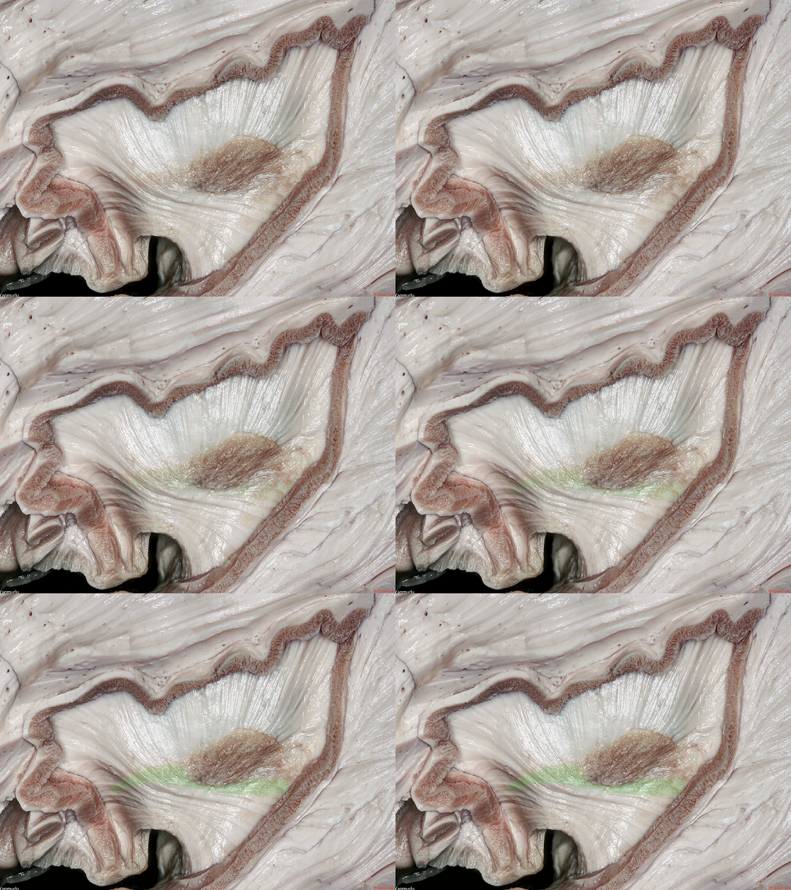
▼腹侧屏状核发出纤维进入位于岛阈的 腹侧外囊(下图),连接杏仁核。
The ventral claustrum spreads out into the ventral external capsule at the limen insula to reach the amygdala.
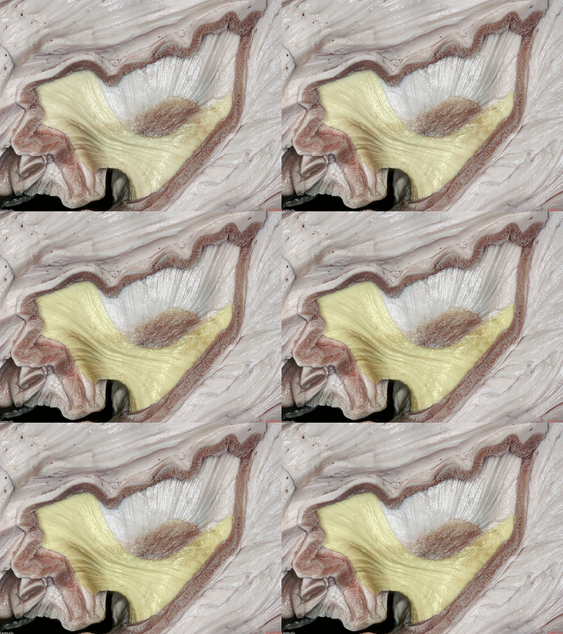
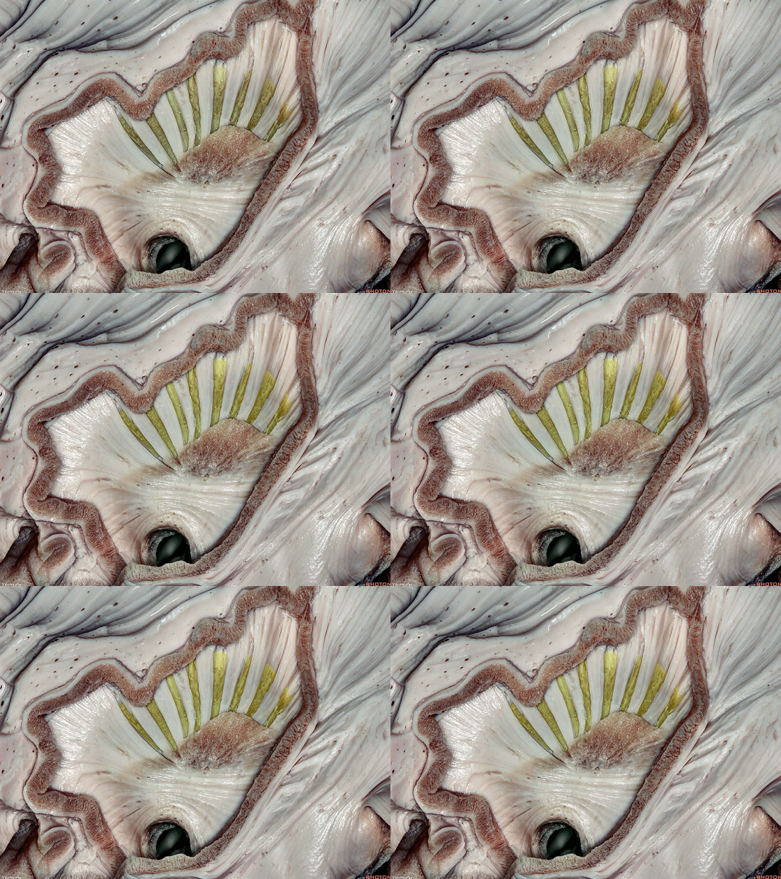
▼屏状皮质纤维 构成了 背侧外囊(下图)
The claustrocortical fibers,which is the dorsal external capsule,
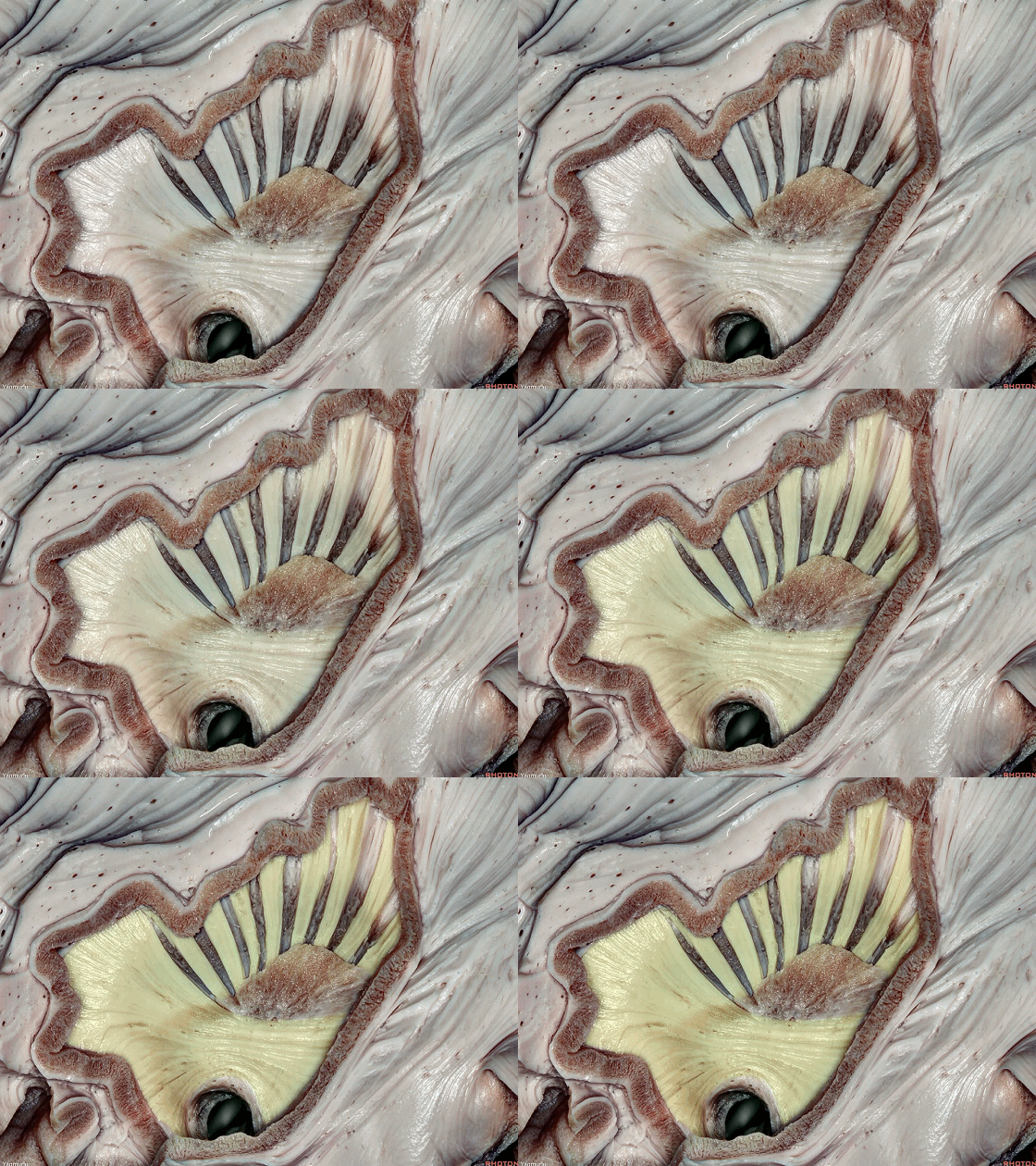
▼背侧外囊 沟通 背侧屏状核(下图)与辅助运动区至顶叶之间的皮层联系。
屏状皮质系统参与视觉、体感和运动信号的整合。
the dorsal external capsule course from the dorsal claustrum to the cortex between the supplementary motor area anteriorly and the parietal lobe posteriorly.The claustrocortical system has been related to the integration of visual, somatosensory and motor informations.
▼下图示 外囊
▼外囊的内侧为壳核(下图)。壳核属于基底节结构,其主要功能为介导运动和学习。
Just medial to the external capsule is the putamen. The putamen is one of the basal ganglia structures. Its main function is to regulate movements and learning.
▼另一基底节结构为苍白球(下图),其位于壳核的内侧。
Another basal ganglia structure is the globus pallidus, which is located just medial to the putamen.
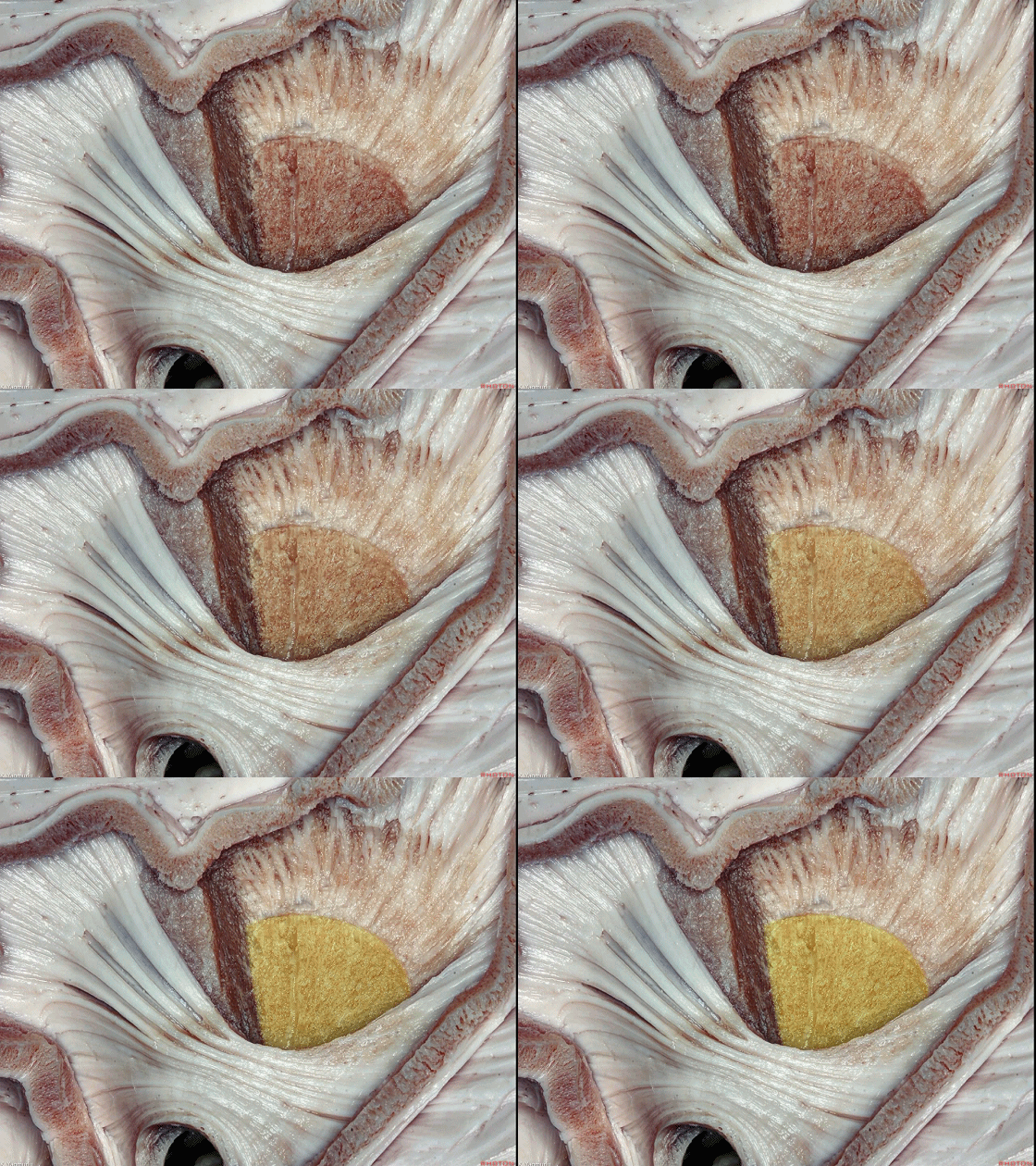
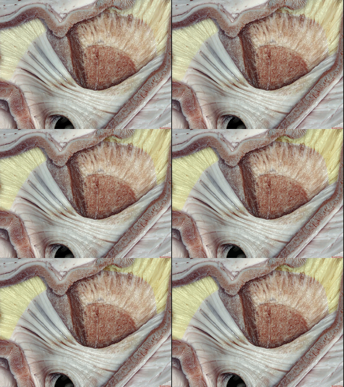
▼下图中,壳核(下图)的后半部已被切除,从而显露出苍白球和内囊。
In this picture, the posterior half of the putamen has been removed to expose the globus pallidus and internal capsule.
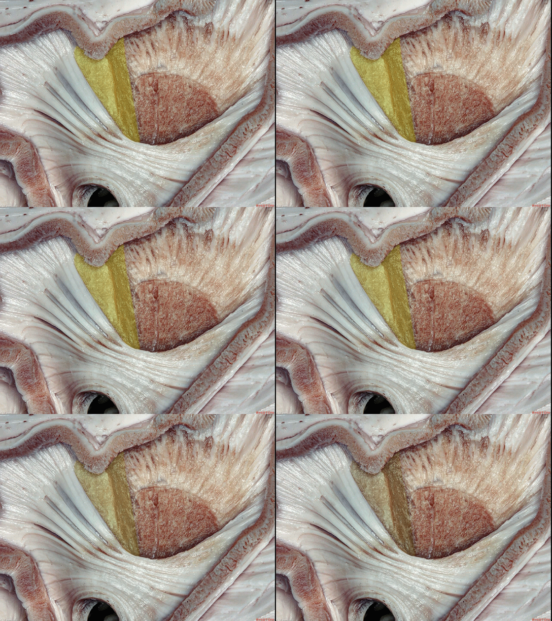
▼这是 内囊。
internal capsule.
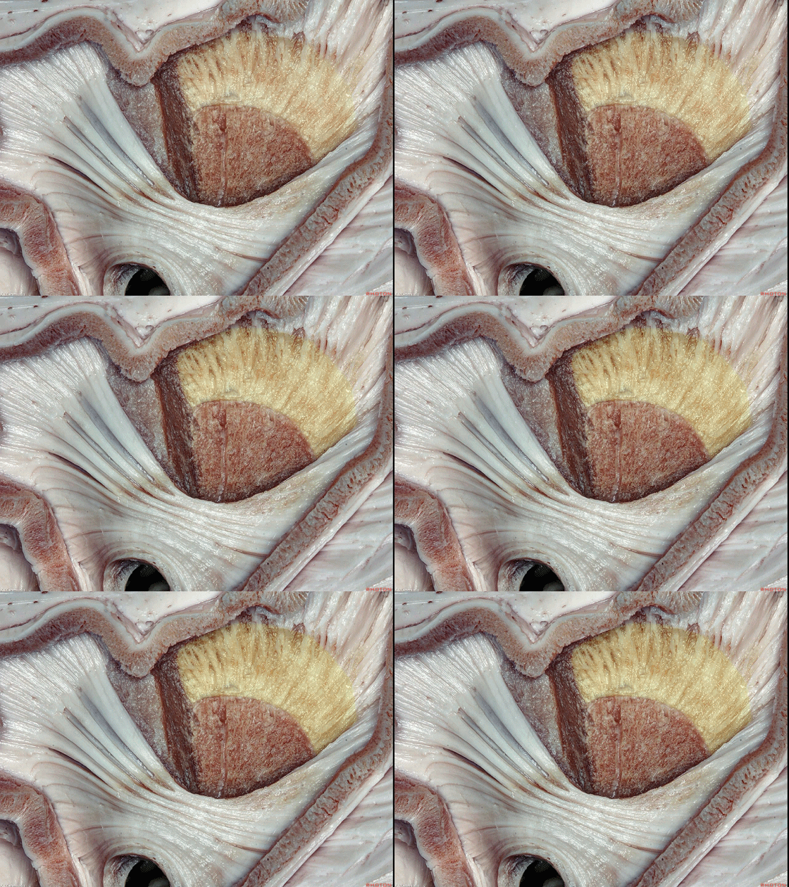
▼这是 外囊。
external capsule.
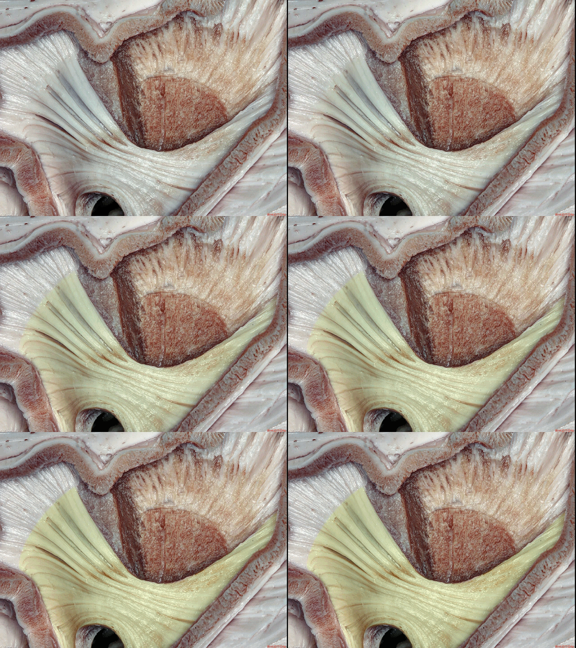
▼外囊和内囊在壳核层面以上共同形成 放射冠(下图)。
The external and internal capsule come together to form the corona radiata above the putamen.
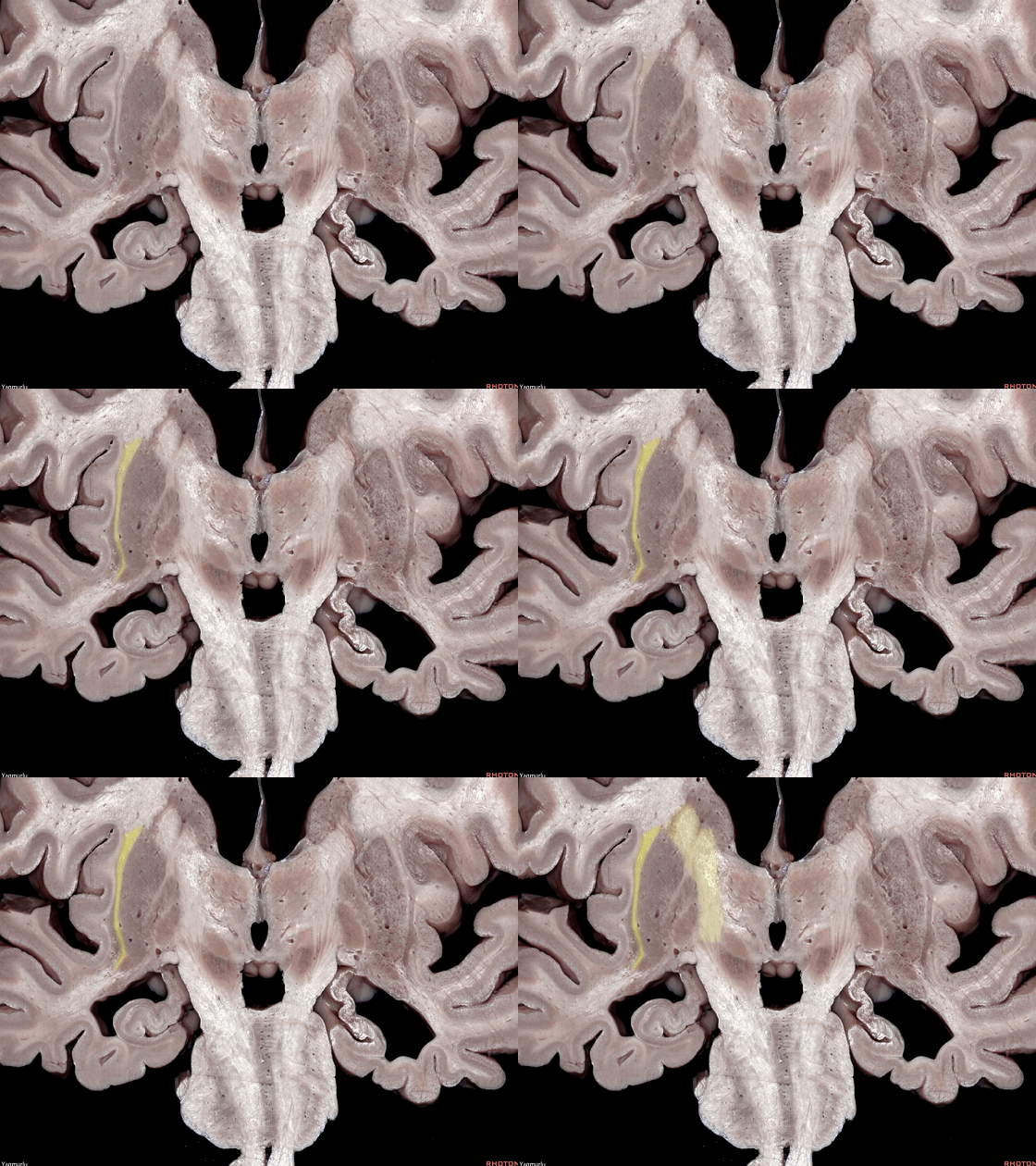
▼在冠状位上,可见 外囊(下图)和 内囊(下图)。
A coronal section, what we can see here is that the external and internal capsules.
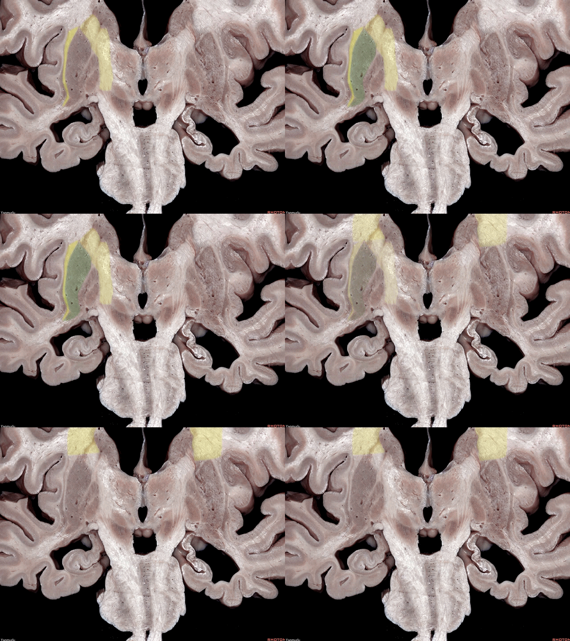
▼外囊 和 内囊 相汇聚在壳核上方形成 放射冠 。
the external and internal capsules come together above the putamen above the putamen to form the corona radiata.
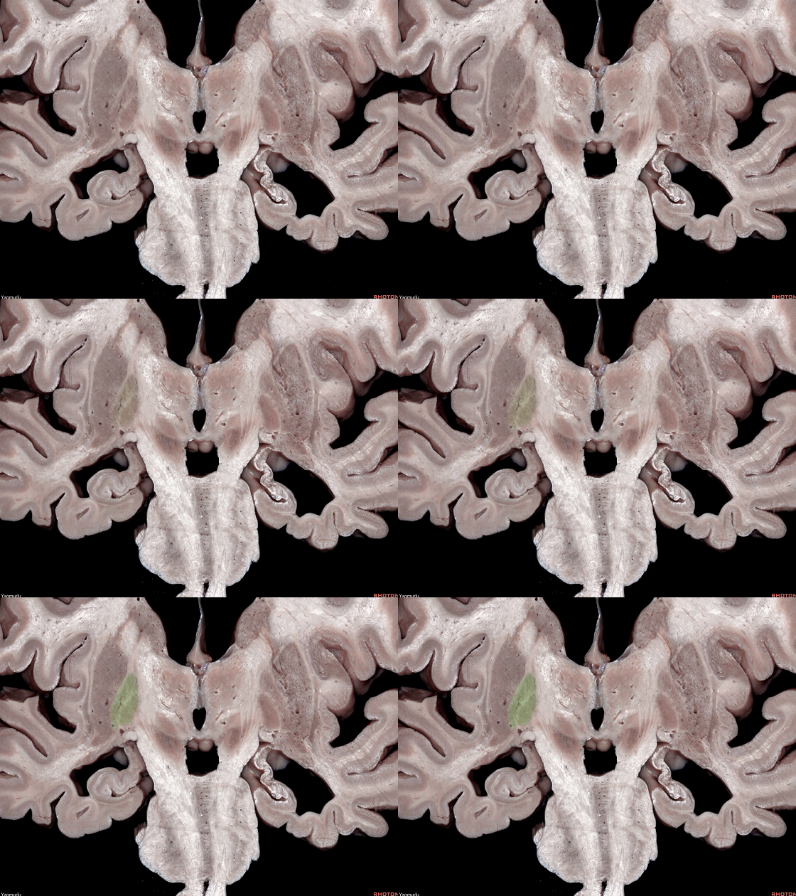
▼另外,苍白球(下图)位于壳核的内侧。
Also, the globus pallidus is situated medial to the putamen.
▼这是 腹侧外囊。
The ventral external capsule.
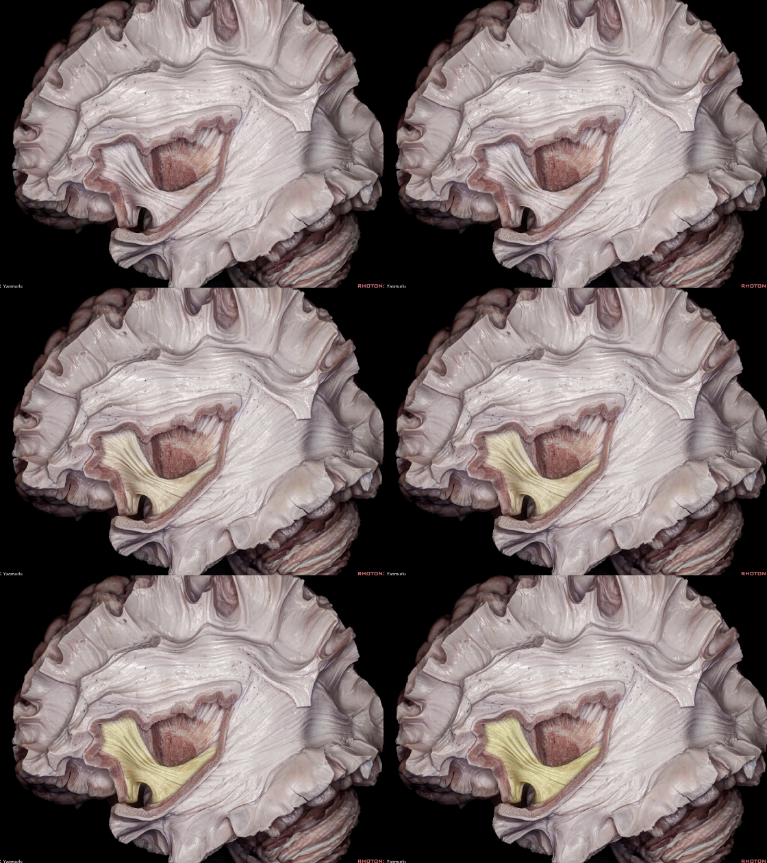
▼腹侧外囊包含 上方的下额枕束 和下方的位于岛阈处的钩束。
The ventral external capsule is formed by the inferior fronto-occipital fasciculus above and the uncinate fasciculus below at the limen insula.
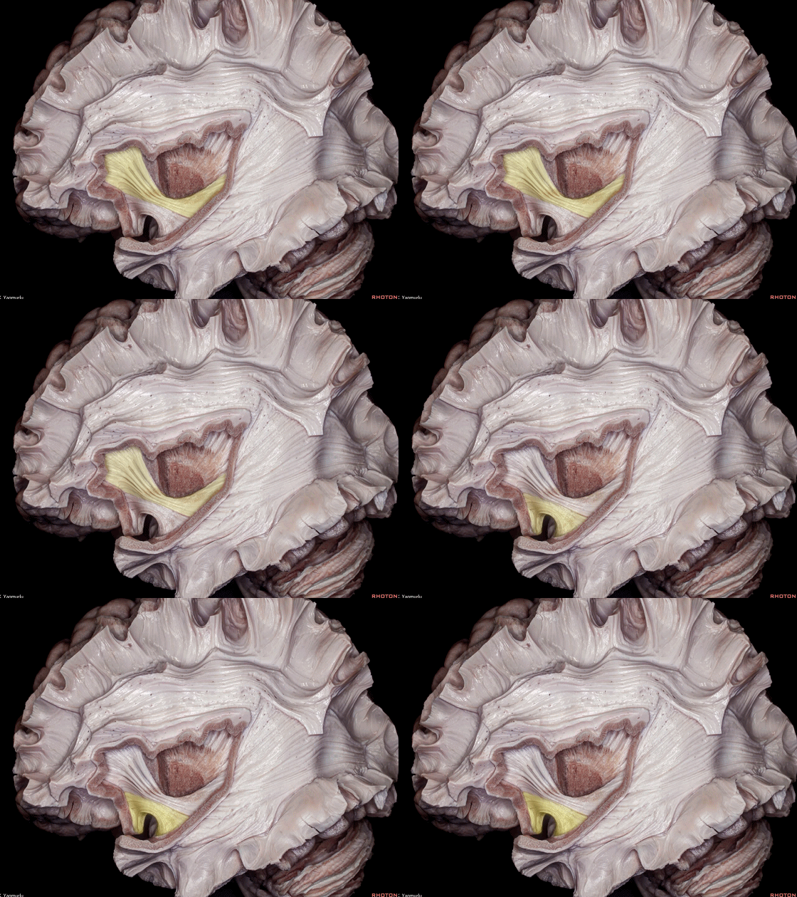
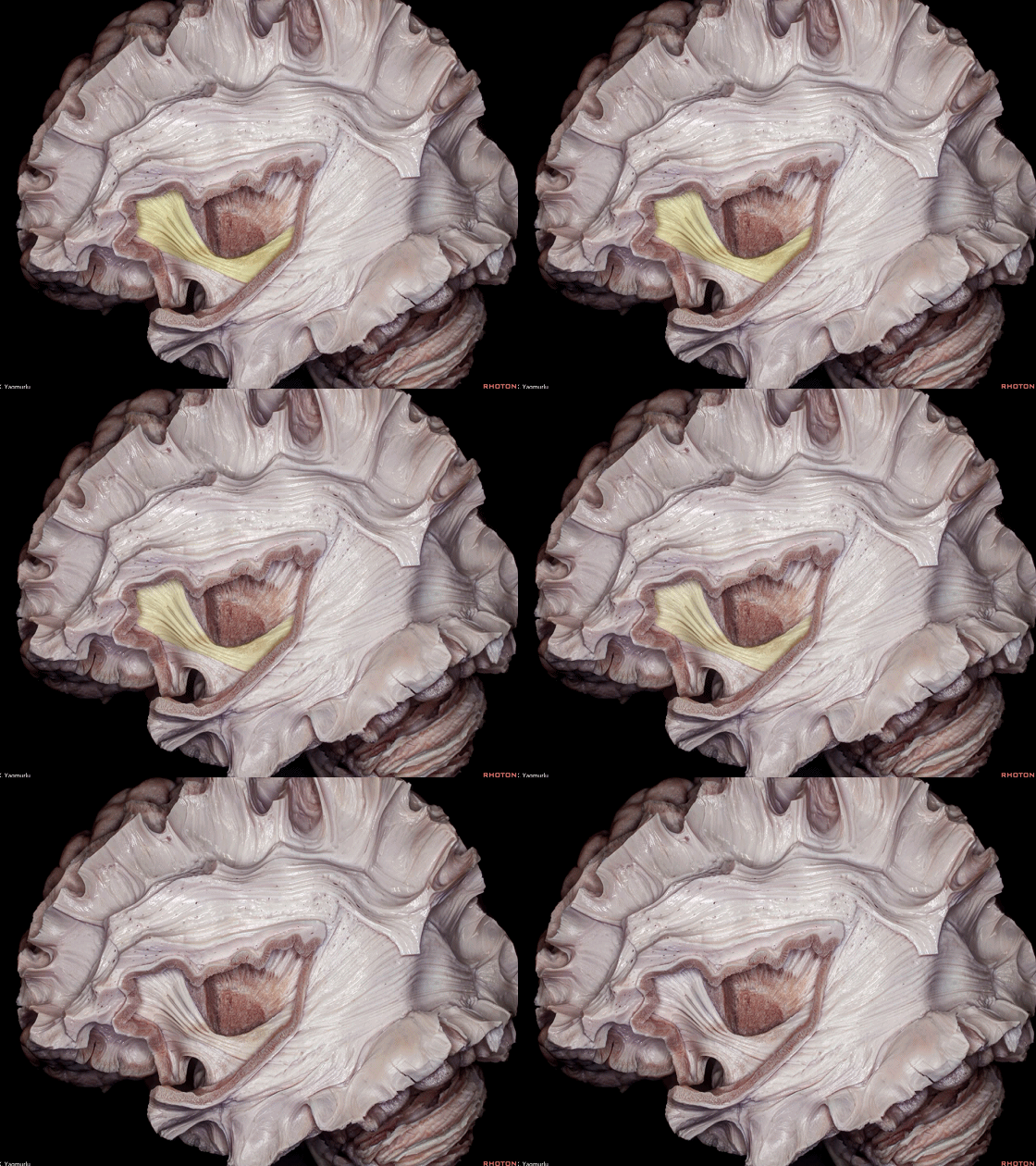
▼下额枕束(下图)连接 额叶 和 枕叶。
The inferior fronto-occipital fasciculus courses between the frontal and occipital lobes.
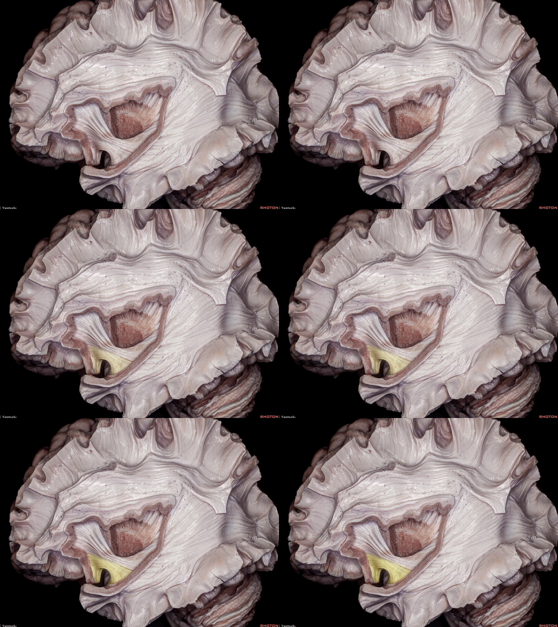
▼钩束(下图)连接 眶额区 与 颞极,形态如钩状。
The uncinate fasciculus connects the orbitofrontal area to the temporal pole, forming a hook shape.
▼去除壳核和下额枕束可完整显露苍白球和前连合。
下图示苍白球。苍白球恰居于前连合后方,参与自主运动的调控。苍白球毁损术常用于某些运动障碍性疾病的治疗。
Removal of the putamen and inferior fronto-occipital fasciculus exposes the entire globus pallidus and anterior commissure.The globus pallidus sits just behind the anterior commissure and plays in the regulation of voluntary movements.The procedure known as pallidotomy is used to treat some movement disorders.

▼下图示 前连合。
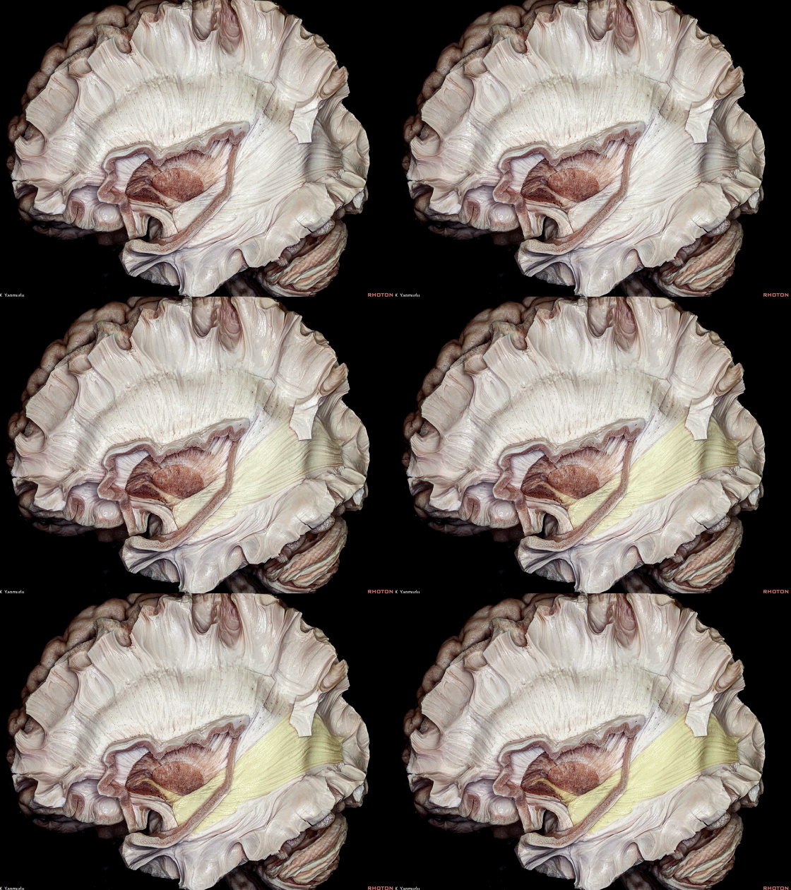
▼前连合位于内囊前支(下图)腹侧面的下方。
The anterior commissure courses below the ventral aspect of the anterior limb of the internal capsule.
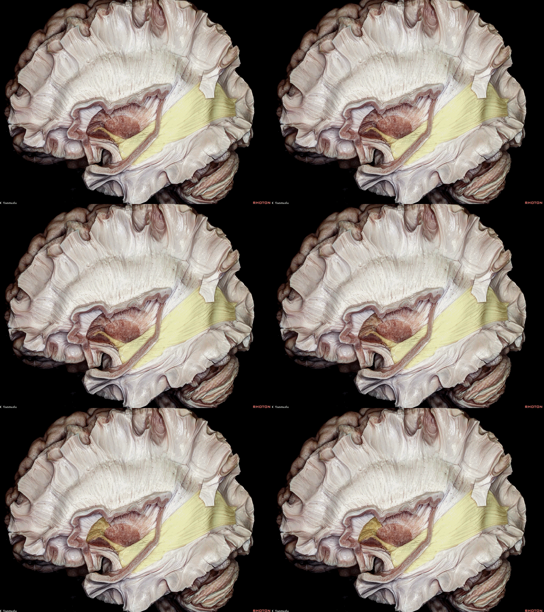
▼在半球内,前连合向尾端、外侧走行穿过苍白球腹侧面的Gratiolet管。其向下行至颞极(下图),走行于钩束后方。
In the hemisphere, it moves caudally and passes laterally through the ventral aspect of the globus pallidus in the "Gratiolet's canal".It courses downward to the temporal pole just behind the uncinate fasciculus.
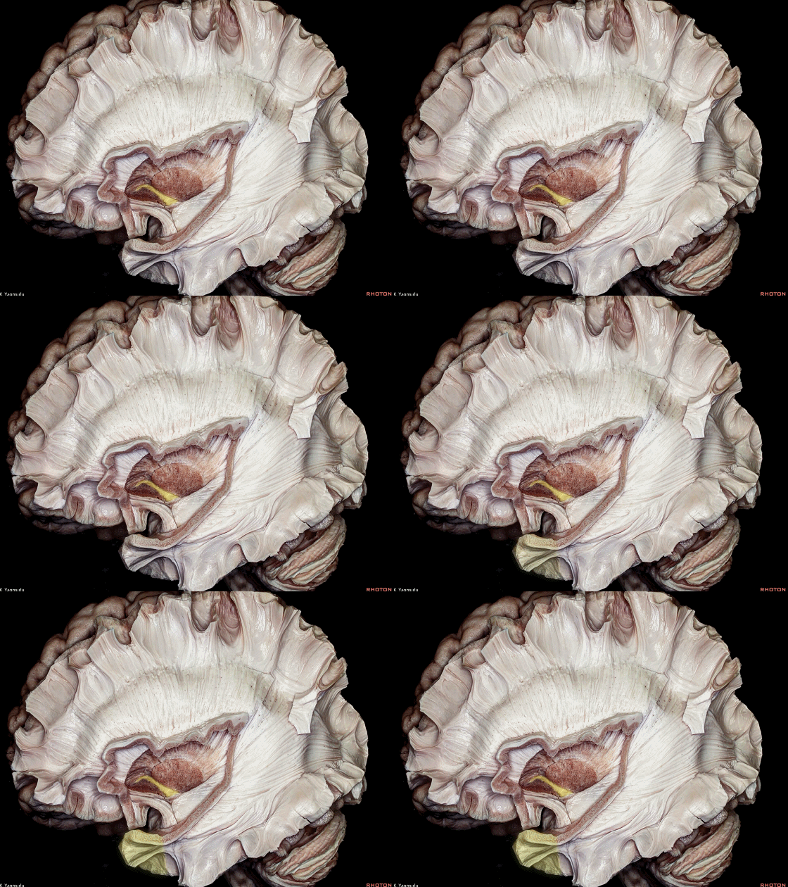
▼下图示上述结构的放大观。
内囊前支、内囊膝部、内囊后支均已显露。
Enlarged view of previous slide. The anterior limb, genu and posterior limb of the internal capsule are observed.
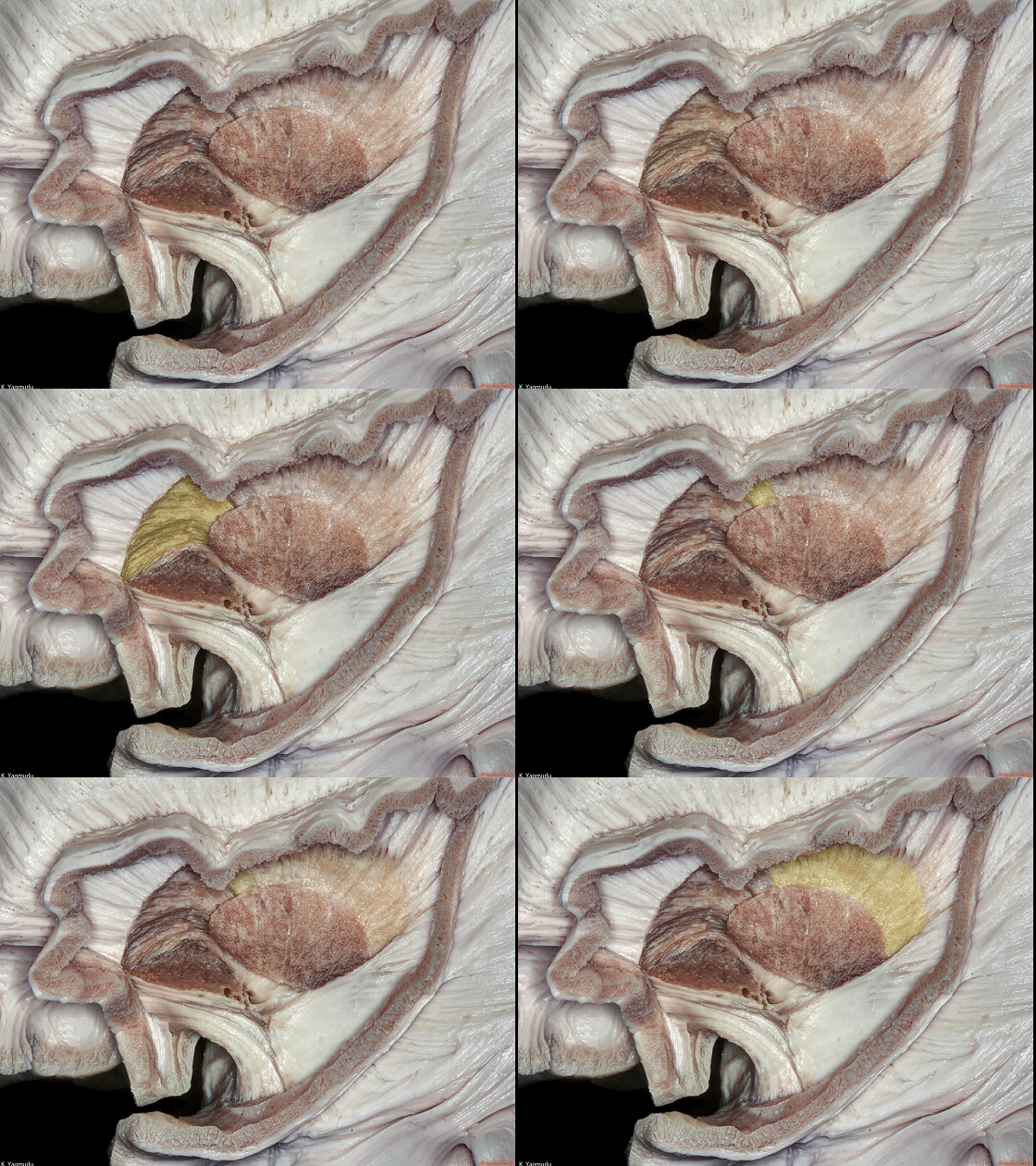
▼无名质(基底前脑 下图)位于前连合的前下方。
The substantia innominata (basal forebrain) is located in front and beneath the anterior commissure.
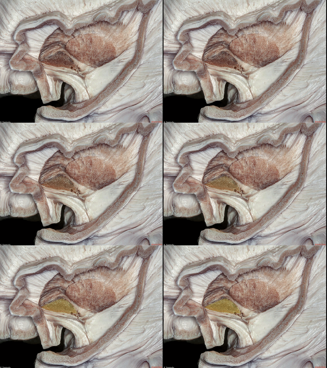
▼这是 前连合。
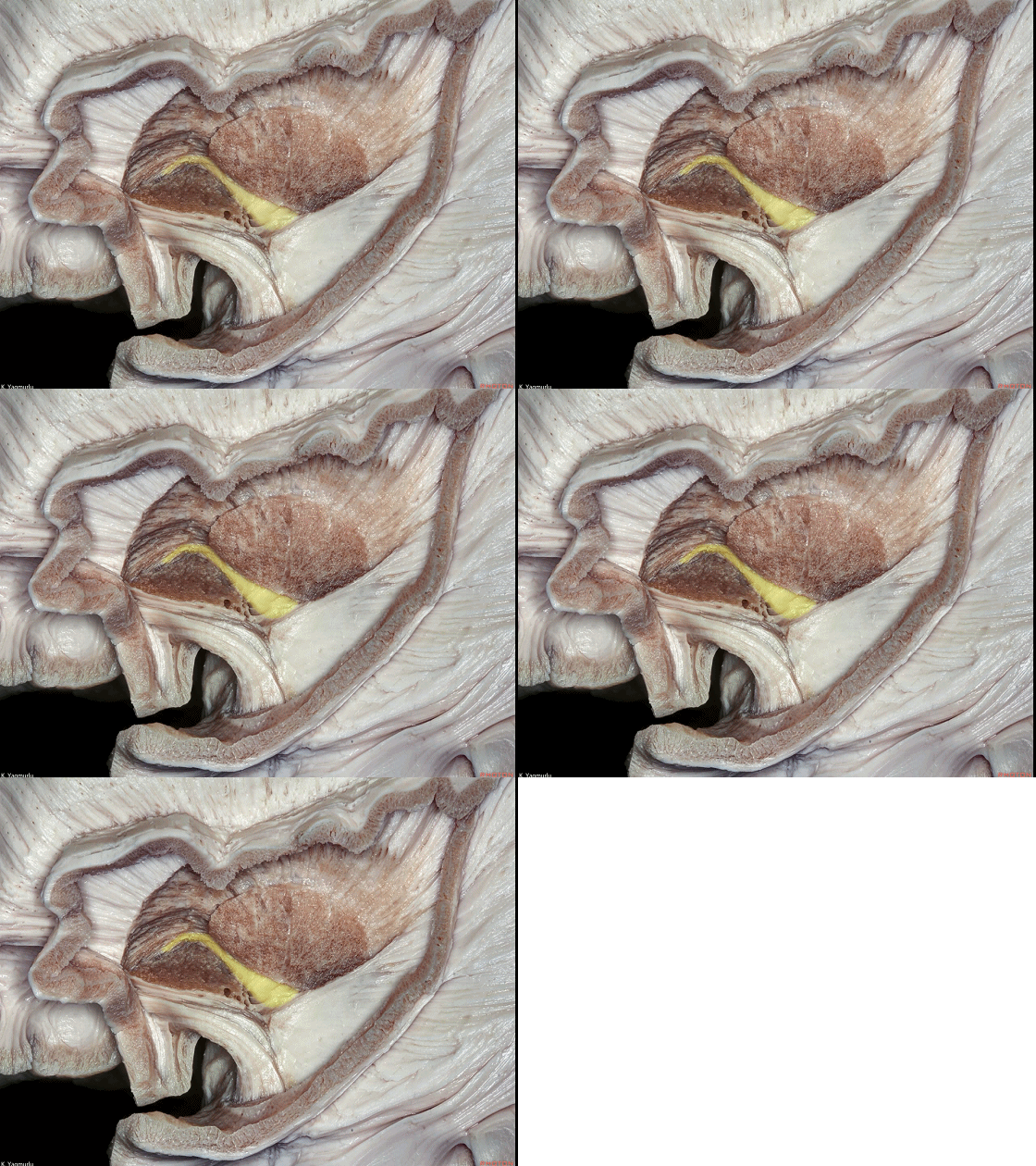
▼这是 腹侧屏状核。
Here is the ventral claustrum.

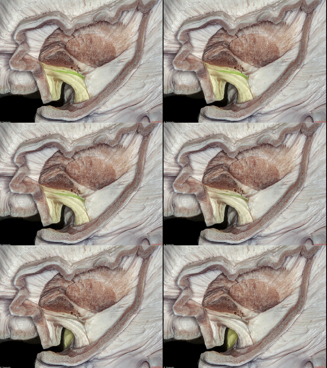
▼腹侧屏状核 纤维展开于 钩束(下图)内连接至 杏仁核(下图)。
the ventral claustrum spreads out in the uncinate fasciculus to reach the amygdala.

▼这是轴位断面观。
内囊前支(下图绿色部分)位于 尾状核头 与 豆状核 之间。
An axial section. The anterior limb of the internal capsule is located between the head of the caudate nucleus and the lentiform nucleus.
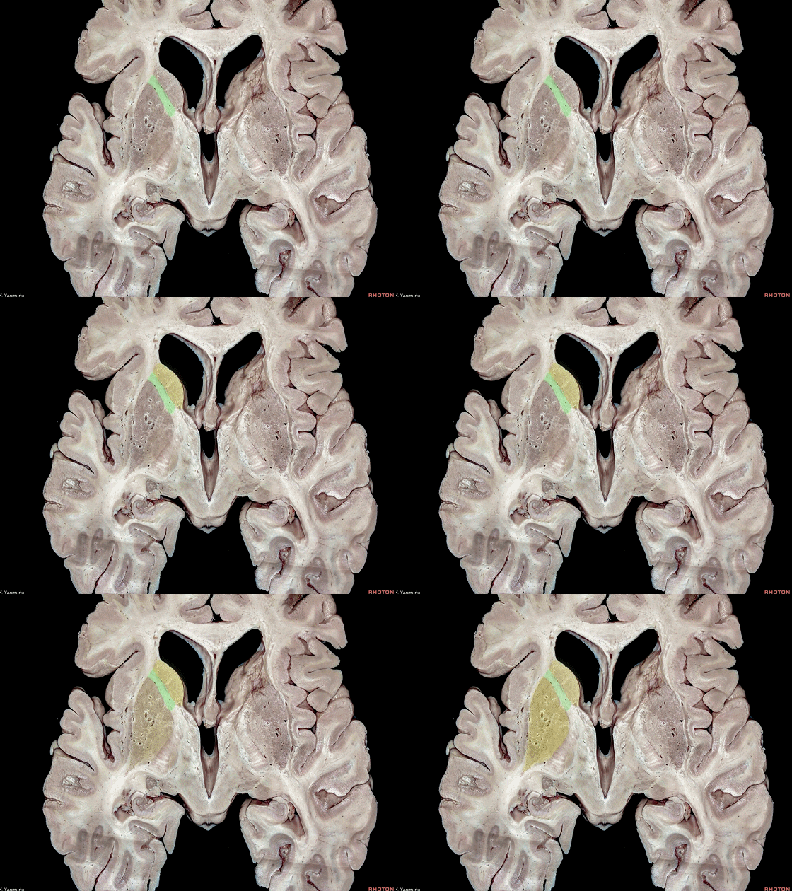
▼豆状核 包括 壳核 和 苍白球。
the lentiform nucleus is composed of the putamen and globus pallidus.
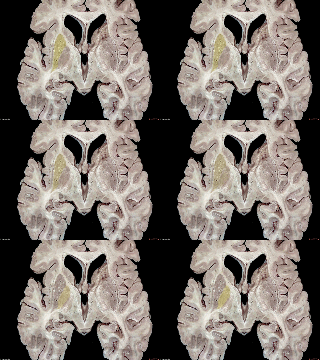
▼内囊后支(下图)位于豆状核和丘脑之间。
The posterior limb of the internal capsule is located between the lentiform nucleus and thalamus.
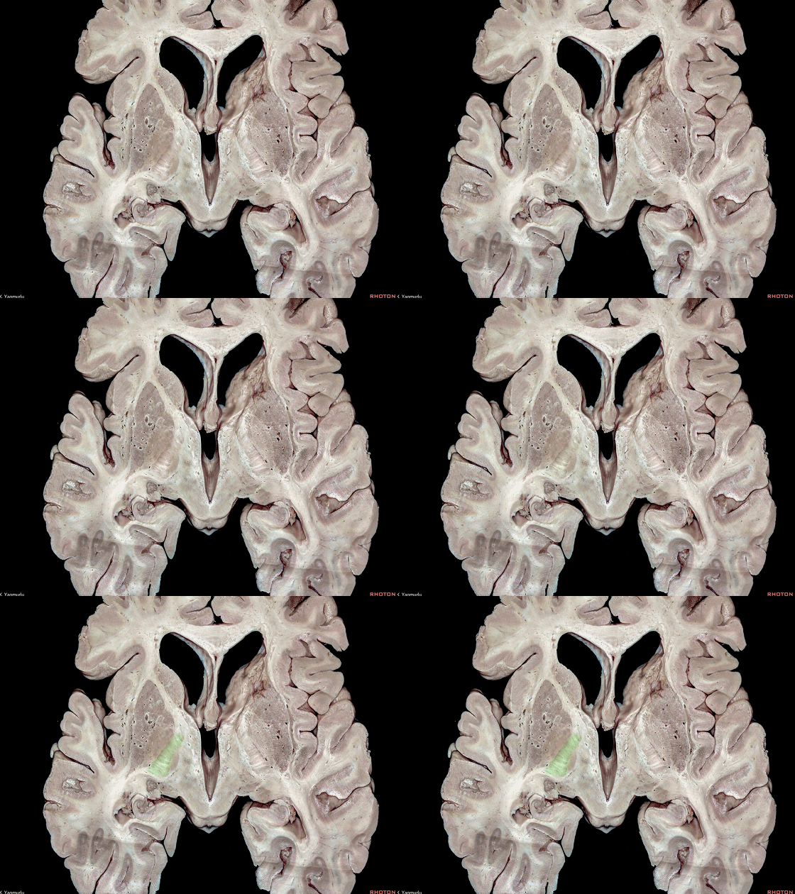
▼内囊膝部(下图)即为 前支 和 后支 交界处。
The genu is the junction of the anterior and posterior limbs.
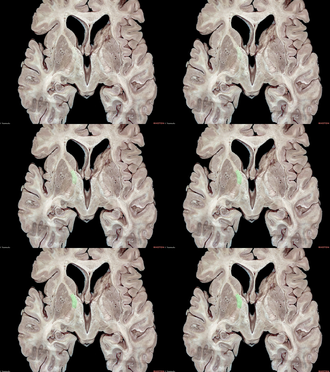
▼这是上外侧观。这是内囊膝部。
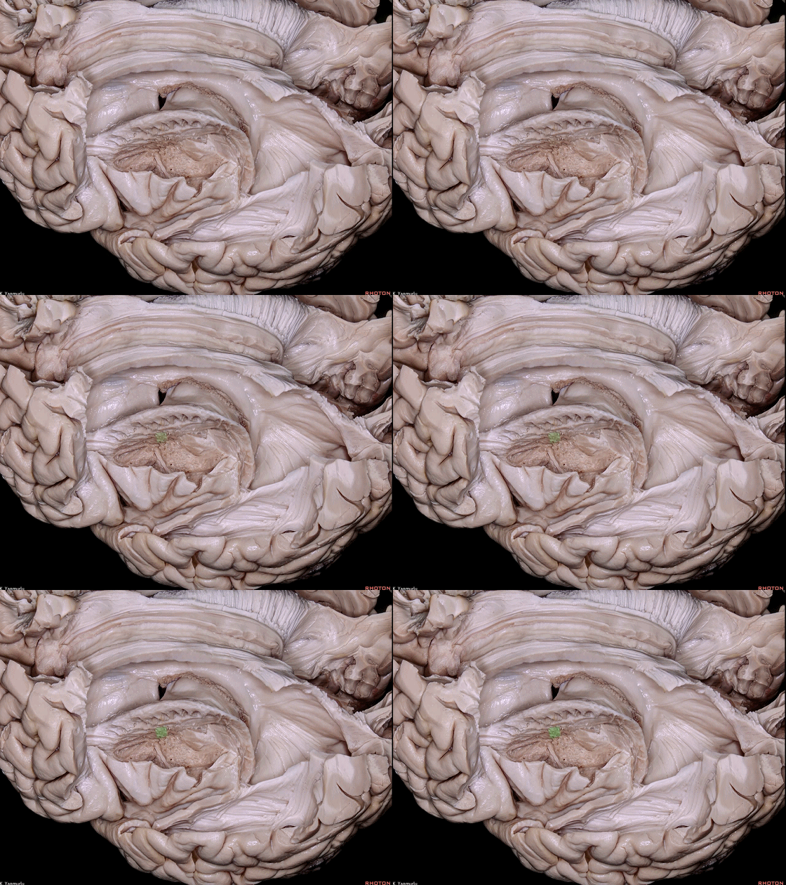
▼内囊膝部位于前连合(下图)后方。
A superolateral view. The genu is located just behind the anterior commissure.
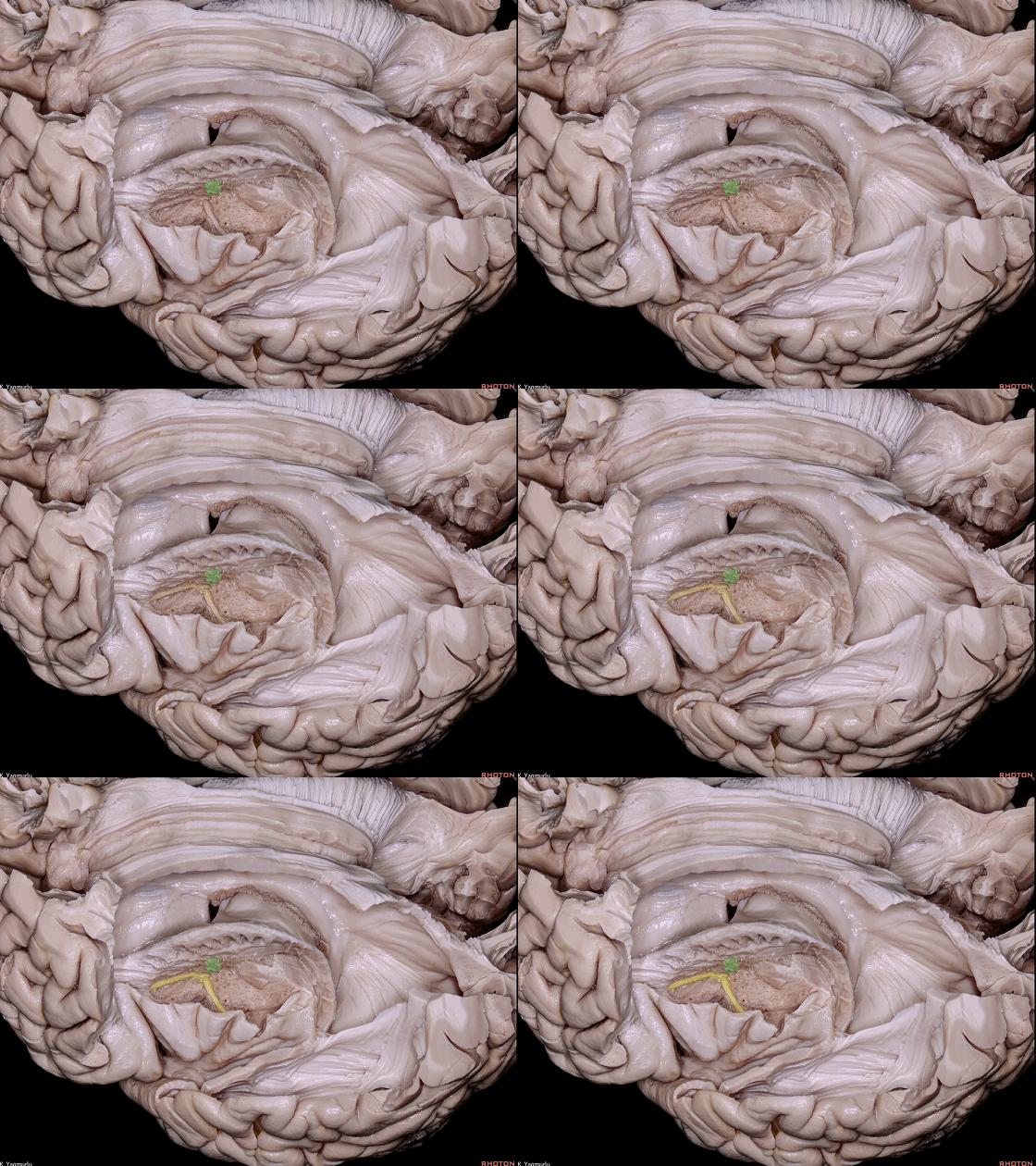
▼在同一冠状层面的结构还有Monro孔(下图) 和 岛后短回。
at the same coronal level as the foramen of Monro and posterior short gyrus.
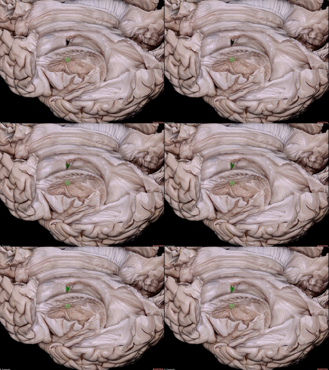
▼这是 岛后短回。
posterior short gyrus.
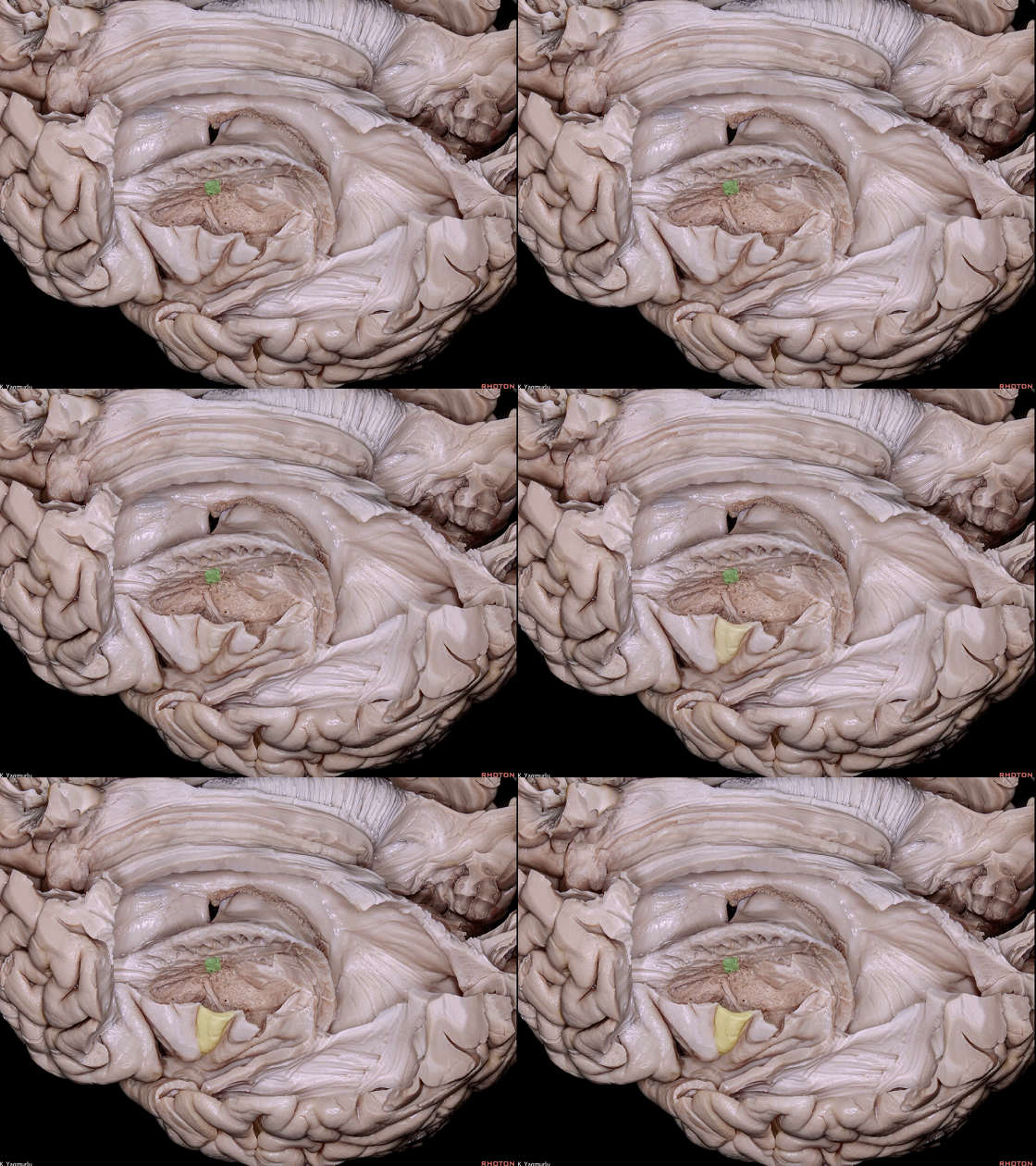
▼去除内囊前支即可暴露 尾状核头(下图)。
The removal of the anterior limb of internal capsule exposes the head of caudate nucleus.
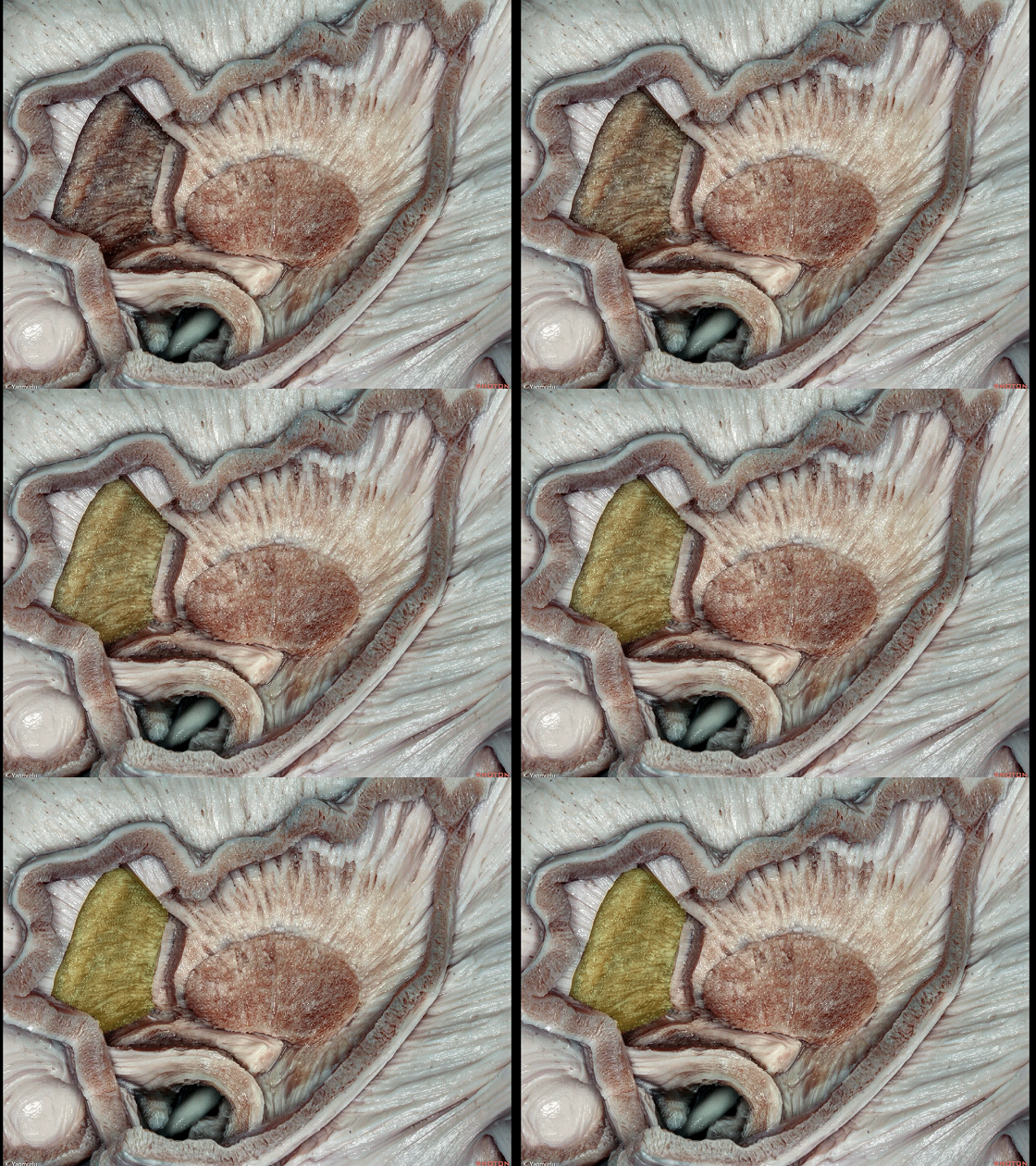
▼这是 腹侧屏状核(下图),发出纤维进入钩束(下图)到达杏仁核。
Here is the ventral claustrum, which spreads out into the uncinate fasciculus to reach the amygdala.
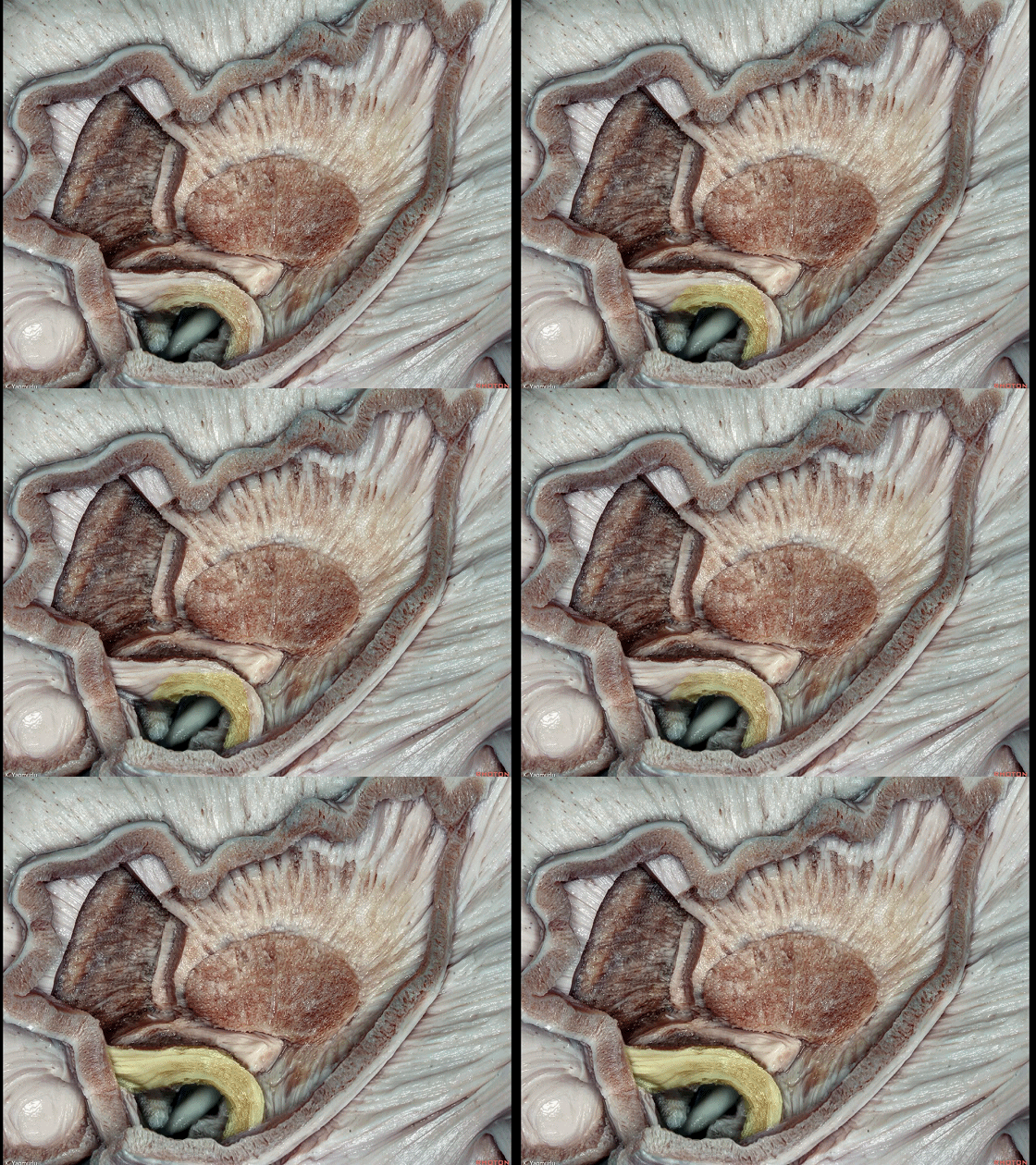
▼钩束(下图)实质上是 额颞长联络纤维通路。
The uncinate fasciculus is a frontotemporal long association fiber pathway.
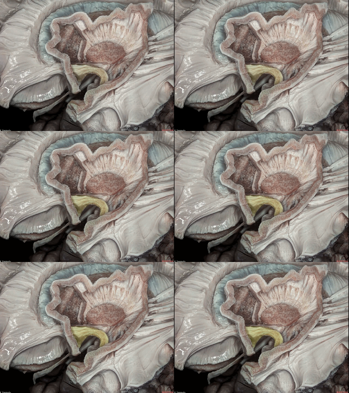
▼钩束分为背外侧部,连于眶额区(下图)。
The uncinate fasciculus has a dorsolateral part, which goes to the orbitofrontal area.
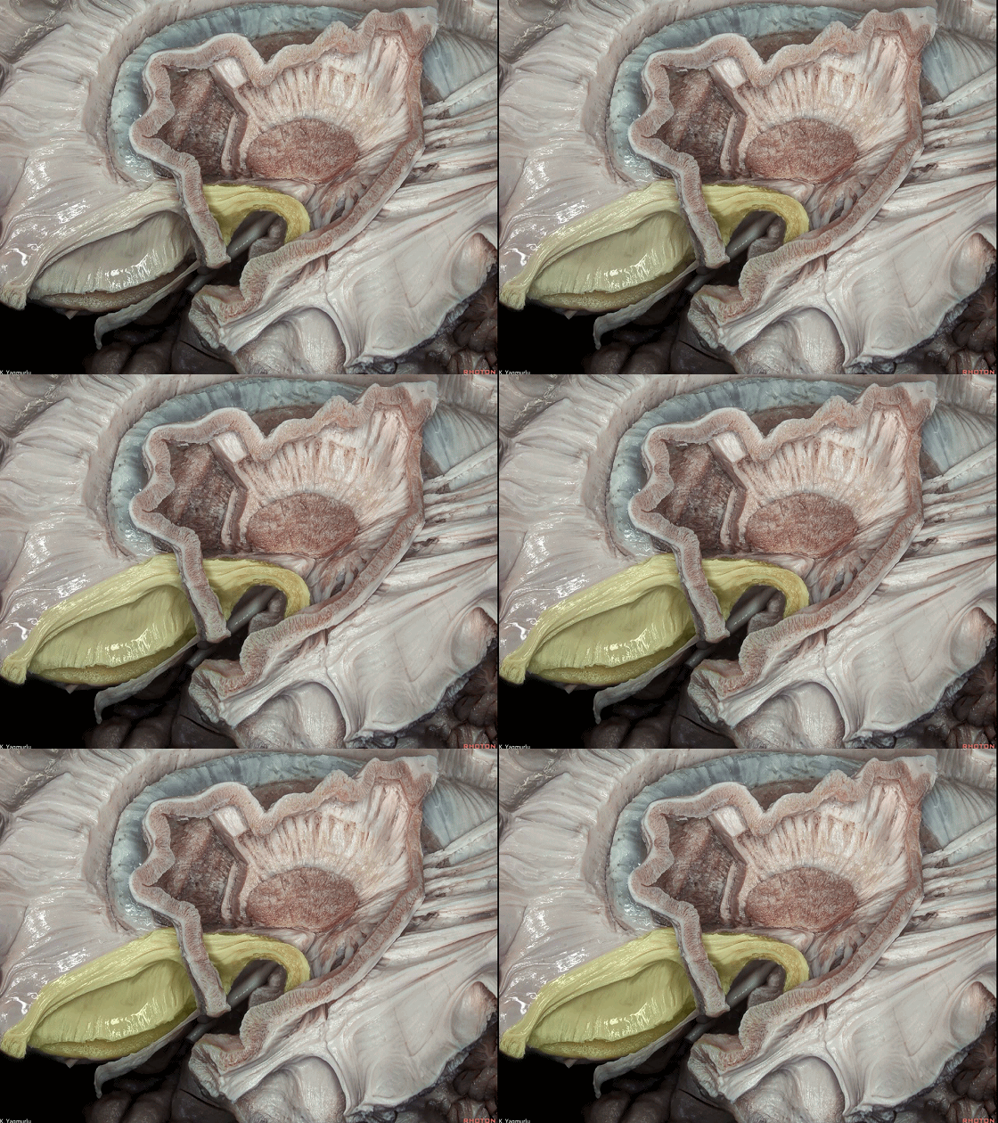
▼腹内侧部 连接 隔区 与 颞极(下图)。
a ventromedial part, which goes to the septal area from the temporal pole.
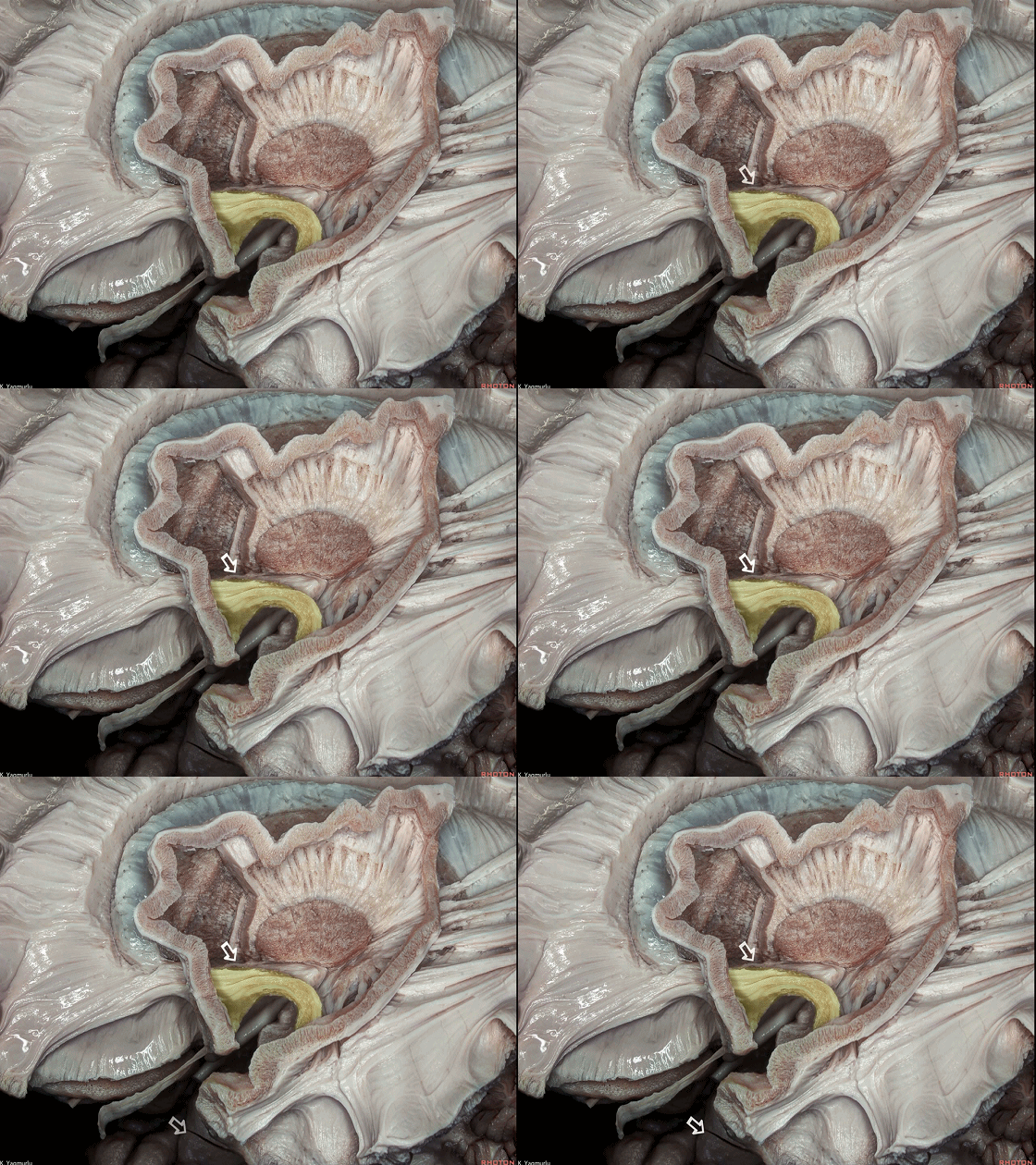
▼临床上,对精神分裂症患者进行DTI检查可发现钩束的形态学改变。此时,行前内侧颞叶切除术时对钩束进行毁损,有可能改善临床精神症状。
原因在于切断钩束,使得其介导的病理信号在 颞叶 与 眶额部皮质 之间的传输得以中断,而眶额部皮质与思维决策有关。
Clinically, some DTI studies in patients with schizophrenia have found morphometric changes in the uncinate fasciculus. At the same time, disruption of the uncinate fasciculus during the anteromedial temporal lobectomy may be associated with psychosocial clinical improvement,because the uncinate fasciculus can no longer convey the pathological information from the temporal lobe to the orbitofrontal cortex, which is involved in decision making.
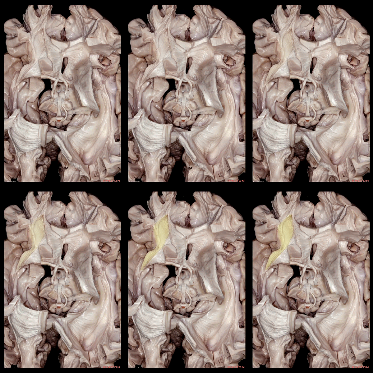
▼这是上面观。
钩束的背外侧部(下图),连接于外侧眶额区。
We are looking from above. The dorsolateral part of the uncinate fasciculus,which projects to the lateral orbitofrontal area.
▼钩束的腹内侧部(下图),连接于隔区以及内侧眶额区。
the ventromedial part, which projects to the septal and medial orbitofrontal areas, can be seen.

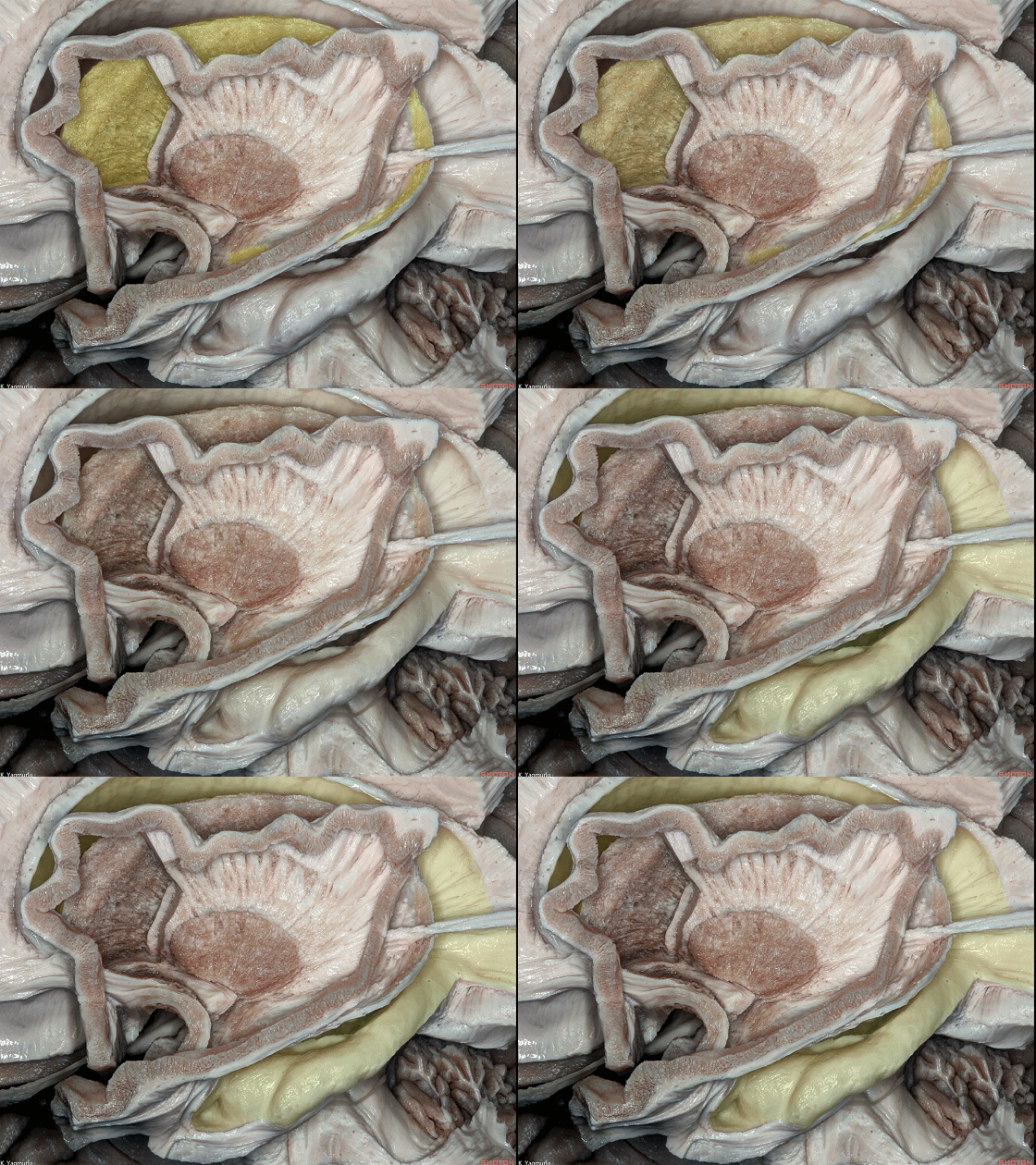
▼尾状核(下图)是呈C形的弓状结构。
The caudate nucleus is an arched C-shaped structure.
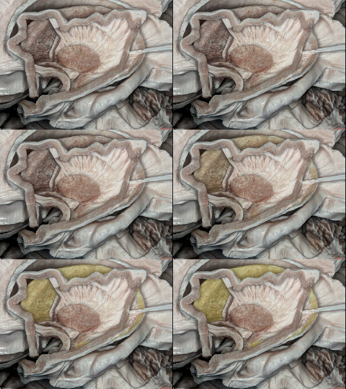
▼尾状核分为头部、体部、尾部。
The caudate nucleus with head, body and tail parts.
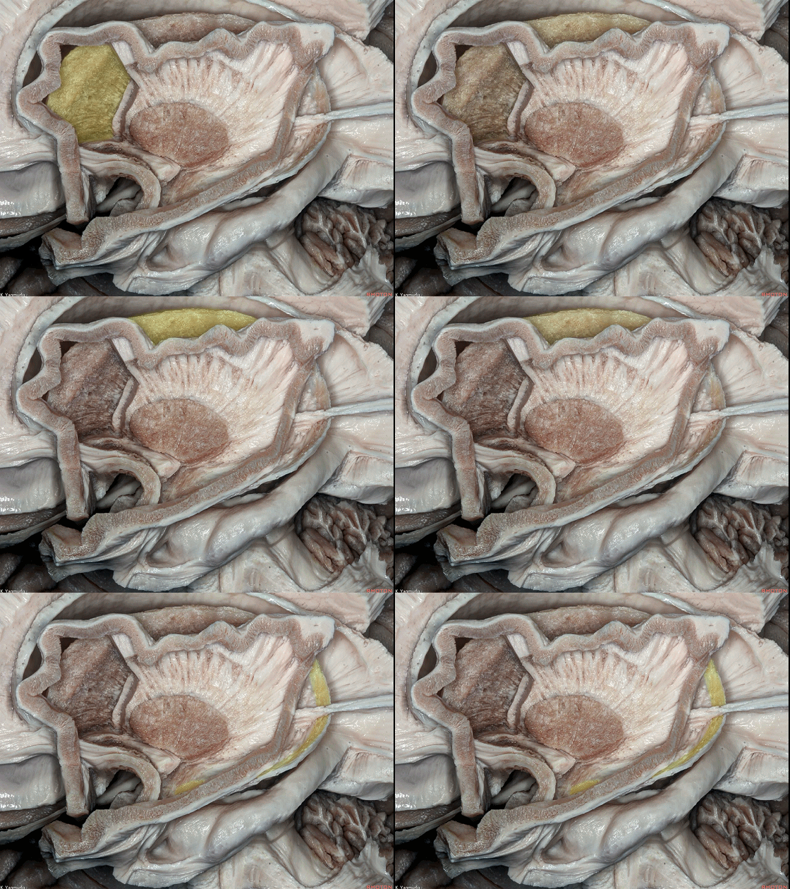
▼尾状核 构成 侧脑室 的外侧壁。
It also forms the lateral wall of the lateral ventricle.
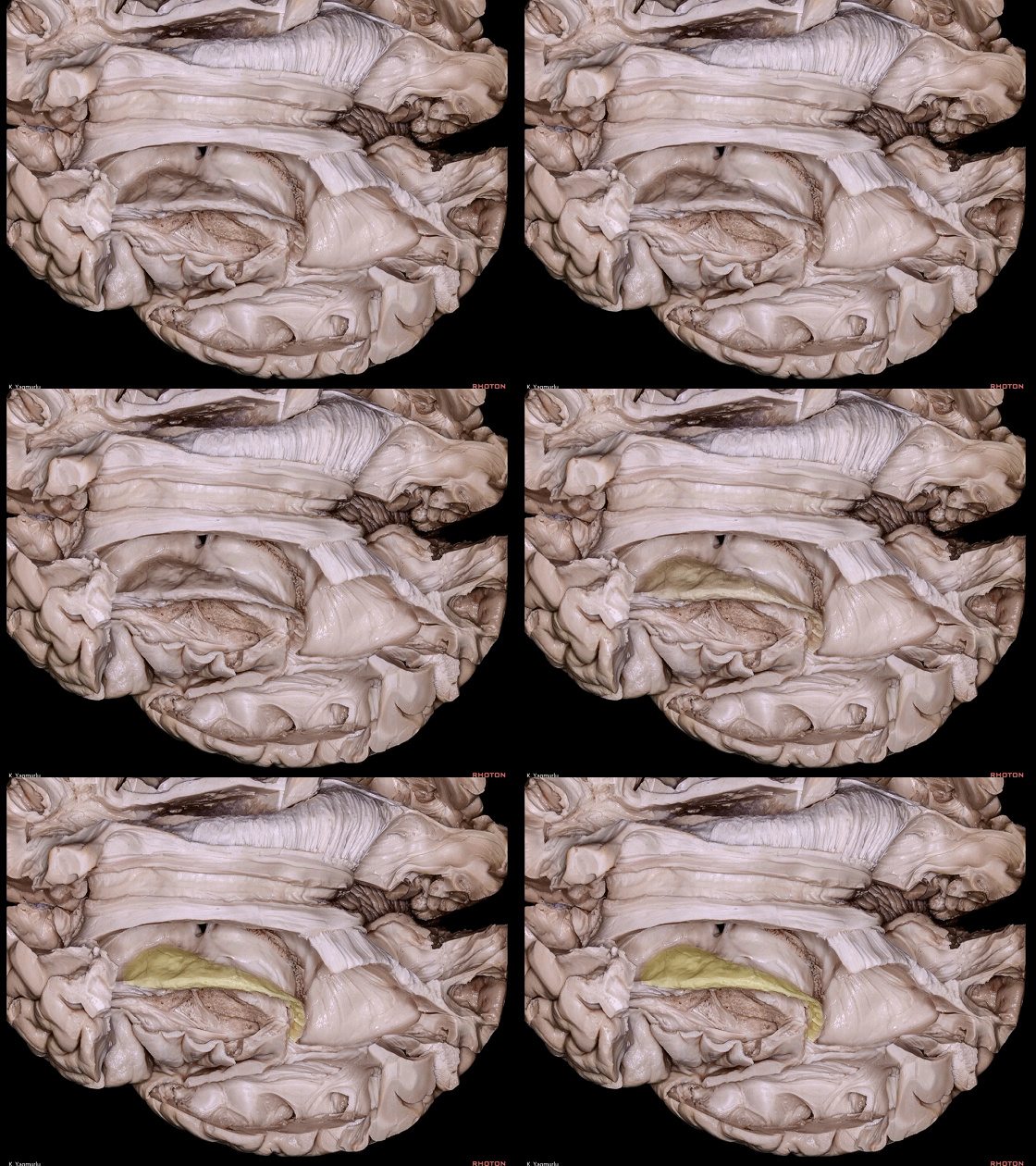
▼这是上面观。下图示尾状核。
We are looking from above.
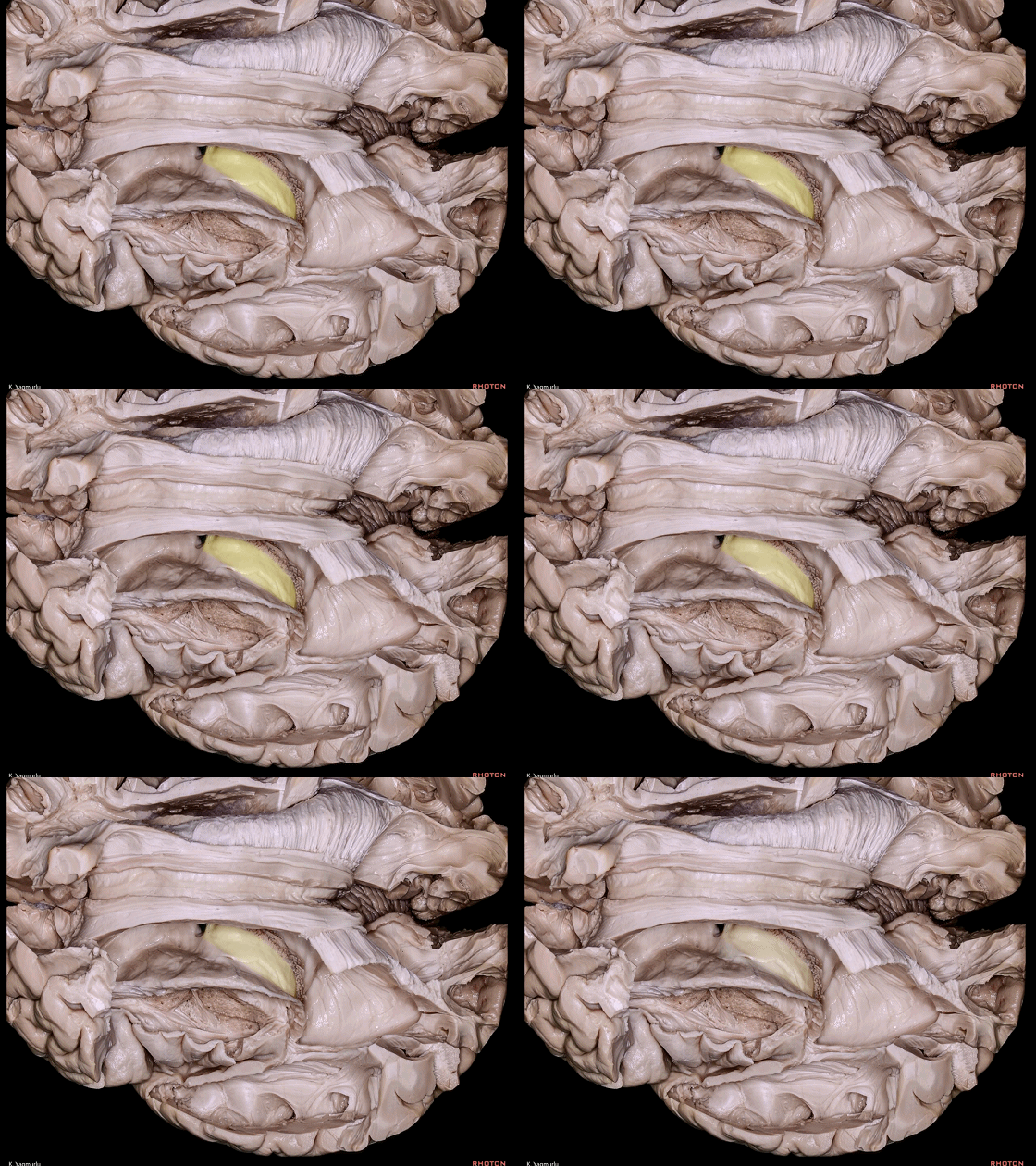
▼尾状核包绕 丘脑(下图)的外侧部。
The caudate nucleus surrounds the lateral part of the thalamus.
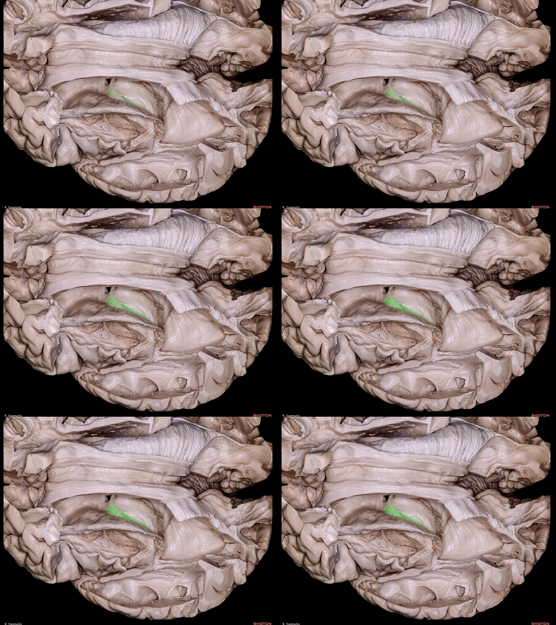
▼终纹(下图)行于尾状核和丘脑之间。
and the stria terminalis travels between the caudate nucleus and thalamus.
▼去除丘脑,可见 尾状核(下图)与 岛界沟(下图)的相对位置关系。
After removal of the thalamus, we can see the position of the caudate nucleus in relation to the insular limiting sulci.
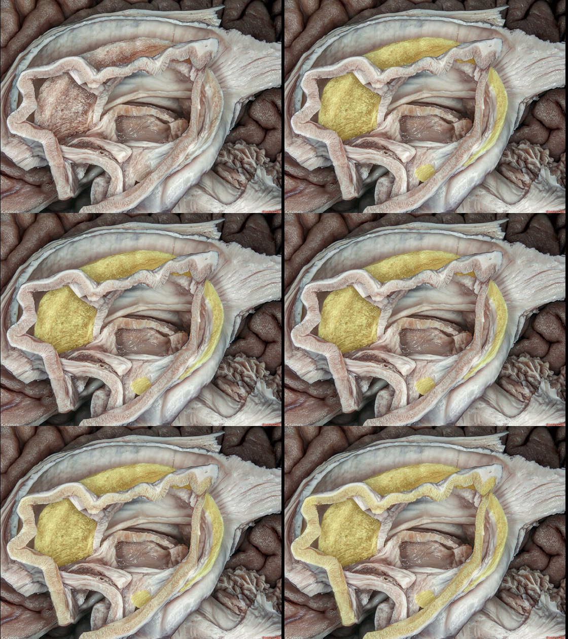
▼尾状核头(下图)位于岛叶的界限(岛界沟)以内。
岛前点(下图绿色部分)位于尾状核头的上方。
The head of the caudate nucleus is positioned inside the insular area, The anterior insular point is positioned above the head of the caudate nucleus.
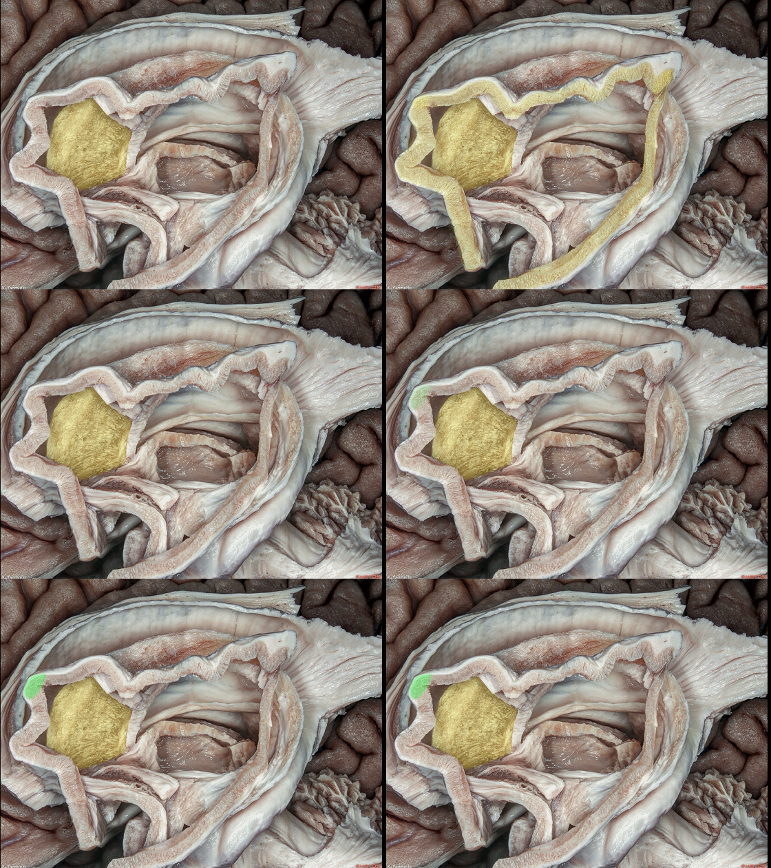
▼尾状核体(下图)位于上界沟(下图)的上方,并行经 岛后点(下图绿色部分)的深面。
The body of the caudate nucleus sits above the superior limiting sulcus,and passes just deep to the posterior insular point.
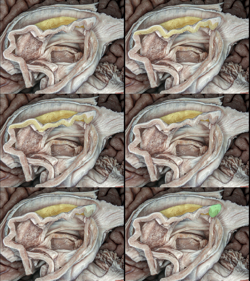
▼岛后点(下图绿色部分)以下的部分,即称为尾状核尾,其位于后界沟的后方。
Below the posterior insular point, it's called the tail of the caudate nucleus, and it travels behind the inferior limiting sulcus.
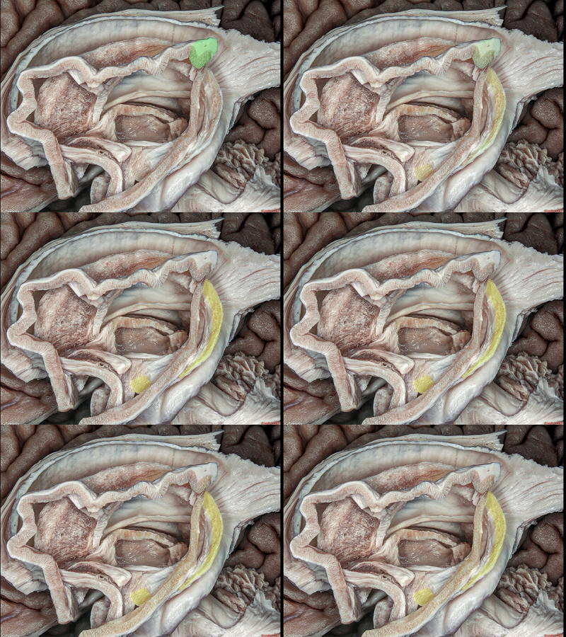
▼下图示 尾状核尾。
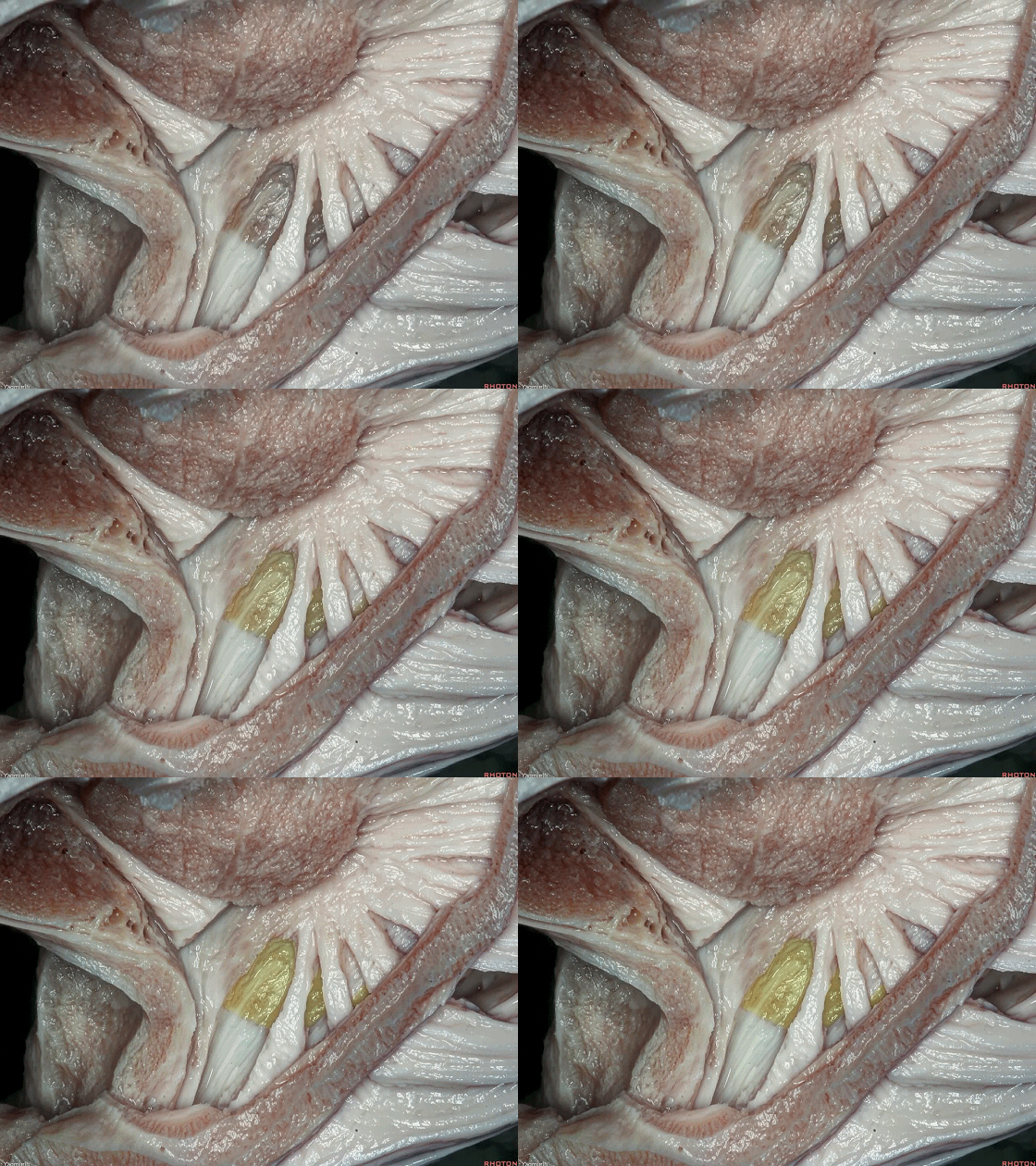
▼尾状核尾 越过 下界沟(下图)随后移行入 杏仁核。
The tail of the caudate nucleus crosses the inferior limiting sulcus before blending into the amygdala.
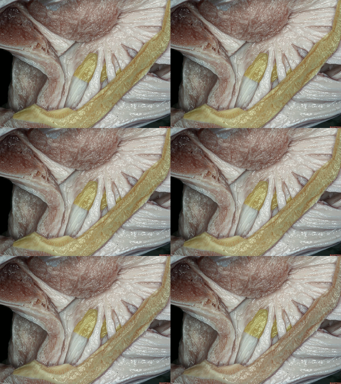
▼下图示 杏仁核。
amygdala.
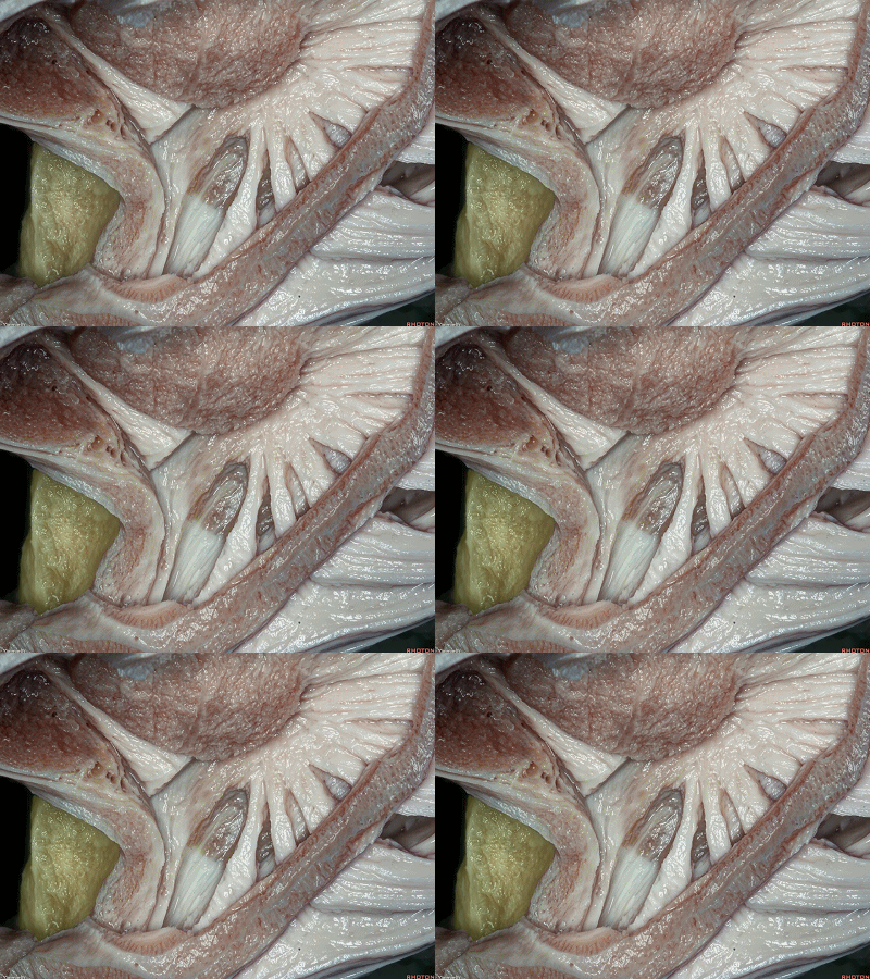

▼接下来我们将介绍其余的基底节结构。
这是 上面观。
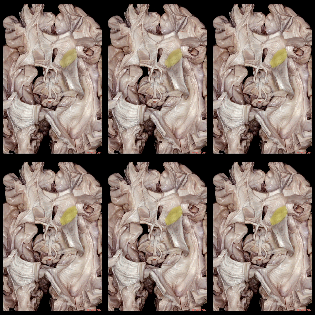
伏核(下图) 位于 尾状核头下方。
Now, we are going to look at rest of the basal ganglia structures. We are looking from above.The nucleus accumbens is located underneath the head of caudate nucleus.
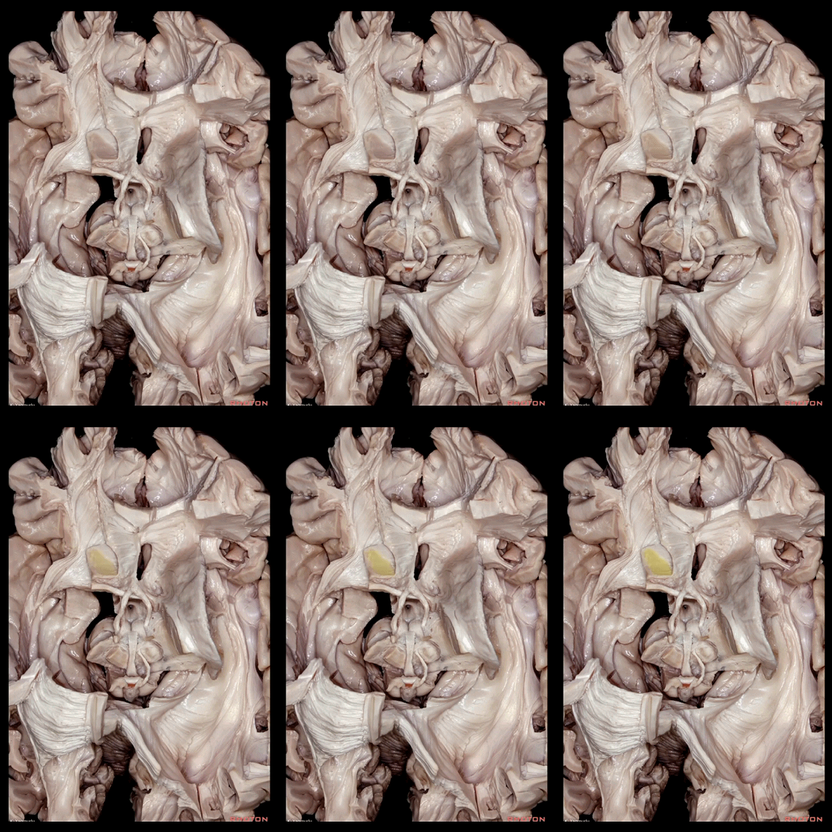
▼下图示尾状核头。
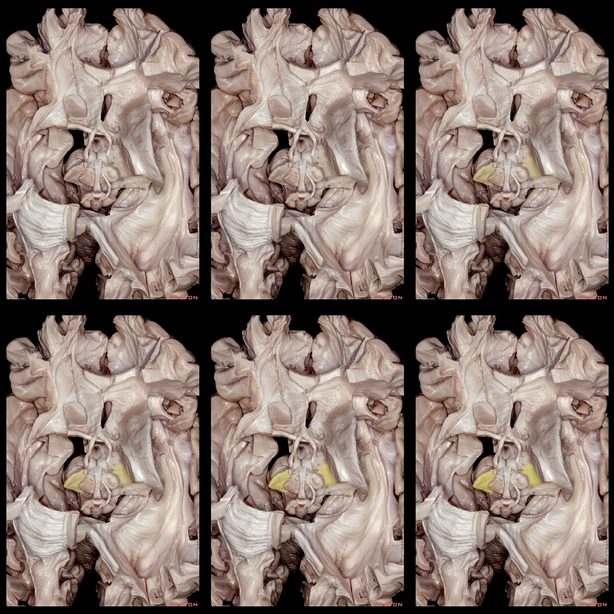
▼底丘脑核(下图)位于间脑内红核的腹外侧。
The subthalamus nucleus is located ventrolateral to the red nucleus in the diencephalon.
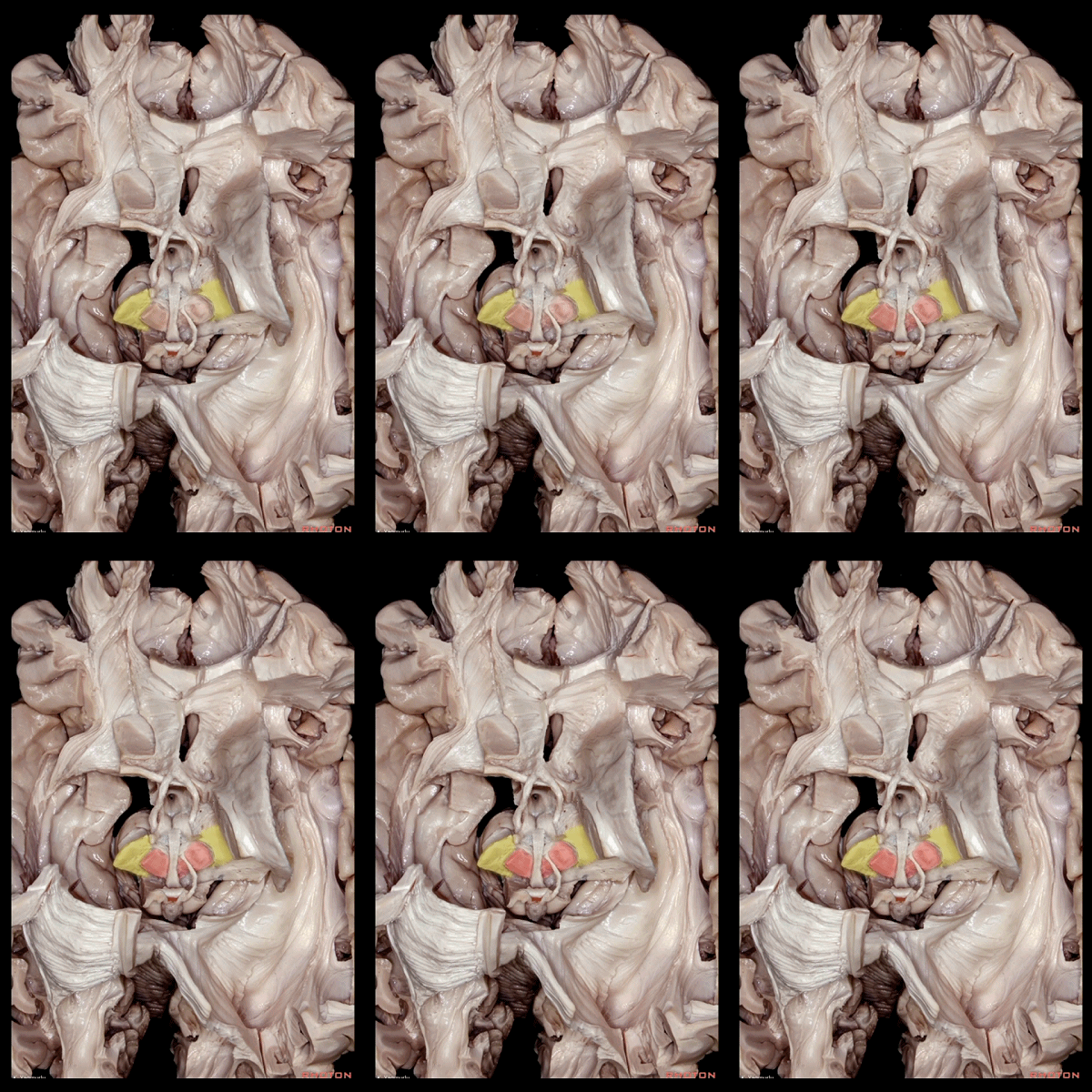
▼下图示 红核。
red nucleus.
▼这是 间脑区域的后面观。
底丘脑核(下图)位于红核的腹外侧,内囊的的背内侧。
Posterior view of the diencephalic area. The subthalamic nucleus is positioned ventrolateral to the red nucleus and dorsomedial to the internal capsule.

▼下图示 红核。
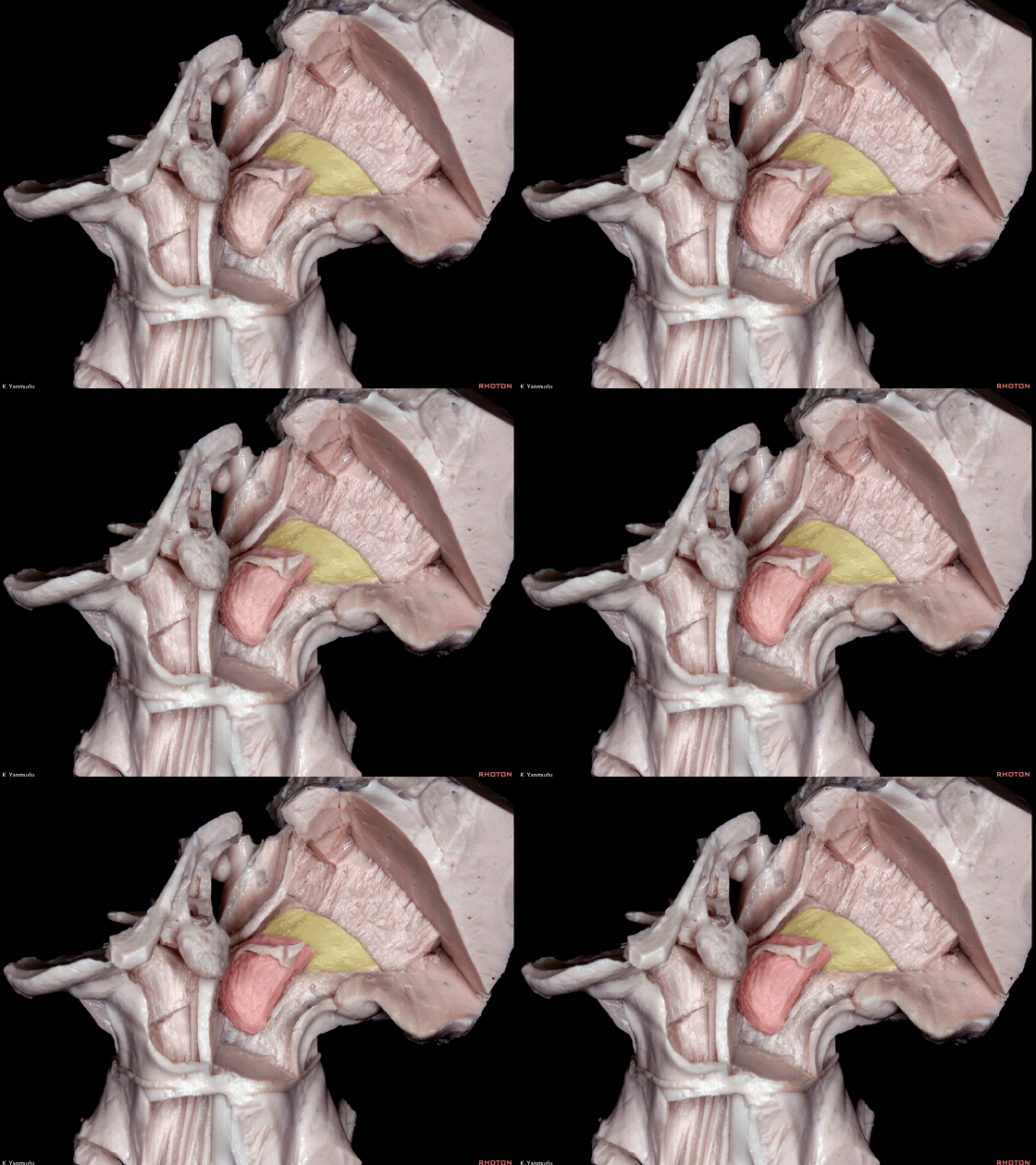
▼下图示 内囊。
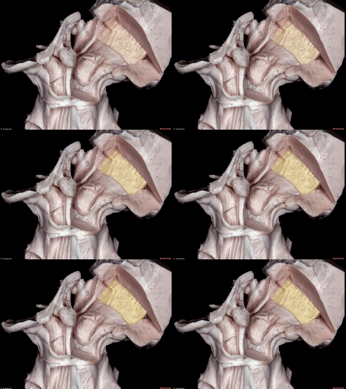
▼这是 左外侧观。
黑质(下图)位于底丘脑核的腹侧面。
Lateral view. The substantia nigra is positioned on the ventral surface of the subthalamic nucleus.
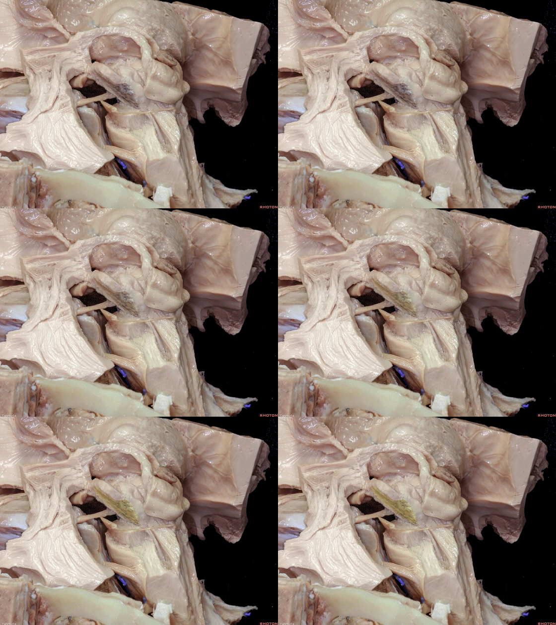
▼这是 底丘脑核。
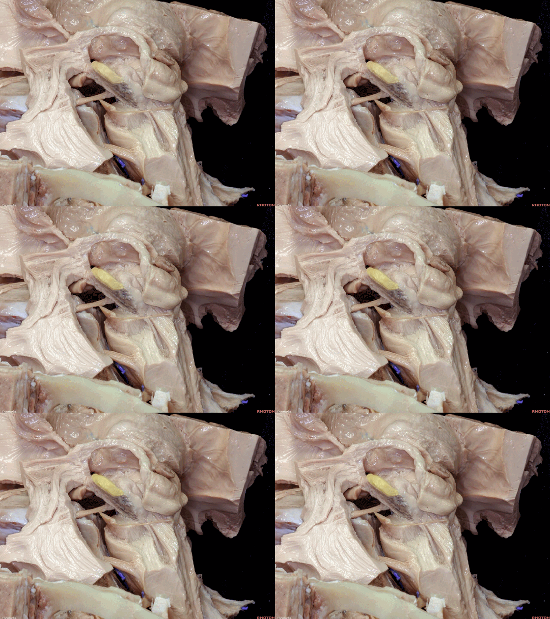
▼这是 红核。
The red nucleus is here.
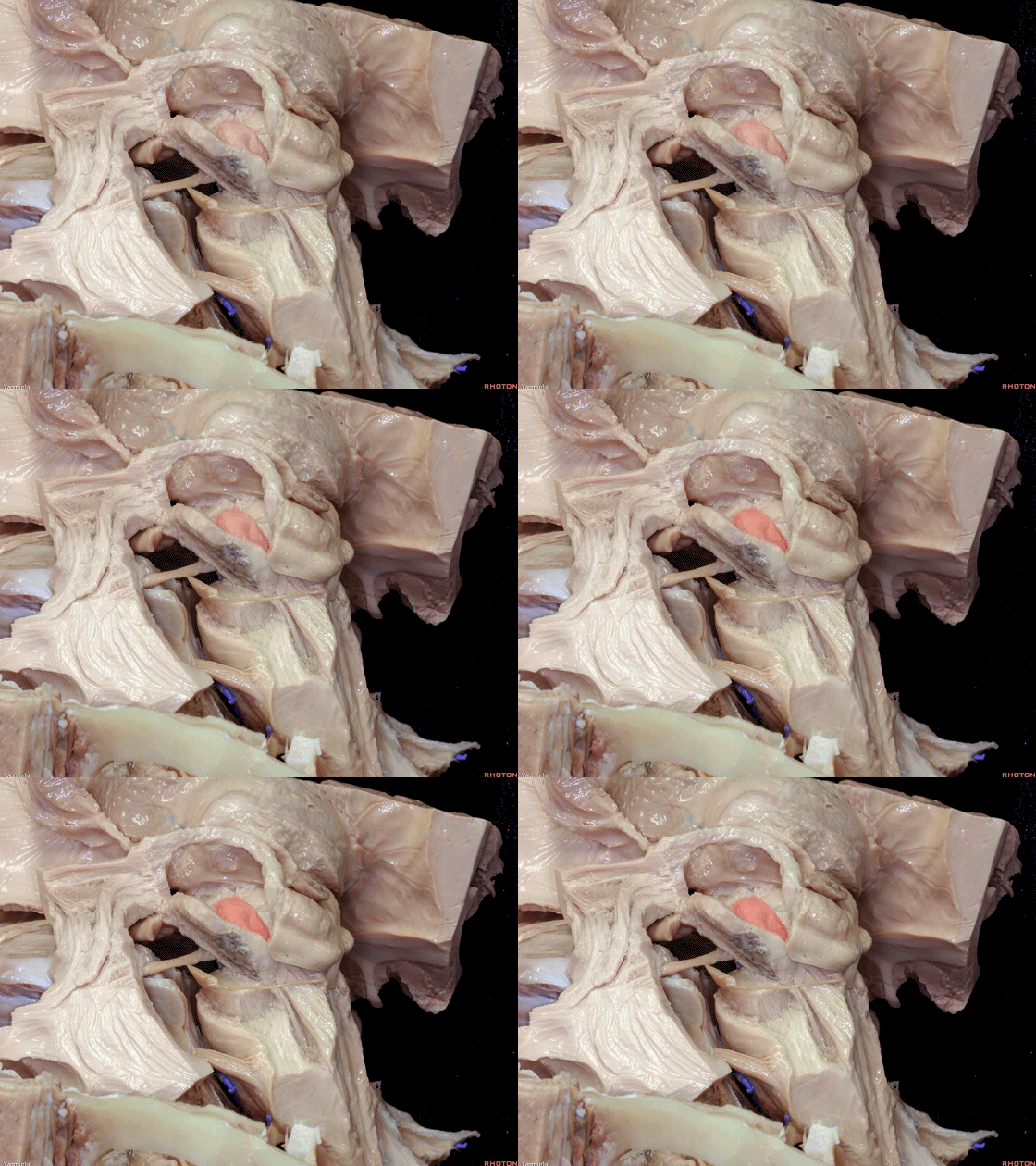
▼基底节与大脑皮层和脑干有广泛联系。
The basal ganglia structures has connections with cerebral cortex and brainstem.


▼接下来展示投射纤维及其与侧脑室的关系。
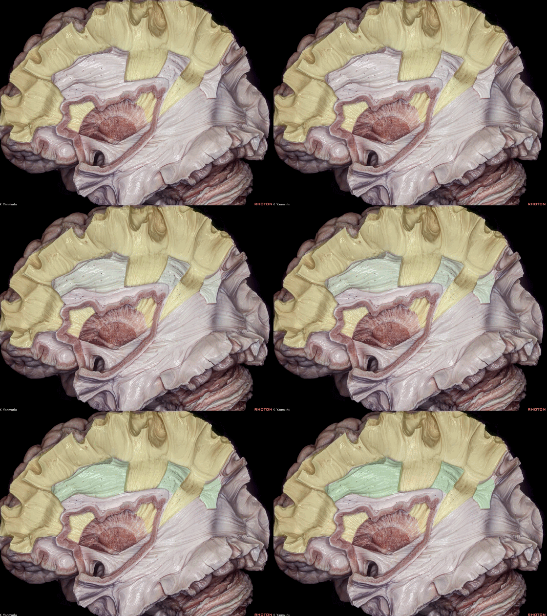
大体上,额顶投射纤维 即称为 放射冠(下图)。
Let's see the projection fibers and their relationship with the lateral ventricle. Basically, the frontoparietal projection fibers can be called as the corona radiata。
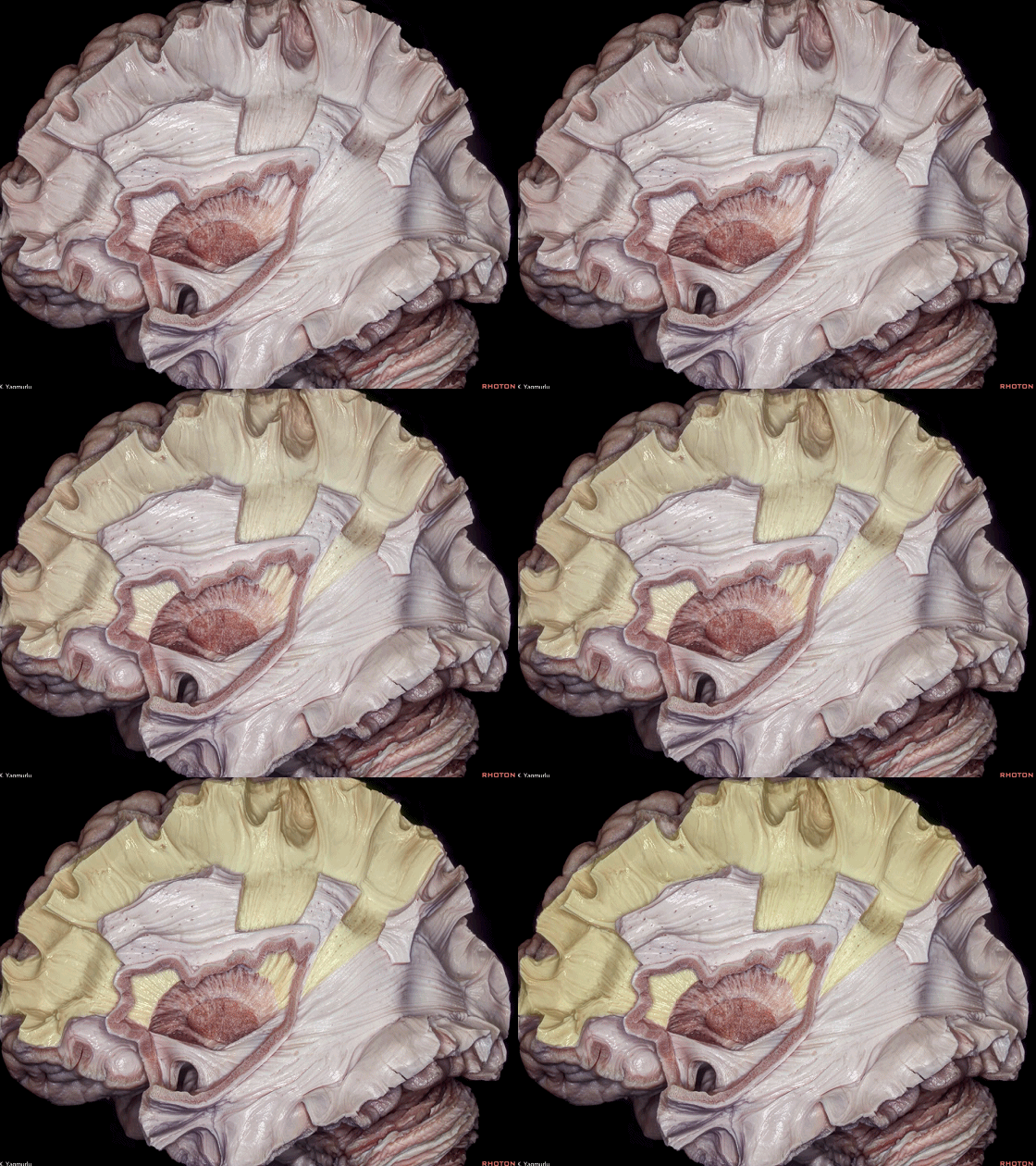
▼放射冠纤维 行于 上纵束(下图)内侧。
The corona radiata fibers run medial to the superior longitudinal fasciculus.
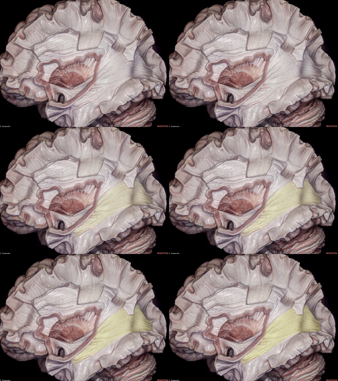
▼枕投射纤维 即称为 矢状层。
the occipital projection fibers can be called as the sagittal stratum.
▼矢状层纤维行于下额枕束(下图)、下纵束、以及前连合枕部延伸部的内侧。
The sagittal stratum fibers run medial to the inferior fronto-occipital fasciculus, inferior longitudinal fasciculus, and occipital extension of the anterior commissure.
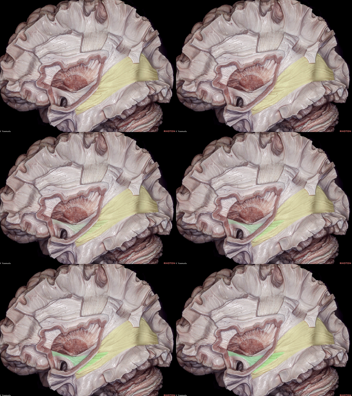
▼这是 下纵束。
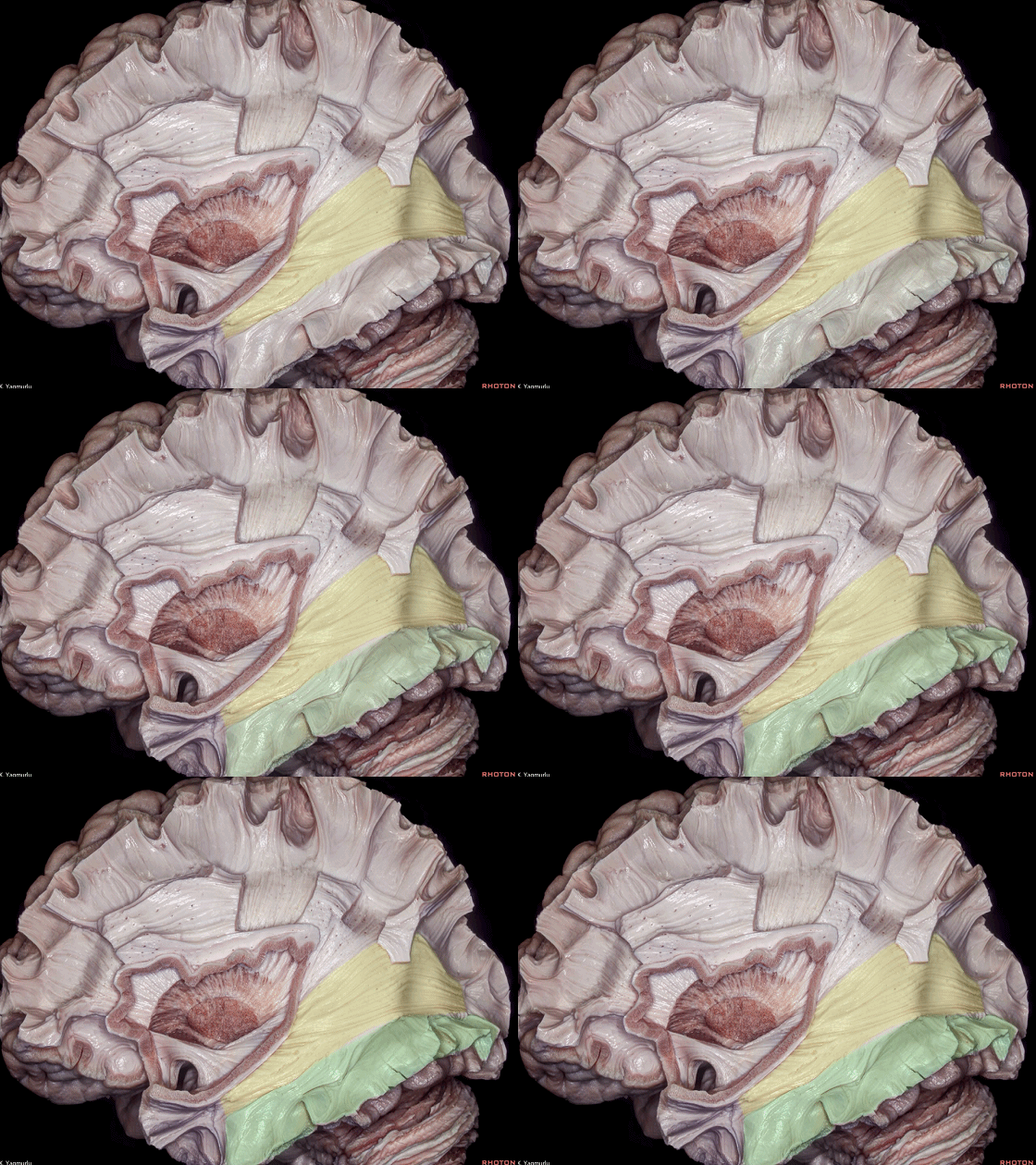
▼这是 前连合。
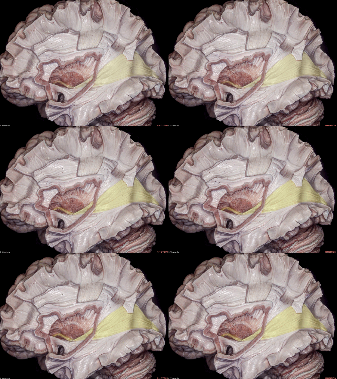
▼去除下额枕束和前连合枕部延伸部即可显露矢状层(下图)。
矢状层包含枕桥纤维和枕丘纤维,后者即所谓的视辐射。
Removal of the inferior fronto-occipital fasciculus and occipital extension of the anterior comissure exposes the sagittal stratum.The sagittal stratum is composed of the occipitopontine fibers and occipitothalamic fibers, which is also called as the optic radiations.
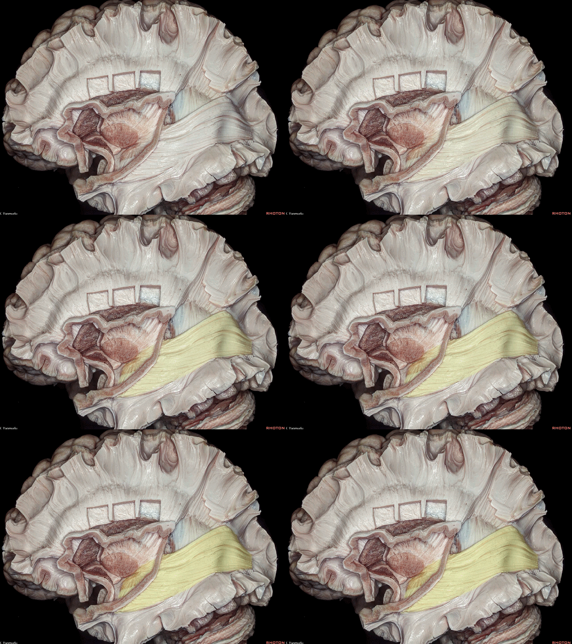
▼这是 视辐射。
optic radiations.
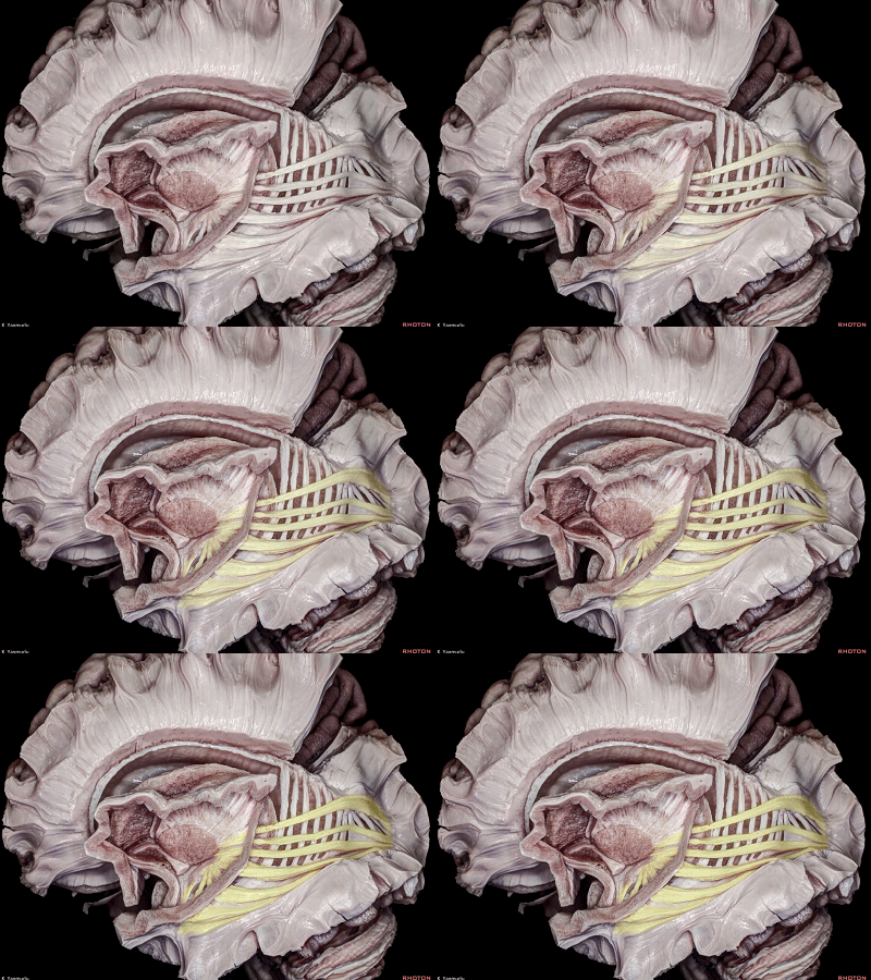
▼视辐射纤维起自外侧膝状体(下图)。
The optic radiation fibers arise from the lateral geniculate body.

▼视辐射行于侧脑室颞角(下图)的顶壁和外侧壁,随后行经侧脑室体部外侧,到达距状沟。
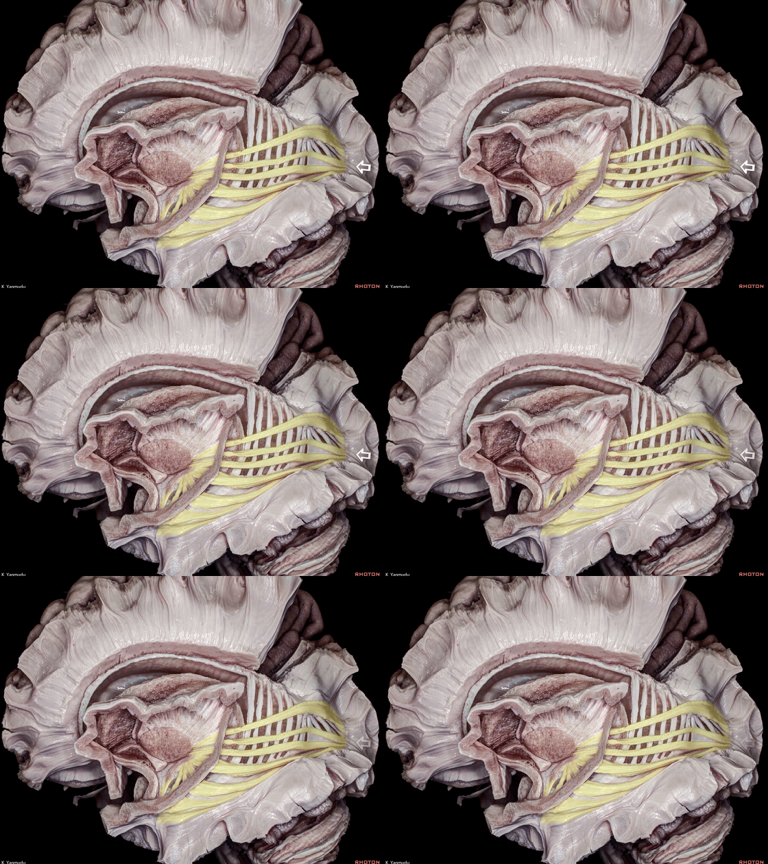
and travel in the roof and lateral wall of the temporal horn, and then lateral to the atrium to reach the calcarine sulcus.
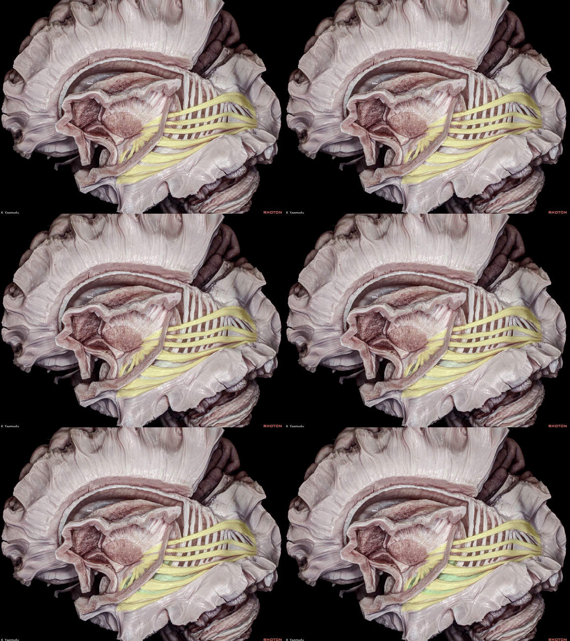
▼这是 侧脑室体部。
值得注意的是,侧脑室体部上三分之一并无视辐射纤维经过。
Notable, the upper one third of the atrium is free from the optic radiation fibers.

▼这是 距状沟位置(箭头处)。
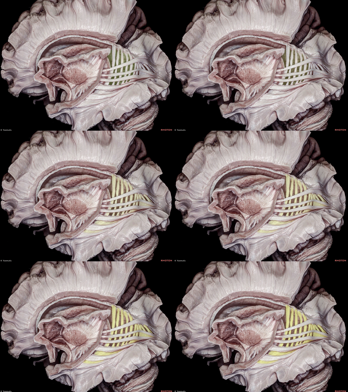
▼视辐射与侧脑室颞角及体部之间被胼胝体毯(下图)相分隔。
The optic radiations are separated from the temporal horn and atrium of the lateral ventricle by the tapetum.
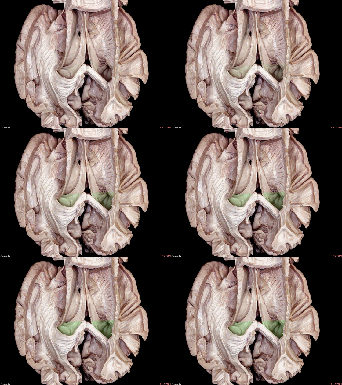
▼这是 侧脑室体部 和 枕角。
The atrium and occipital horn of the lateral ventricle.
▼侧脑室体部和枕角,其外侧由矢状层(下图)和 胼胝体毯部 纤维覆盖,其内侧由大钳覆盖。
The atrium and occipital horn of the lateral ventricle are covered laterally by the sagittal stratum and tapetal fibers,and medially by the forceps major.
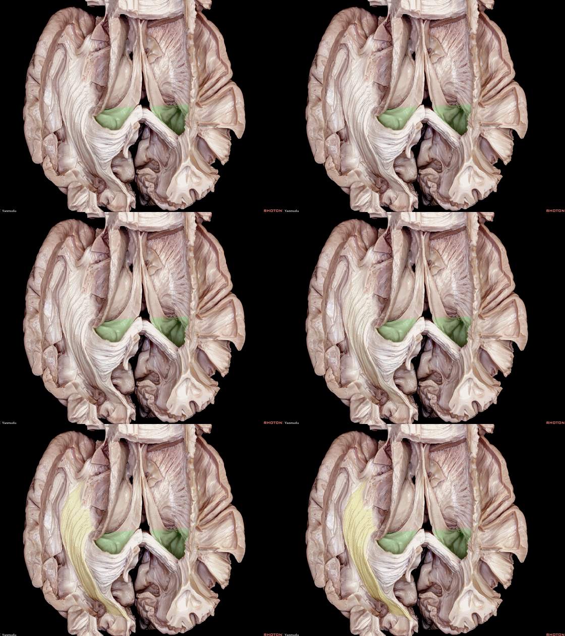
▼这是 胼胝体毯部。
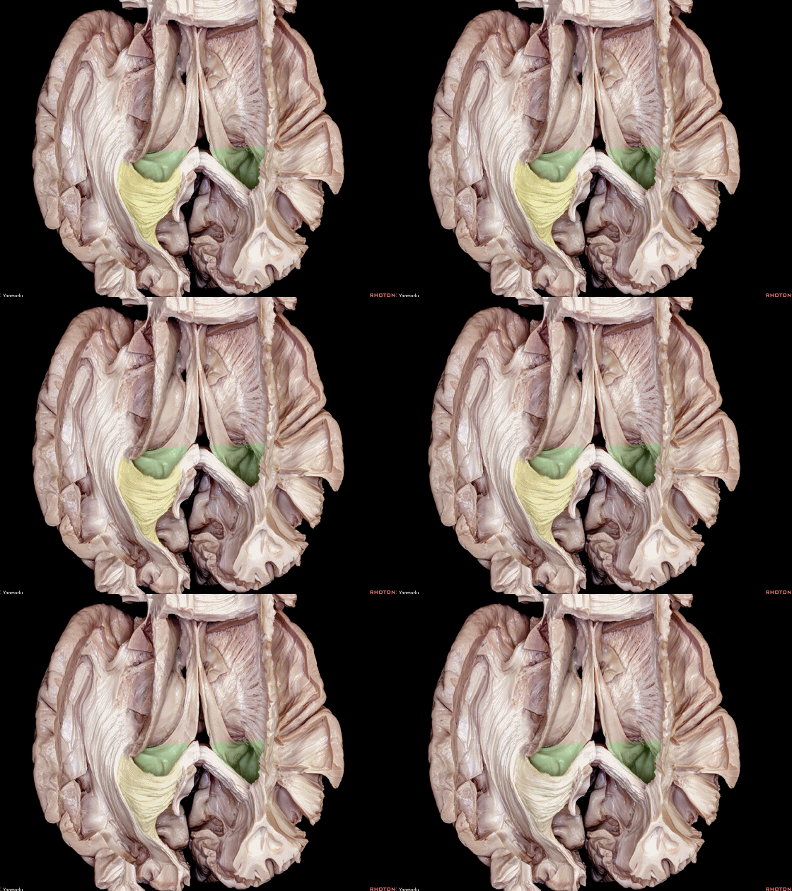
▼这是 胼胝体大钳。
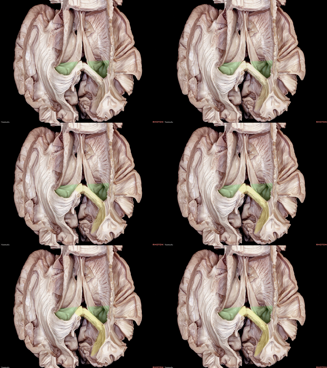
▼这是 侧脑室体部。
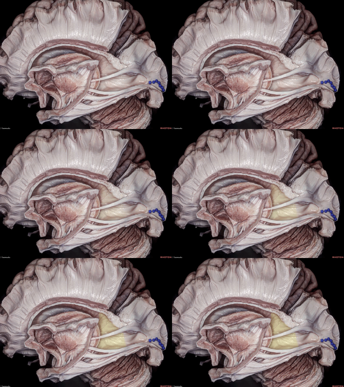
▼在侧脑室体部的内侧壁,可见胼胝体隆起(下图)覆盖着大钳纤维。
In the medial wall of the atrium, we see the bulb of the corpus callosum overlying the fibers of the forceps major.
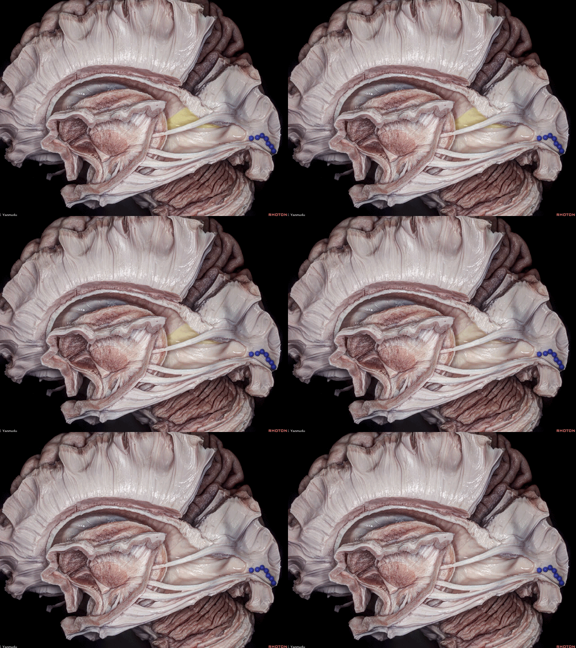
▼在侧脑室体部的内侧壁,还可见 距状隆起(下图)覆盖着距状沟深部。
and the calcar avis overlying the deep end of the calcarine sulcus.
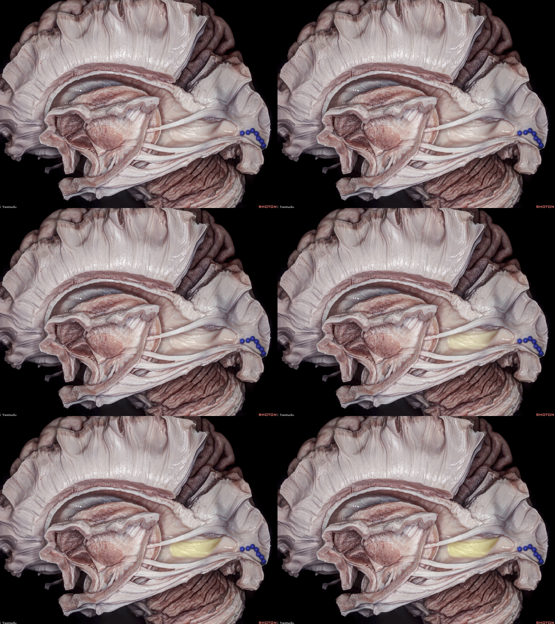
▼视辐射的前组纤维称为 Meyer袢(下图)。
The anterior bundle of optic radiations are called Meyer's loop.
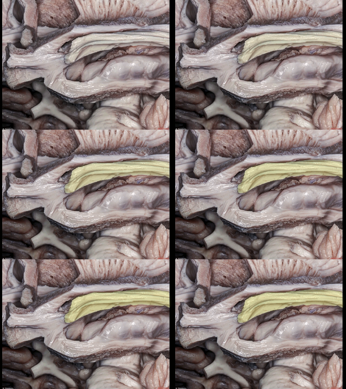
▼Meyer袢向前延伸至 颞角尖端(下图)。
and it extends up to the tip of the temporal horn.
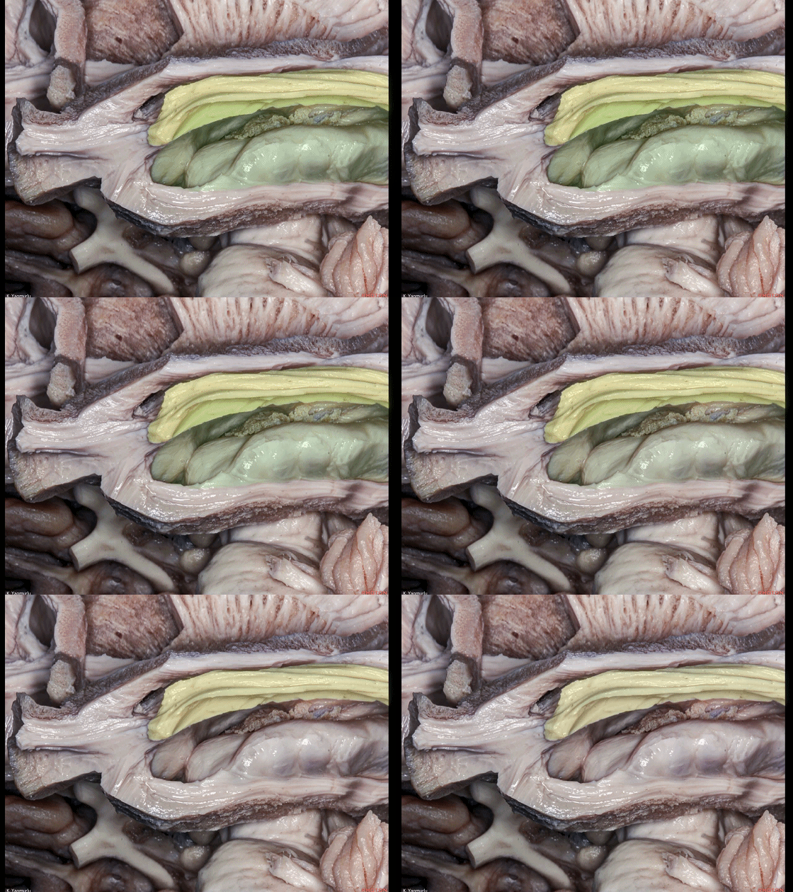
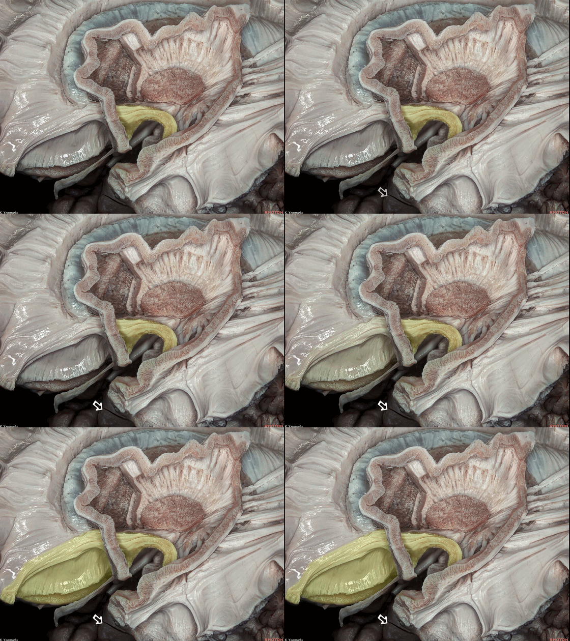
▼杏仁核(下图)构成颞角前壁。
The amygdala forms the anterior wall of the temporal horn.
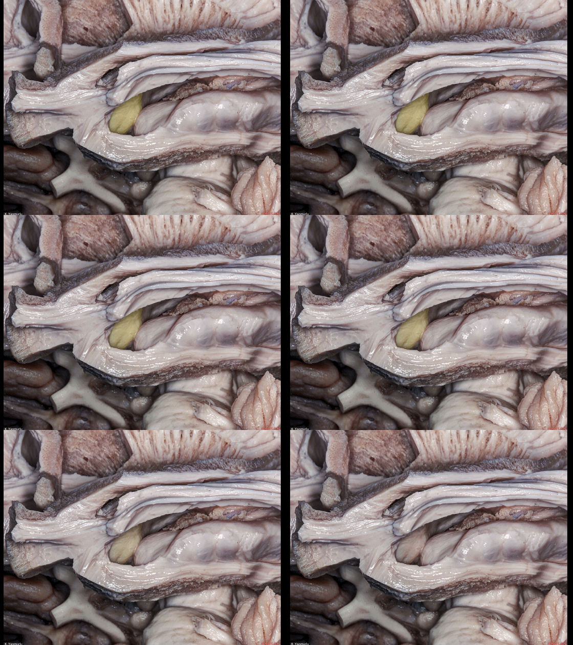
▼海马(下图)位于颞角底壁。
The hippocampus is located in the floor of the temporal horn.
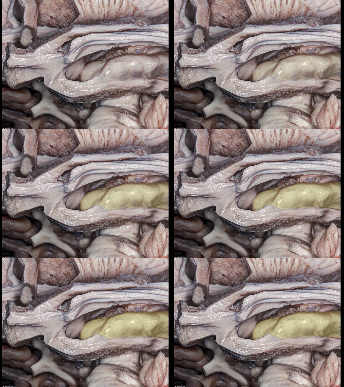
▼去除丘脑即显露 第三脑室(下图)。
Removal of the thalamus exposes the third ventricle.
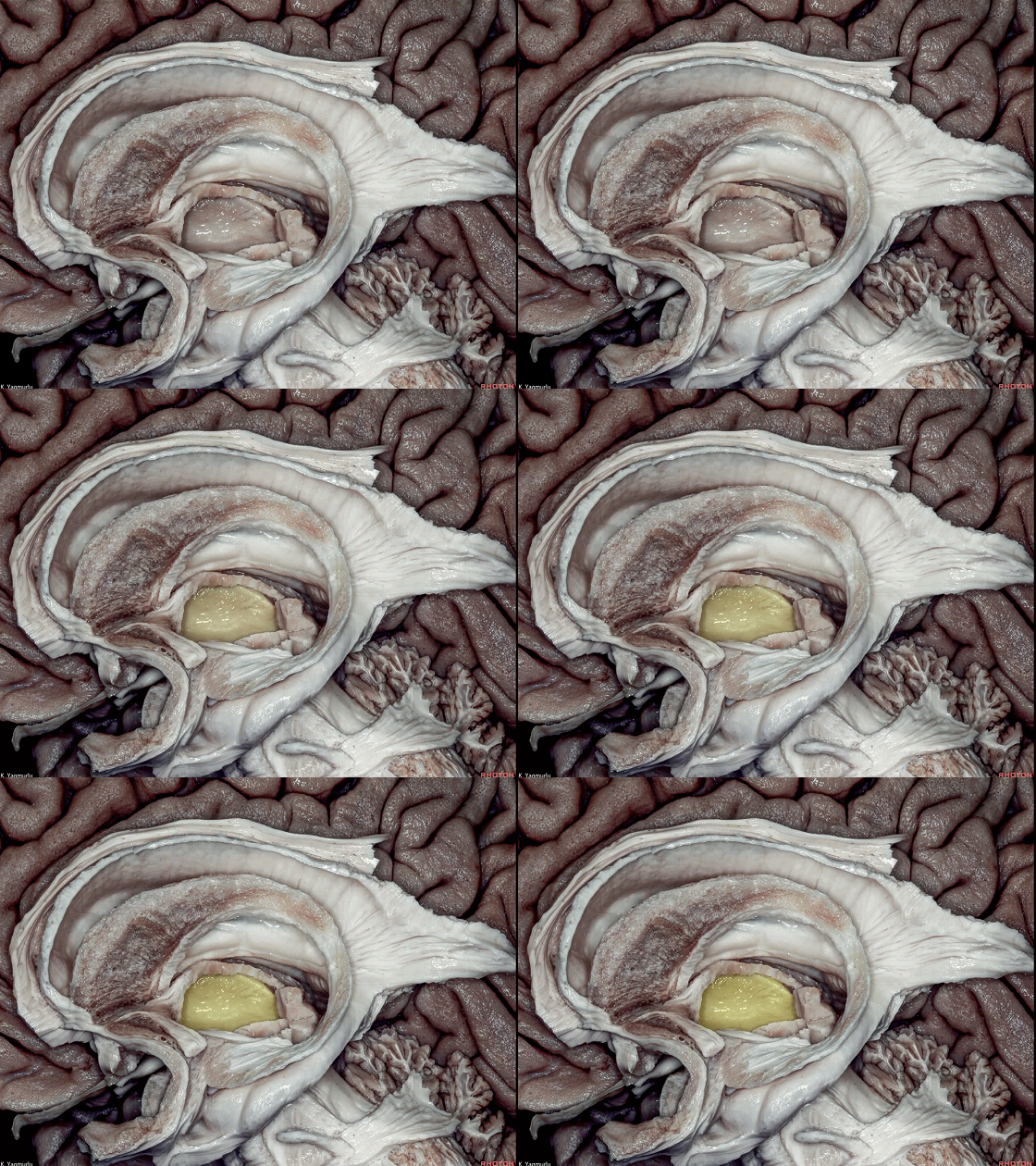
▼终纹(下图)起自 隔区。
The stria terminalis arises from the septal area.
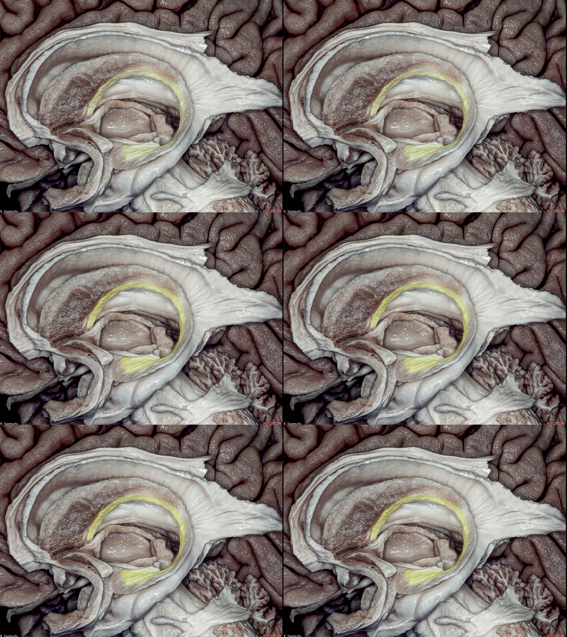
▼隔区(下图)位于前连合附近的中线处。
which is located around the anterior commissure in the midline.
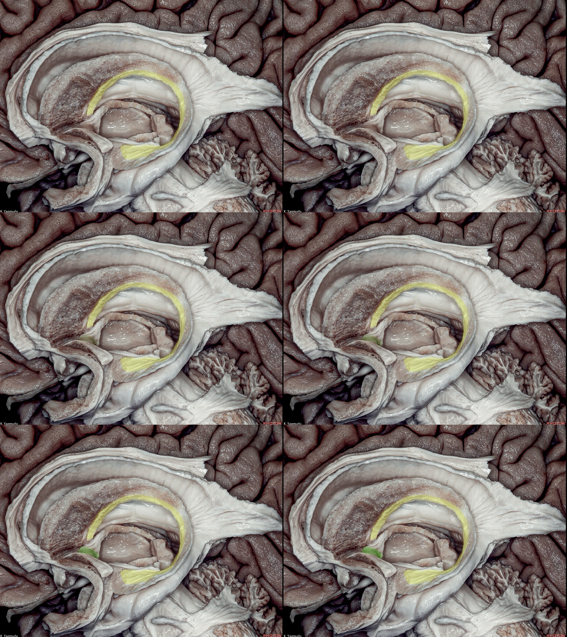
▼下图示 前连合。
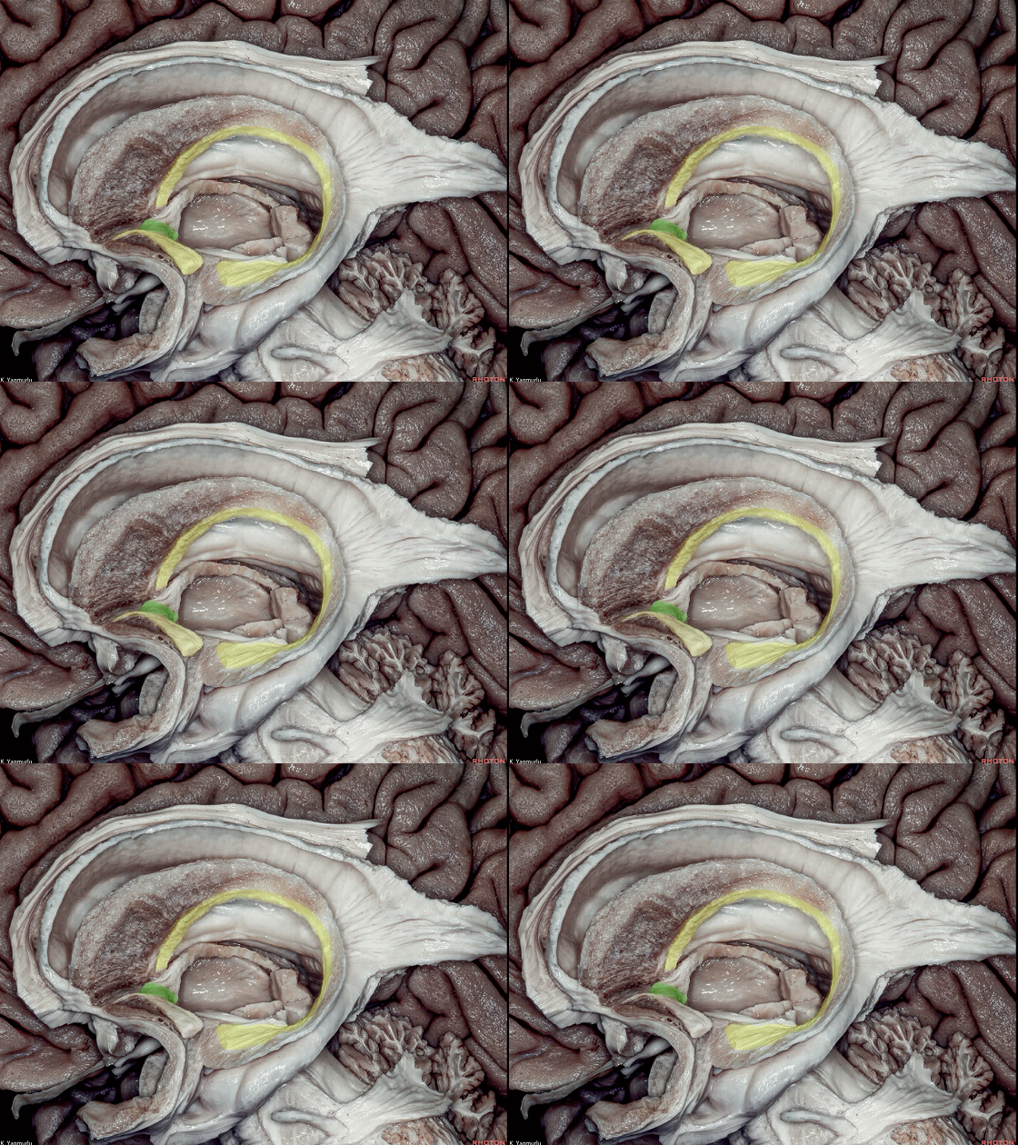
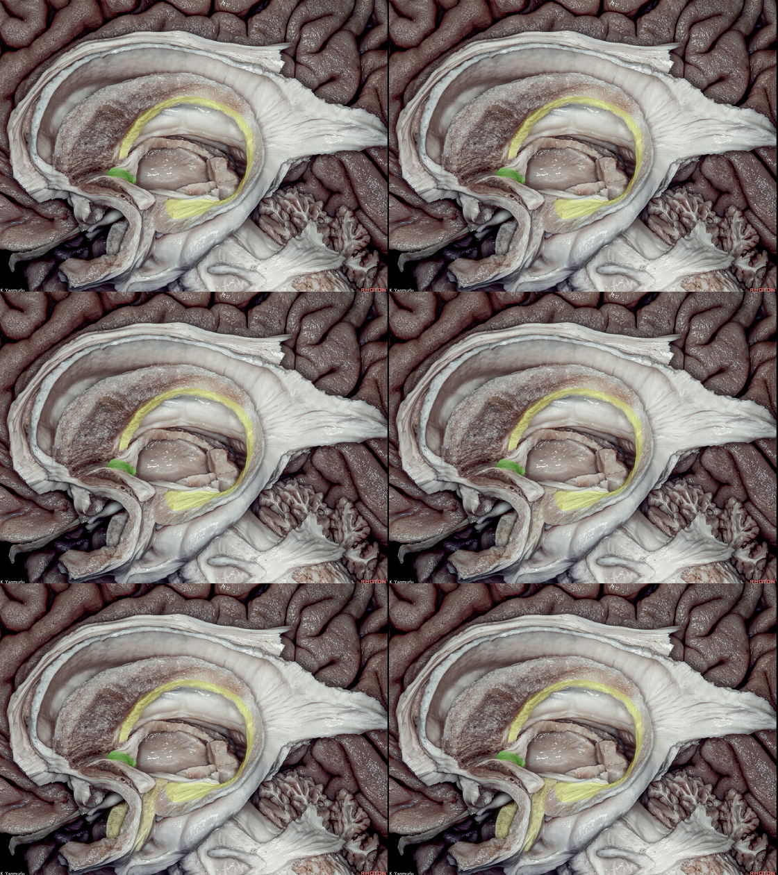
▼终纹包绕丘脑并移行入杏仁核(下图)。
and wraps around the thalamus to blend into the amygdala.
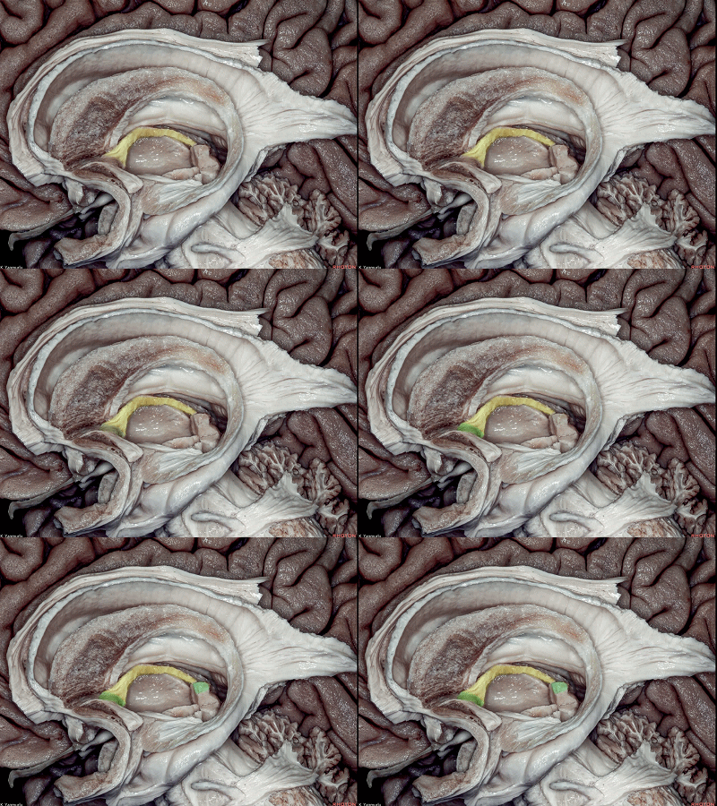
▼丘脑髓纹(下图黄色部分)从隔区(下图前方绿色)延伸至 松果体缰(下图后方绿色)。
The stria medullaris thalami extends from the septal area to the habenula.

▼接下来探讨额叶的皮层下白质解剖。
额叶外侧面的界限,后方为 中央沟(下图),前方为半球上界。
Let’s take a look at the subcortical white matter anatomy of the frontal lobe. The lateral surface of the frontal lobe is bordered posteriorly by the central sulcus and superiorly by the superior hemispheric border.
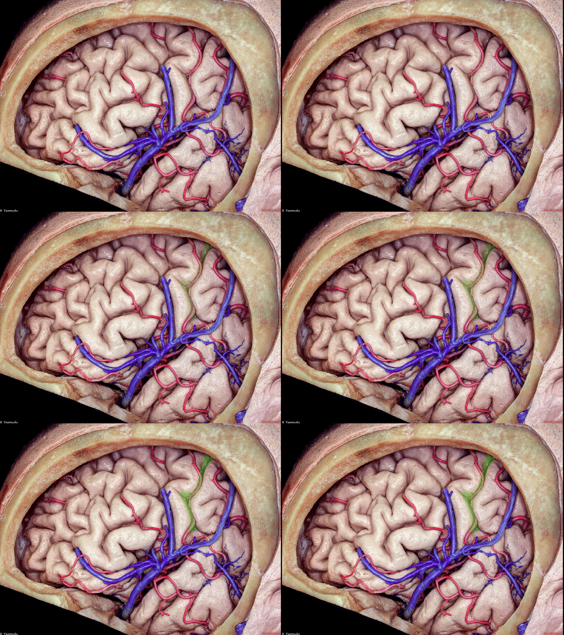
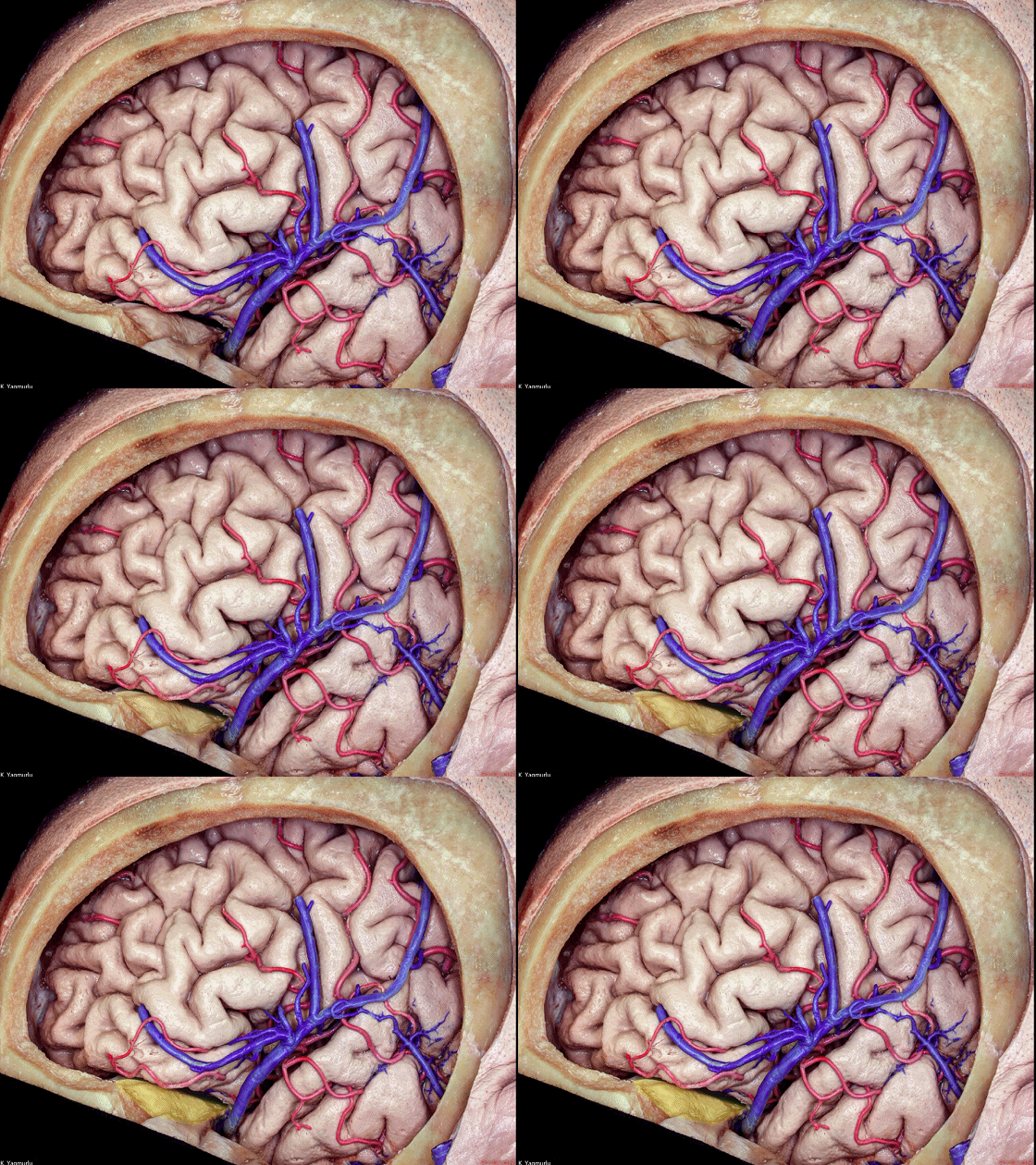
▼额叶外侧面的下界,其前部面向眶顶(下图),后部隔着外侧裂而面向颞叶。
The lower border of the lateral surface of the frontal lobe has an anterior part that faces the orbital roof, and a posterior part, the sylvian border that faces the temporal lobe across the sylvian fissure.
▼额叶的皮层下白质结构包含投射纤维(下图)、联合纤维、短和长联络纤维。
The subcortical white matter organization of the frontal lobe includes the projection,commissural,short and long association fiber pathways.
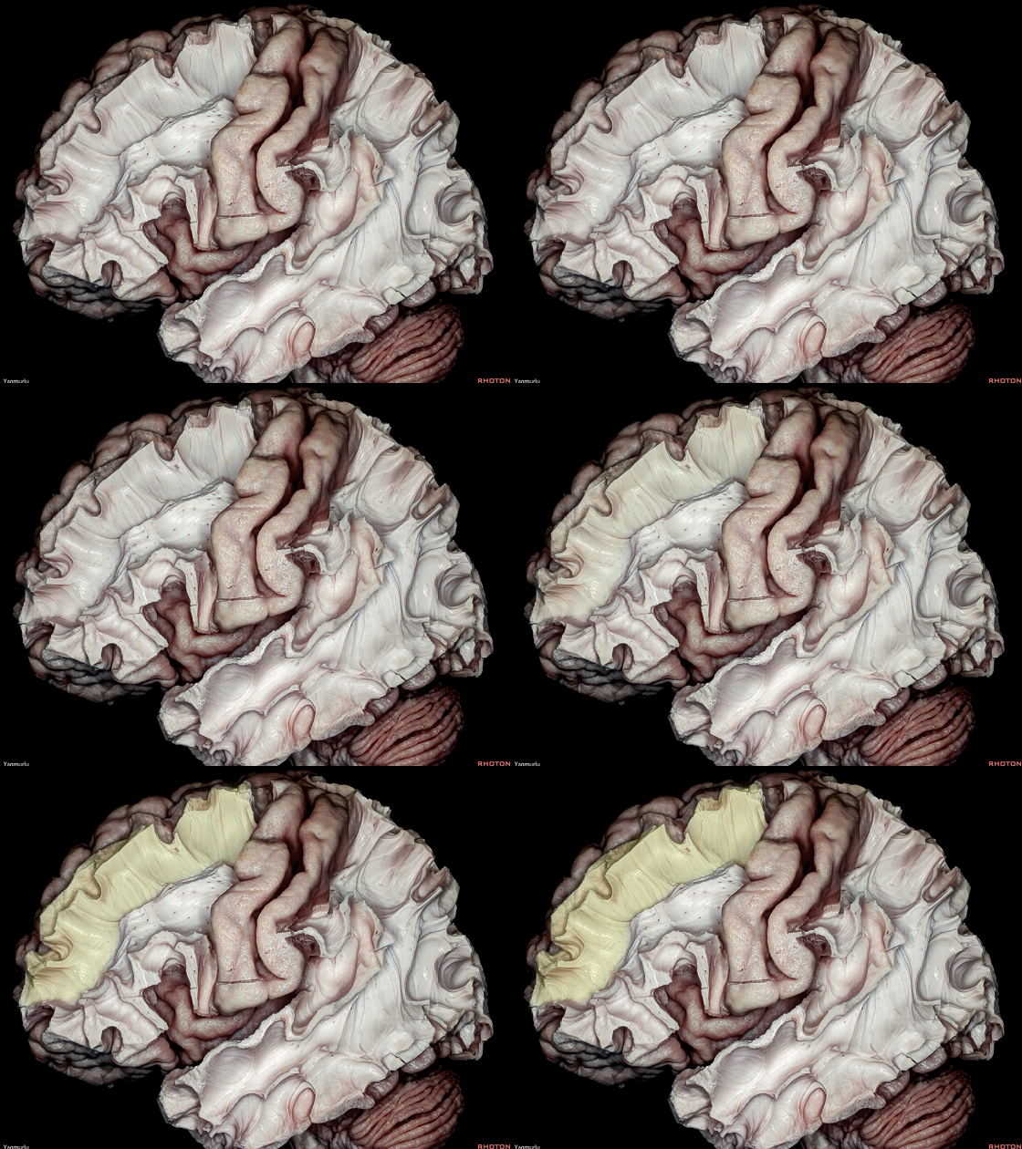
▼这是 短联络纤维。
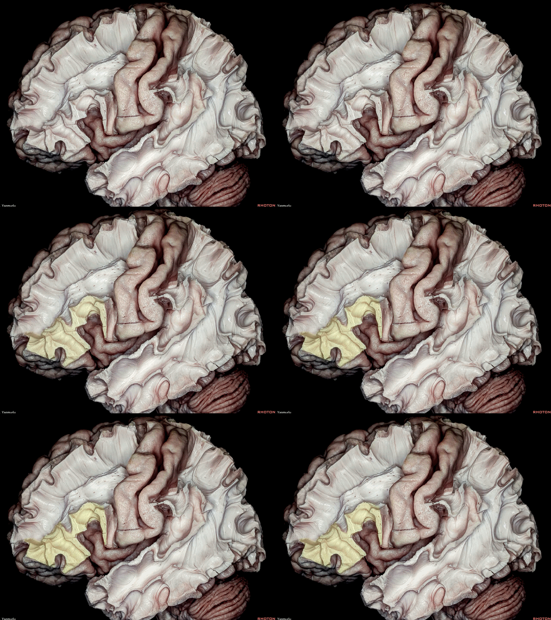
▼这是 长联络纤维。
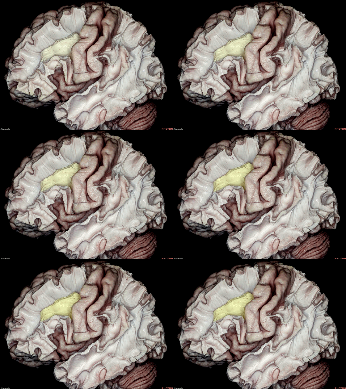
▼上纵束 II (下图)位于额中回内。
The superior longitudinal fasciculus II is situated within the middle frontal gyrus.
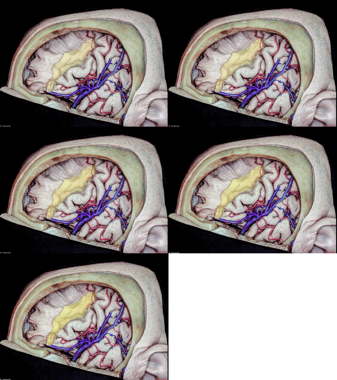
▼上纵束 II 内侧为 放射冠(下图),含有额叶投射纤维。
Medial to the SLF II are the corona radiata, containing the frontal projection fibers.
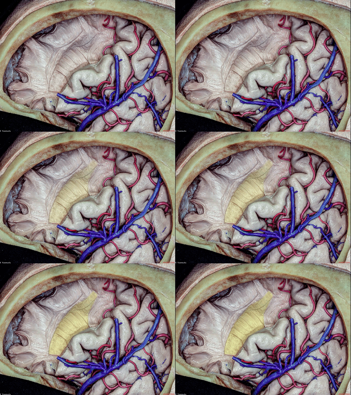
▼继续深入,这是位于额上回层面的胼胝体纤维。
In further dissection, the callosal fibers at the level of the superior frontal gyrus.
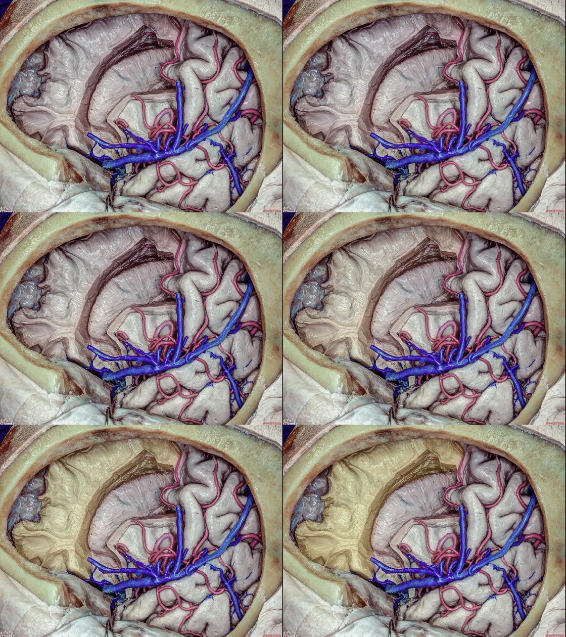
▼这是 侧脑室额角和体部、以及位于额中回和额下回层面的岛叶区域。
the frontal horn and body of the lateral ventricle as well as the insular area at the level of middle and inferior frontal gyri.
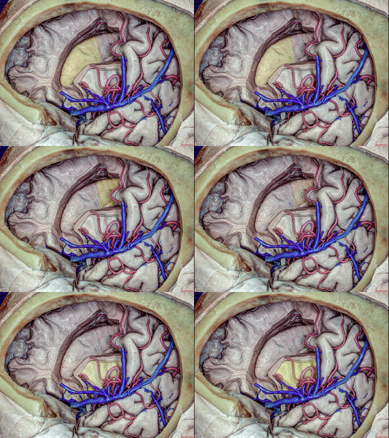
▼这是岛前点,为术中的重要标志。
Here is the anterior insular point, which is an important landmark for surgery.
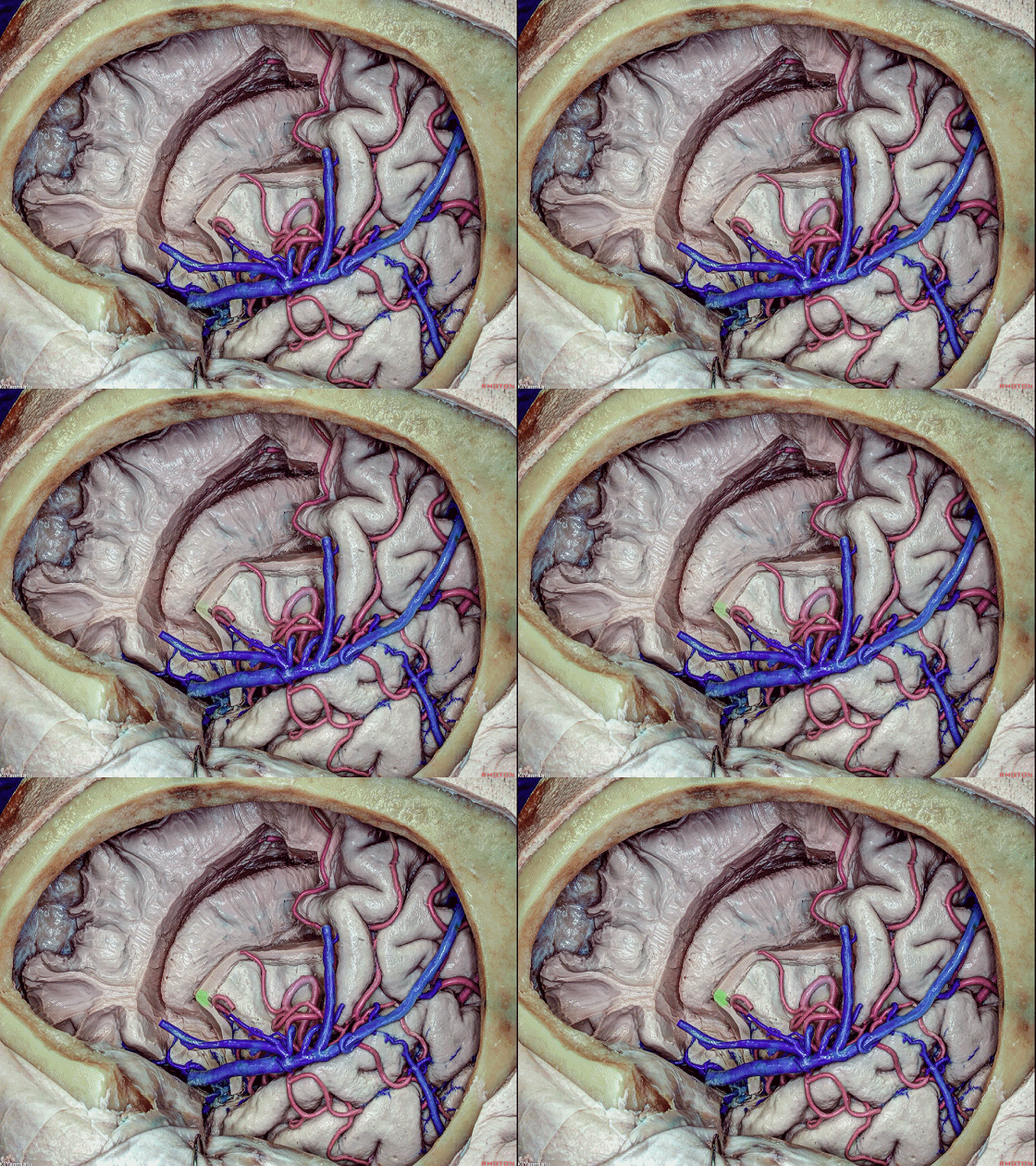
▼我们打开了侧脑室的室管膜层。岛前点 对应 侧脑室额角(下图)。
The ependymal layer of the ventricle was opened. The anterior insular point corresponds to the frontal horn of the lateral ventricle.
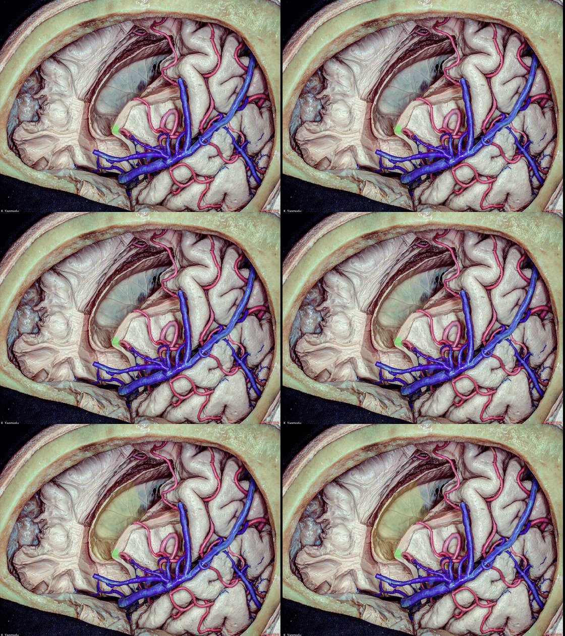
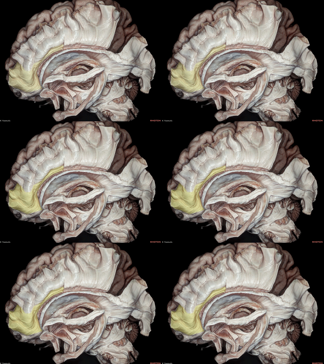
▼这是 扣带,属于边缘系统的一部分。
The cingulum, which is a part of the limbic system.
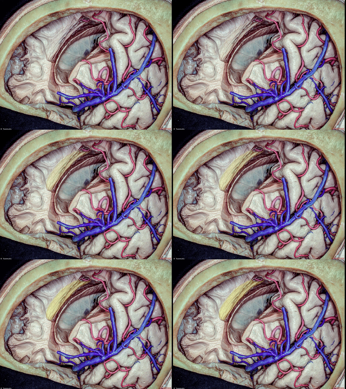
▼扣带 走行于 胼胝体纤维(下图)的内侧。
The cingulumruns medial to the callosal fibers.

▼另一相似的标本。显示的是胼胝体纤维和扣带之间的关系。下图示 胼胝体纤维。
A similar dissection in another specimen. The relationship between the callosal fibers and cingulum can be seen.
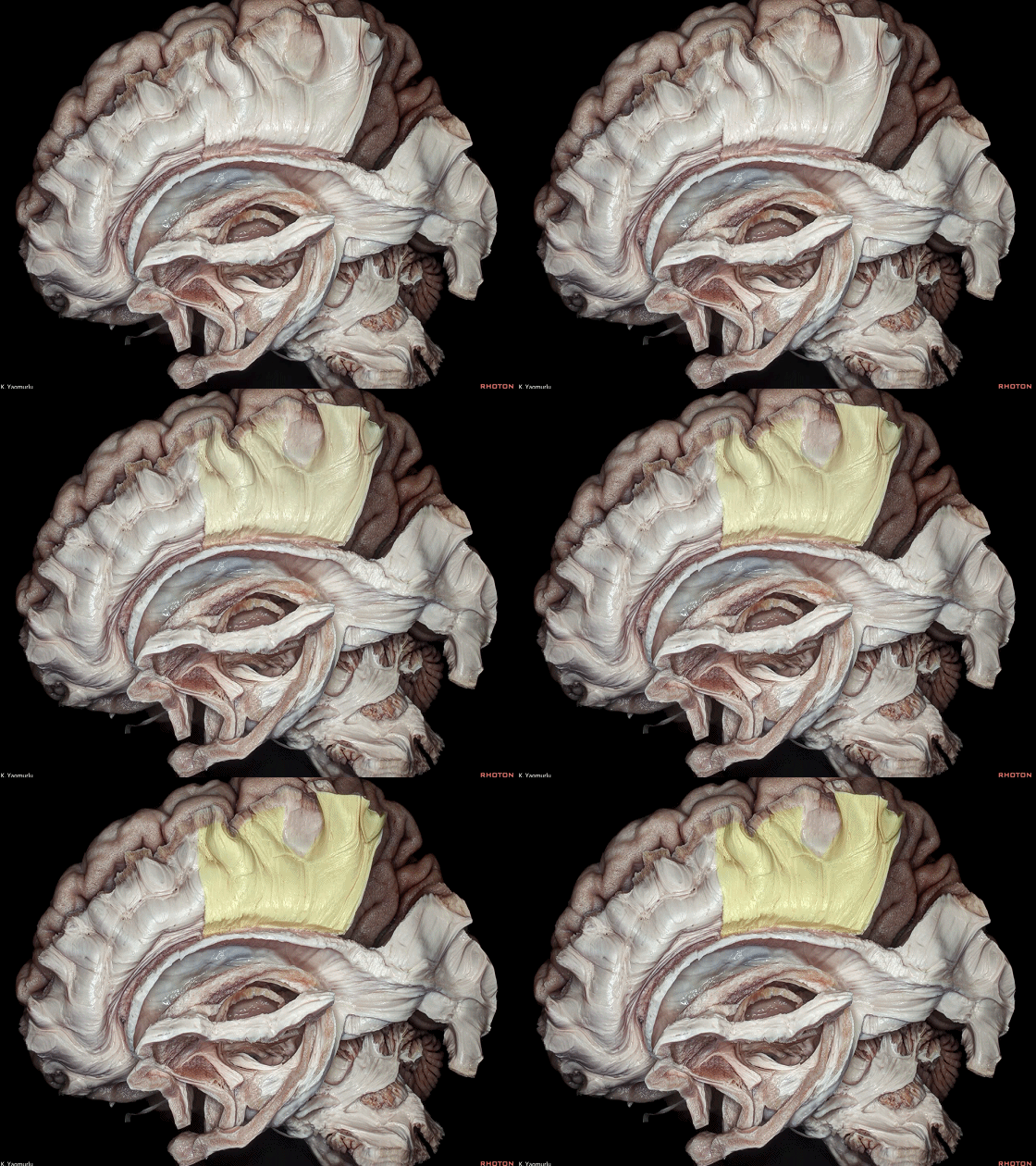
▼下图示 扣带。
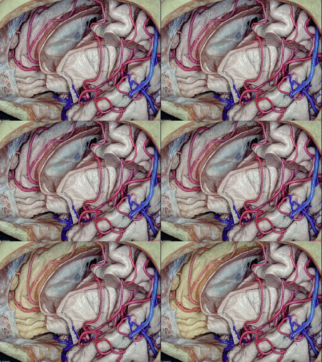
▼去除半卵圆中心即可显露 对侧额叶(下图)。
The removal of the centrum semiovale exposes the contralateral frontal lobe.
▼进一步对核心区的解剖可显露 下额枕束(下图)和 壳核。
The further dissection of the central core area exposes the inferior fronto-occipital fasciculus and putamen.
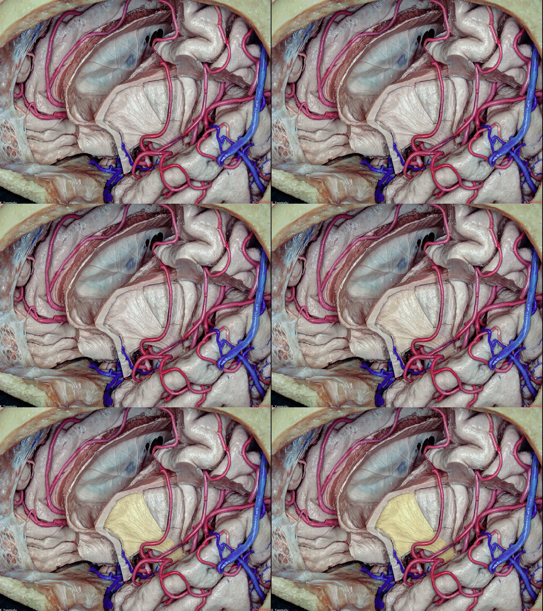
▼这是 壳核。
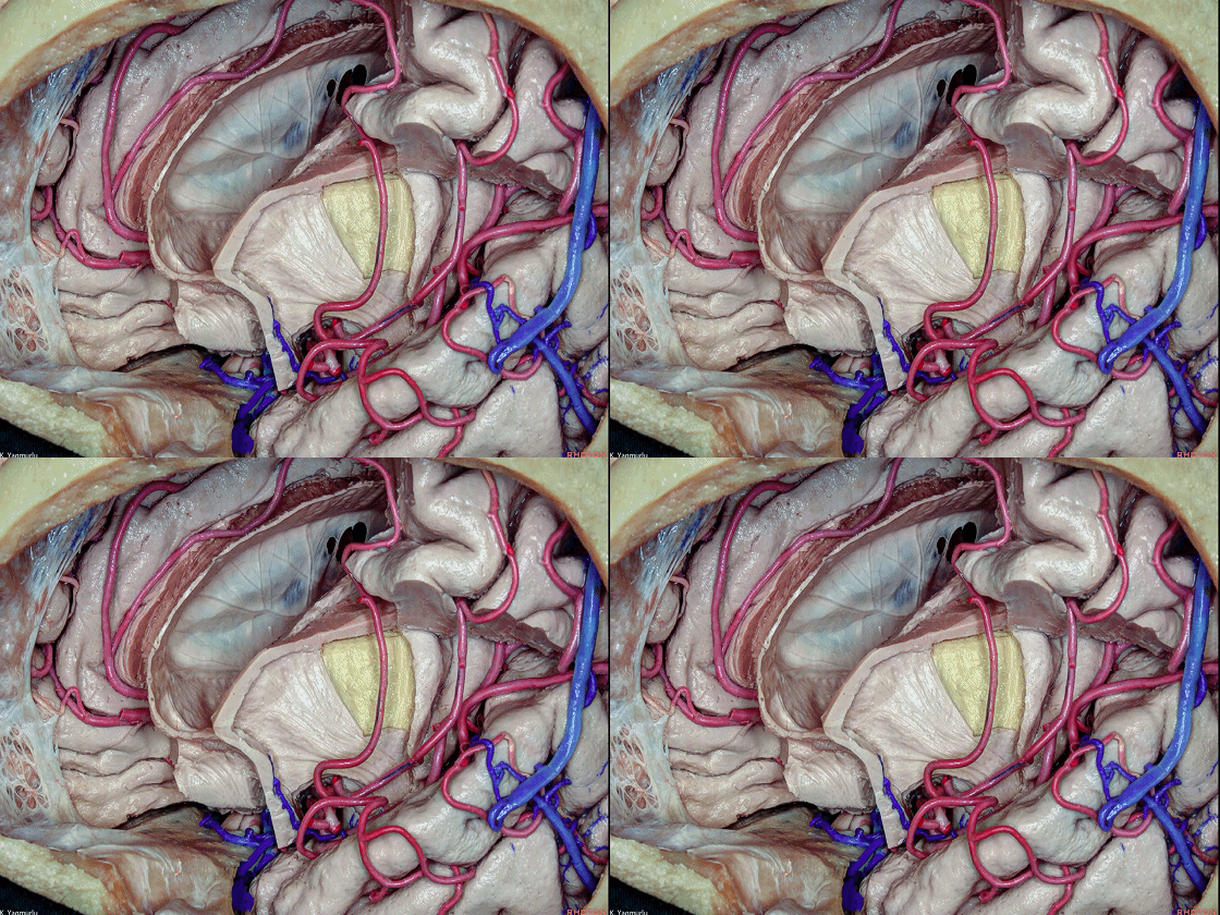
▼以上是关于额叶的解剖。
After seeing the frontal lobe.

![]()
▼接下来展示颞叶的内部解剖,按照从外到内的顺序。
下图中,颞叶皮层和短联络纤维已被去除以显露长联络纤维。
中纵束(下图)和下纵束(下图)都起自颞极。
let's take a look at the internal anatomy of the temporal lobe in order from the lateral to medial. The temporal cortex and short association fibers was removed to expose the long association fibers.The both middle and inferior longitudinal fasciculi originate from the temporal pole.
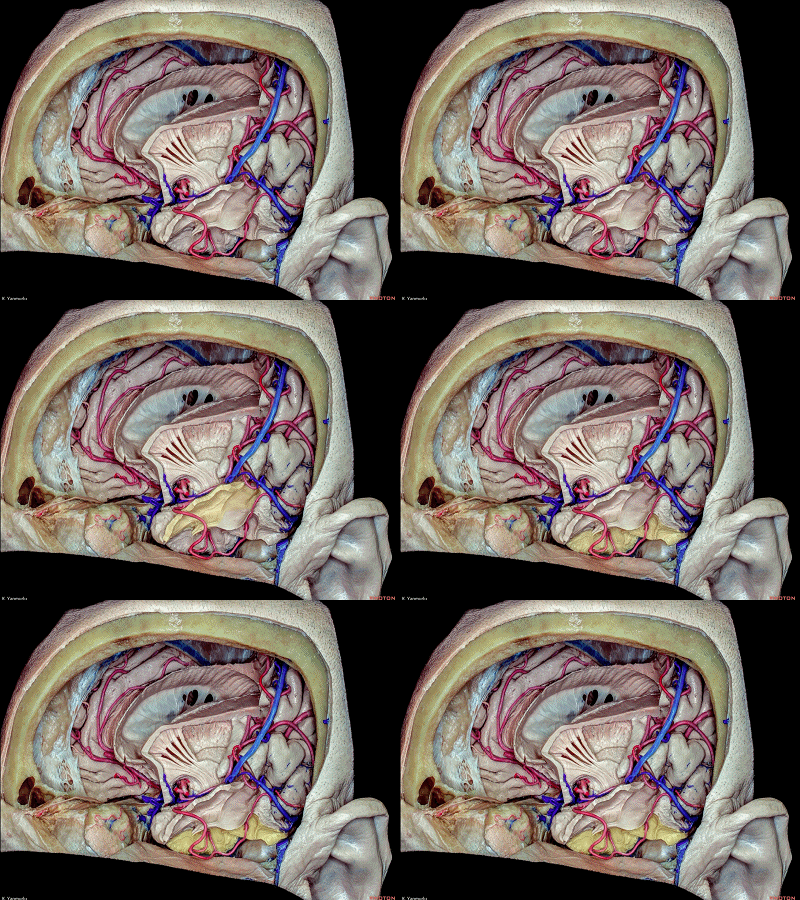
▼中纵束(下图)行于颞上回内。
The middle longitudinal fasciculus courses within the superior temporal gyrus.
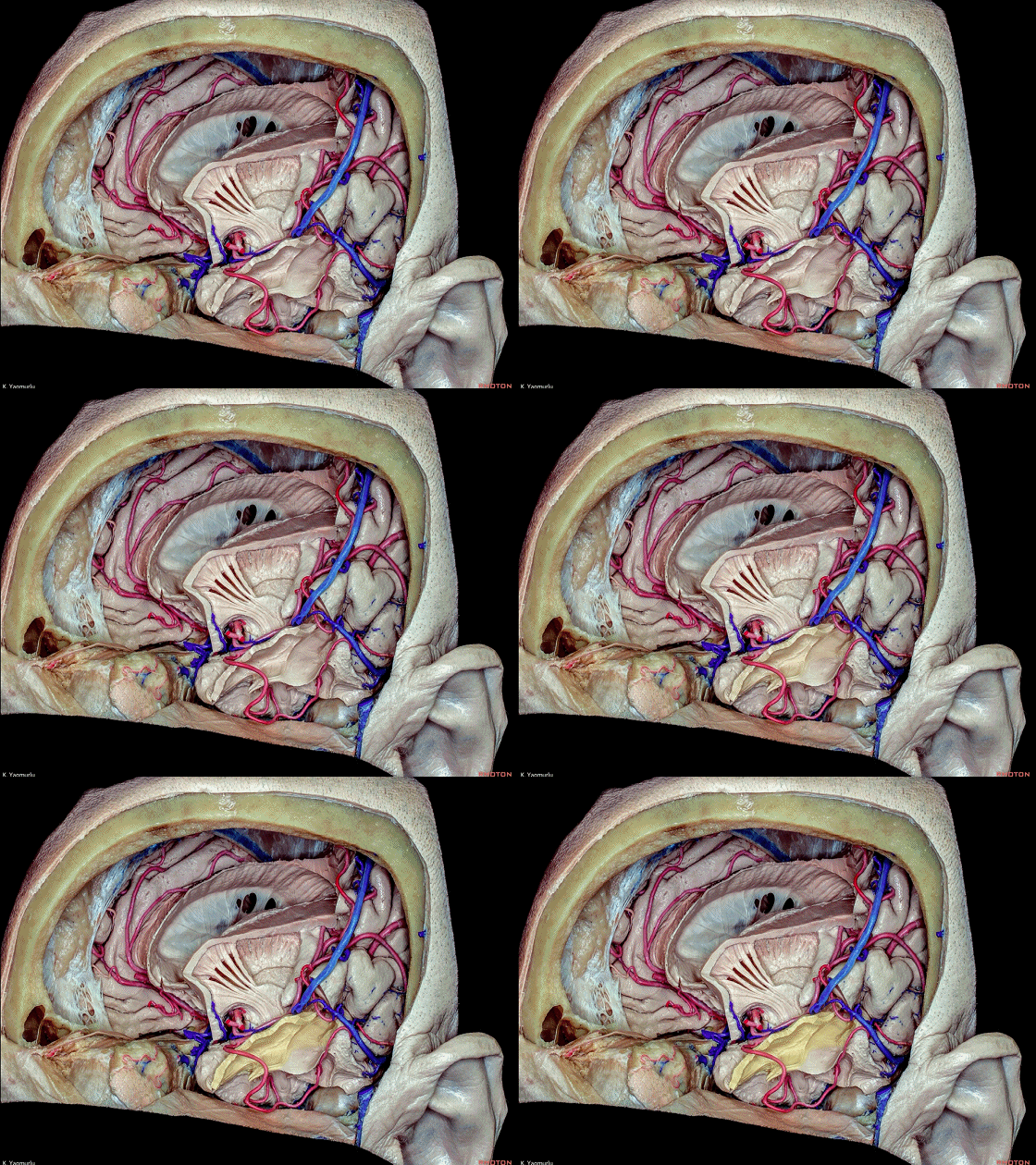
▼下纵束(下图)行于颞下回内。
while the inferior longitudinal fasciculus courses within the inferior temporal gyrus.
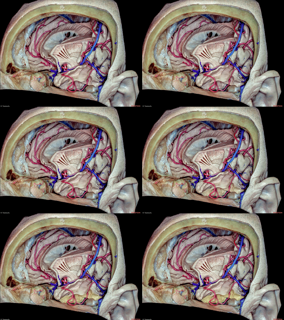
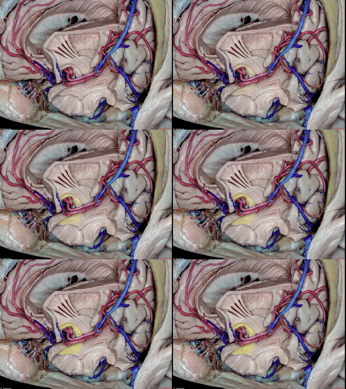
▼进一步解剖。可见 钩束。
Further dissection. The position of the uncinate fasciculus.
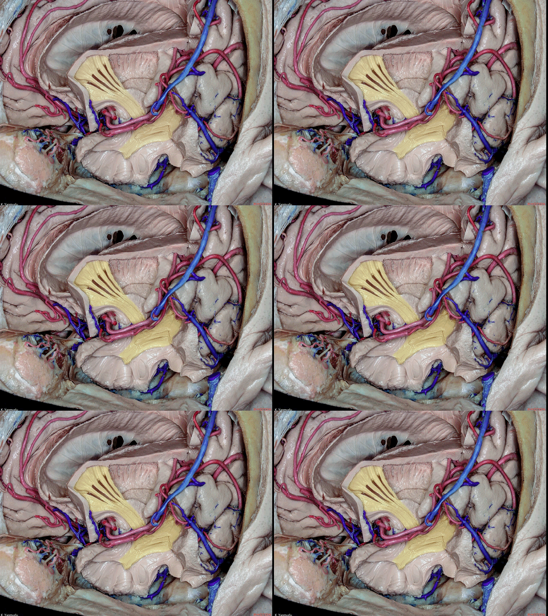
▼这是 下额枕束。
inferior fronto-occipital fasciculus.
▼去除钩束、下额枕束、前连合的延伸部,可显露 视辐射纤维(下图),其起自外侧膝状体。
The removal of the uncinate fasciculus, inferior fronto-occipital fasciculus and extensions of the anterior commissure exposes the optic radiations fibers which arise from the lateral geniculate body.
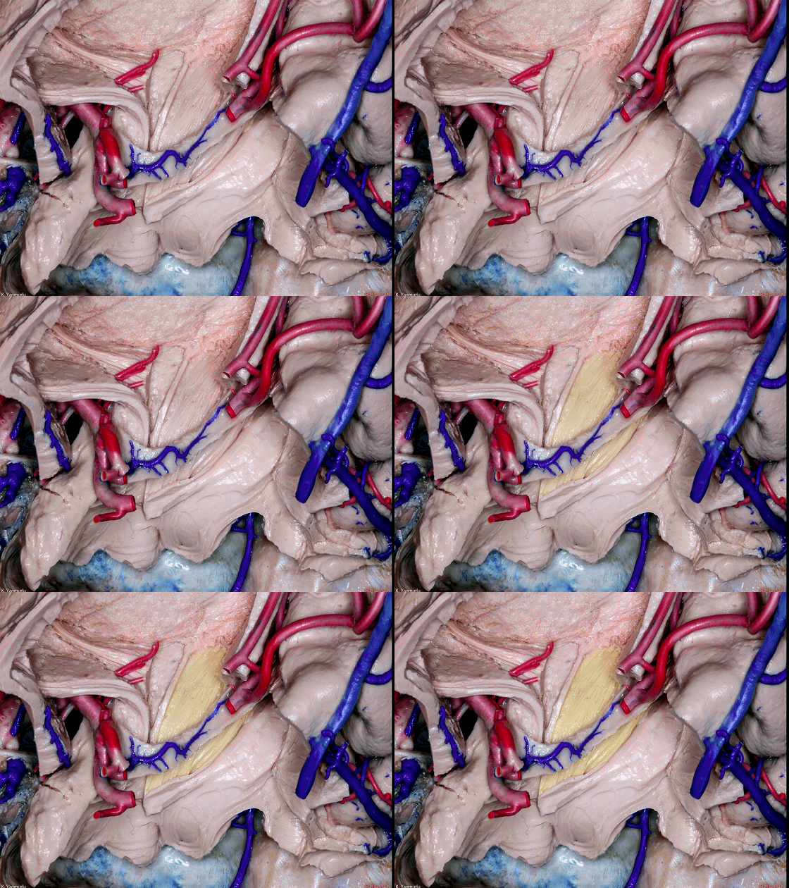
▼视辐射纤维行于下界沟(下图)内侧,构成侧脑室颞角的顶壁和外侧壁。
the optic radiations fibers pass medial to the inferior limiting sulcus, to form the roof and lateral wall of the temporal horn of the lateral ventricle.
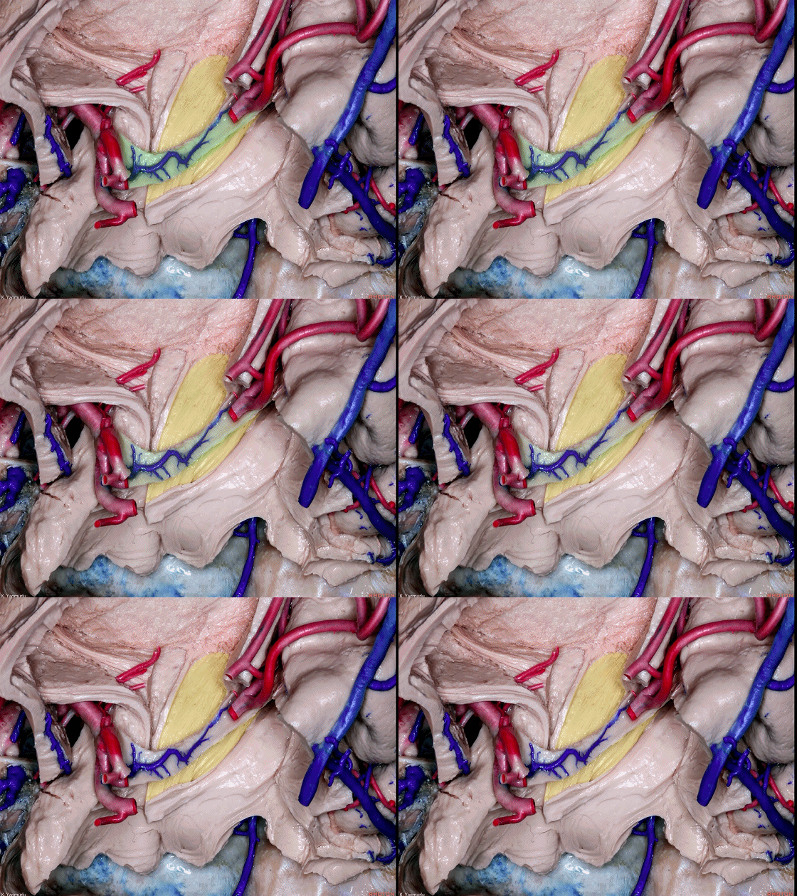
▼这是 外侧膝状体。
lateral geniculate body.
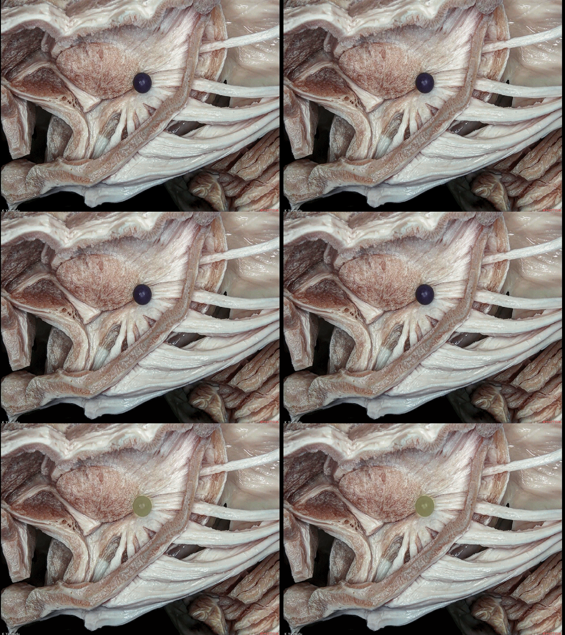
▼视辐射(下图)发出自外侧膝状体。
the optic radiation fibers arise in the lateral geniculate body.
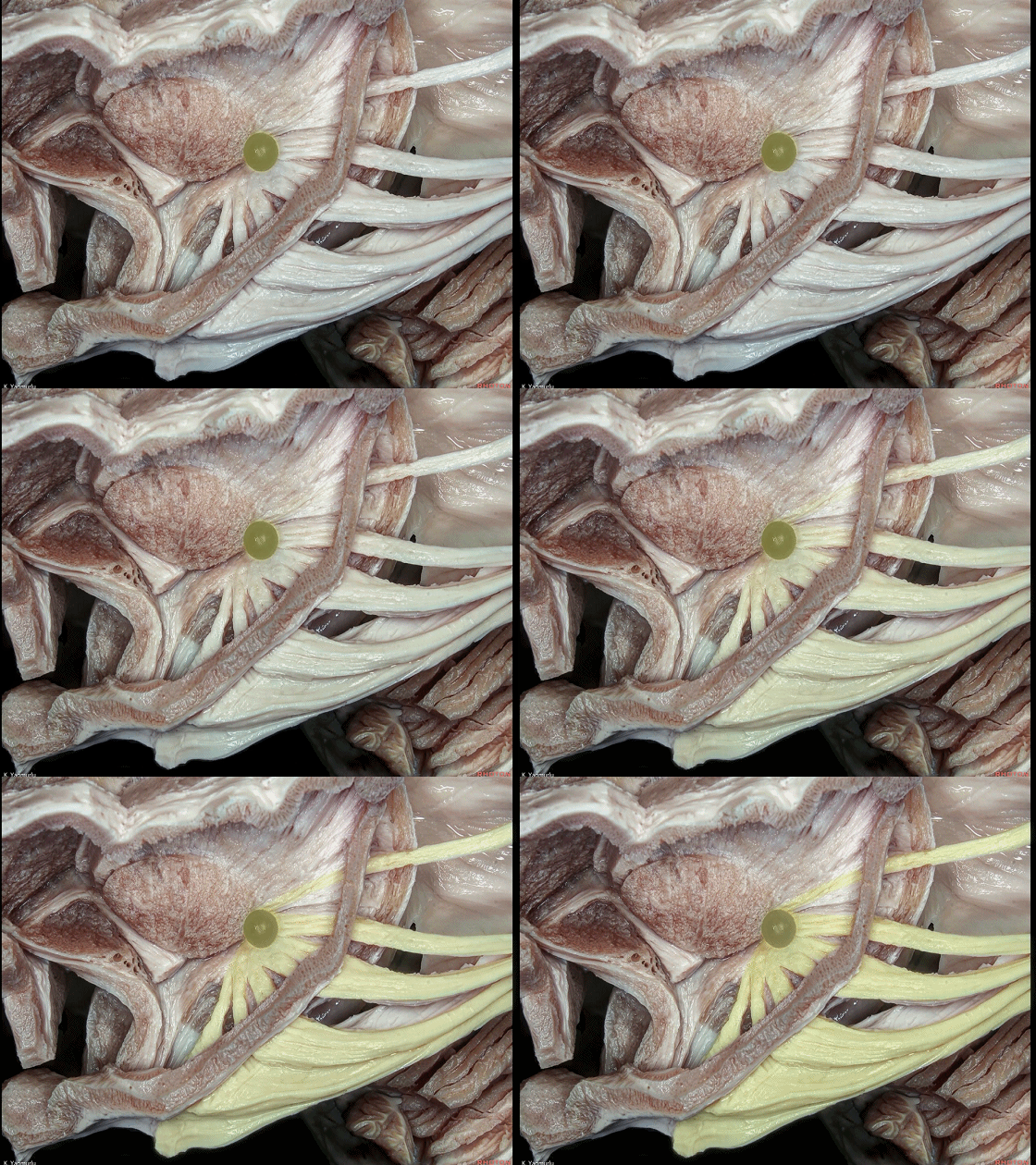
▼视辐射纤维行于下界沟和尾状核尾之间。下图示 下界沟。
the optic radiation fibers pass between the inferior limiting sulcus and tail of the caudate nucleus.
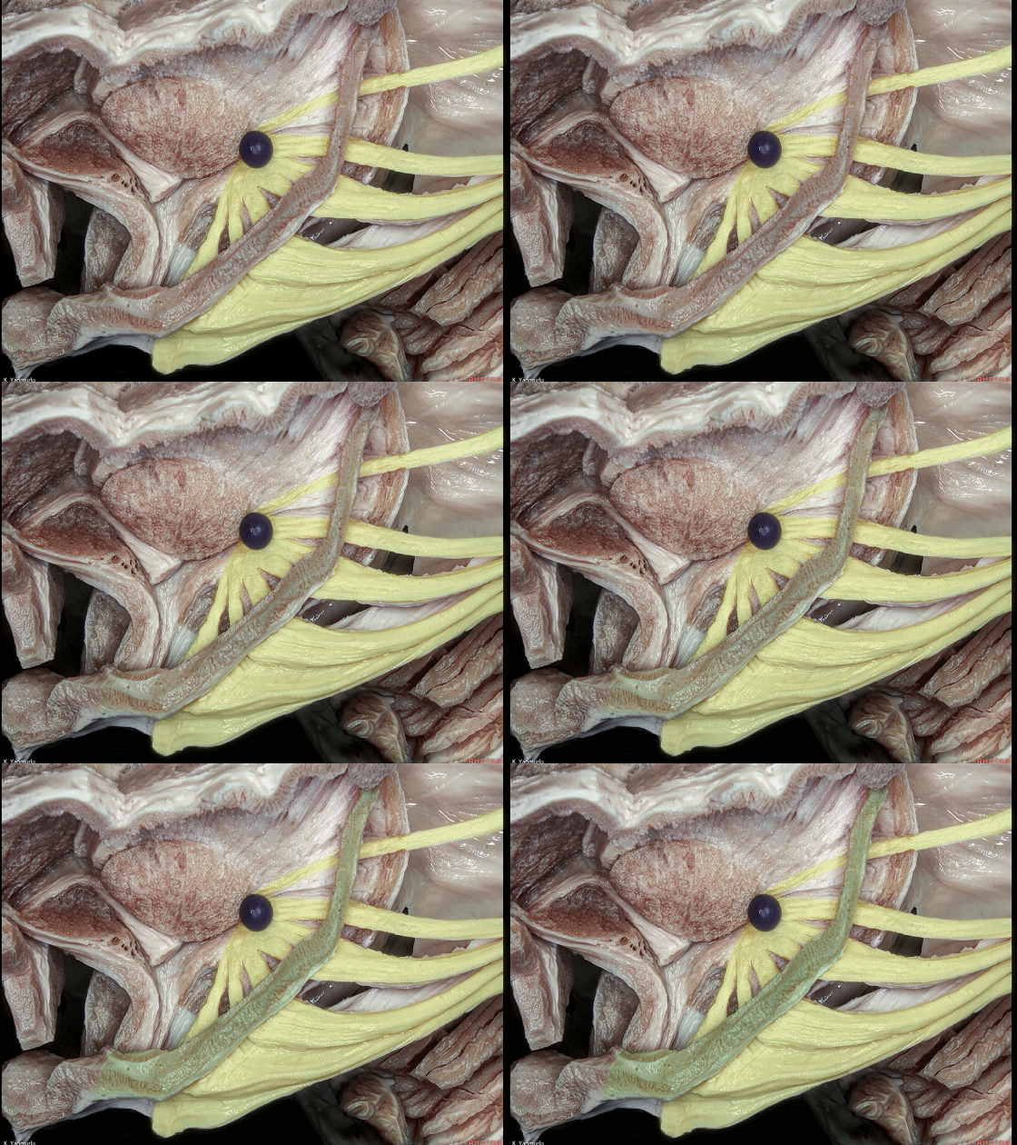
▼下图示 尾状核尾。
tail of the caudate nucleus.
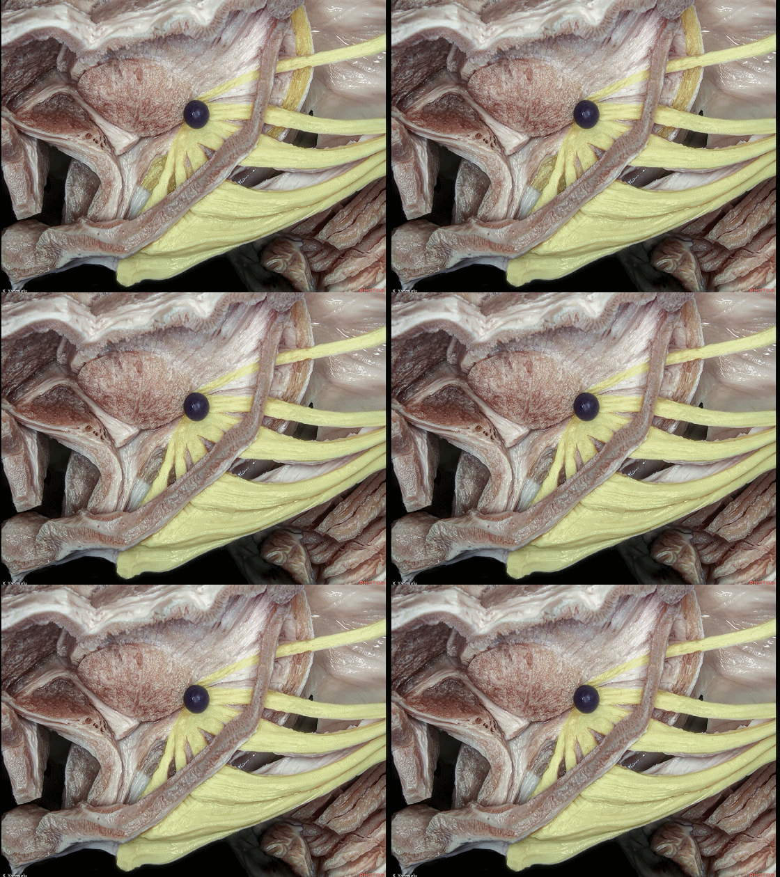
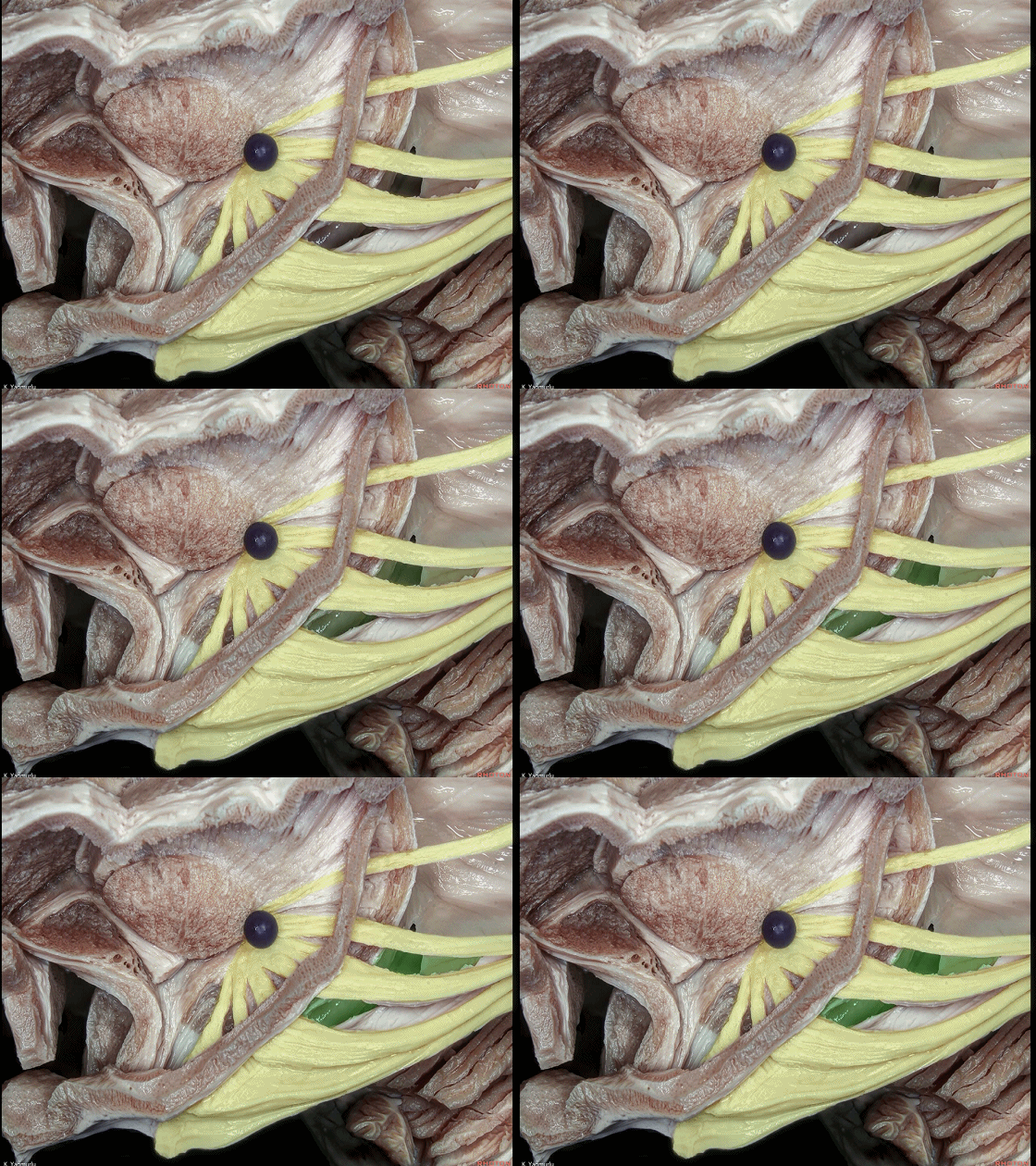
▼视辐射纤维 构成 颞角 的 顶壁 和 侧壁(下图)。
the optic radiation fibers cover the roof and lateral wall of the temporal horn.
![]()
▼最后让我们来探讨边缘系统。
边缘系统为一组相互联系的皮层与皮层下区域,负责将内脏信息、情感反应整合于认知和行为。
Finallly let's look at the limbic system. The limbic system is a group of interconnected cortical and subcortical areas that links visceral states and emotion to cognition and behavior.

▼边缘皮质包括海马旁回、海马结构(暂时未显露)、以及扣带回。
下图示海马旁回,形似拉伸的C形。海马旁回与哺育、行为、注意力等有关。
The limbic cortex is composed of the parahippocampal gyrus,hippocampal formation, which is hidden,and cingulate gyrus.The parahippocampal gyrus is an elongated, C-shaped gyrus, The parahippocampal gyrus is important for nursing, behavior and attention.

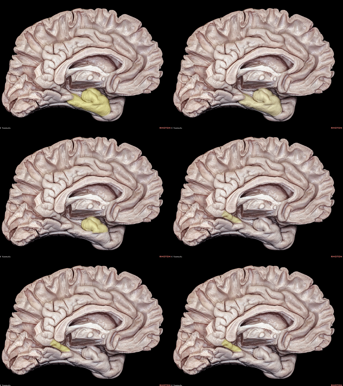
▼海马旁回位于钩回(下图)和扣带回峡部(下图)之间,后者为舌回与海马旁回相移行的部位。
The parahippocampal gyrus is between the uncus and isthmus of the cingulate gyrus, where the lingual gyrus merges into it.
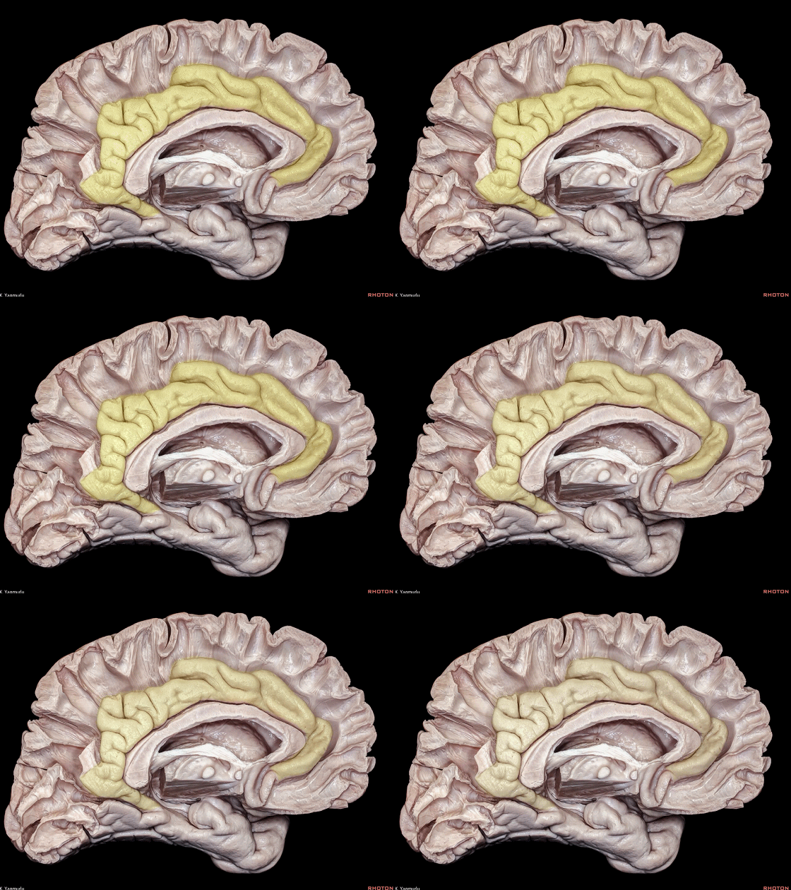
▼这是 扣带回。
cingulate gyrus.
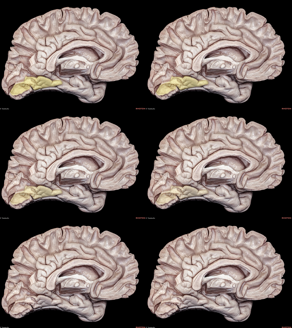
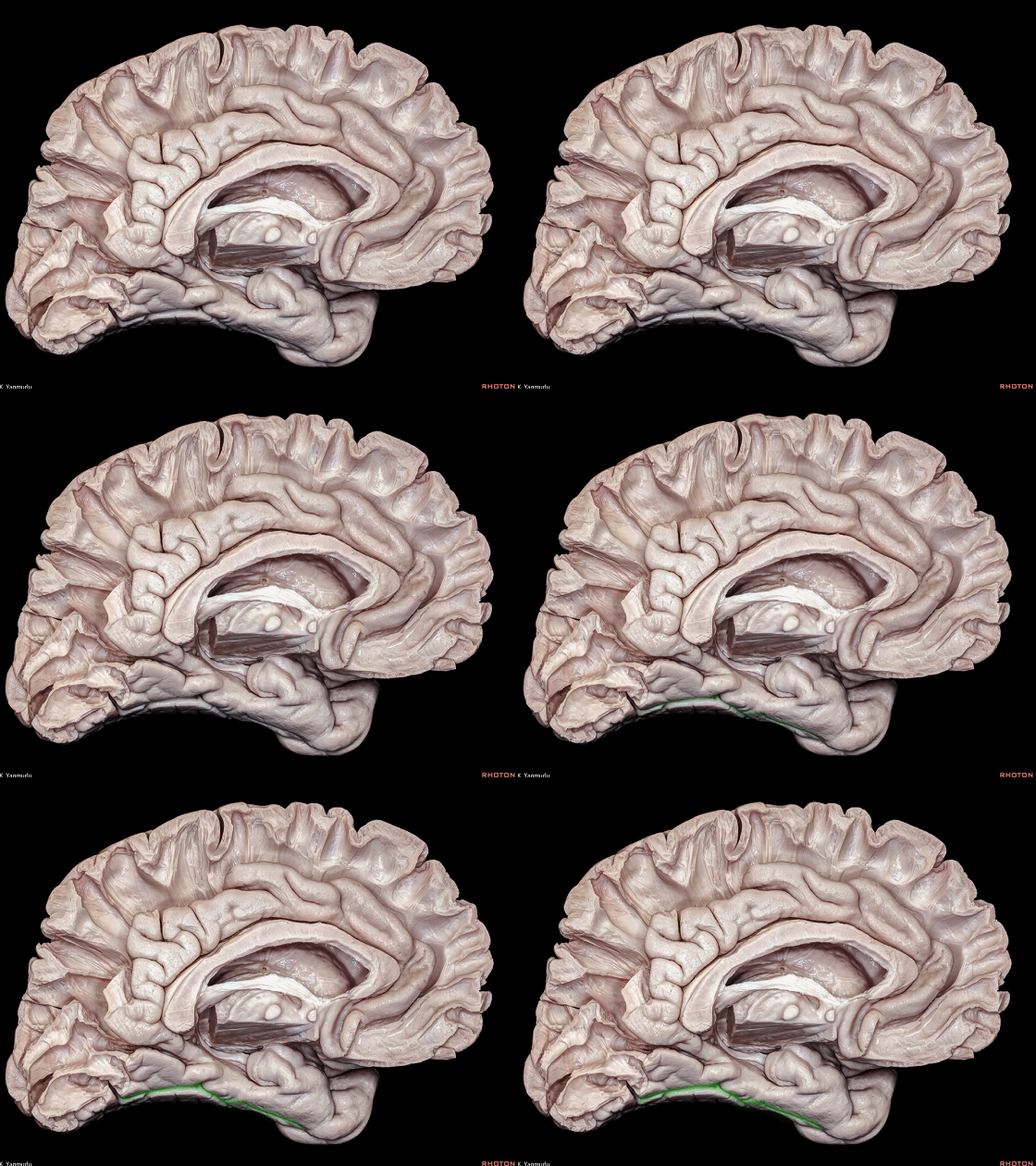
▼这是 舌回。
▼在外侧,有侧副沟(下图)将 海马旁回 与 梭状回 分隔。
Laterally the collateral sulcus seperates it from the fusiform gyrus.
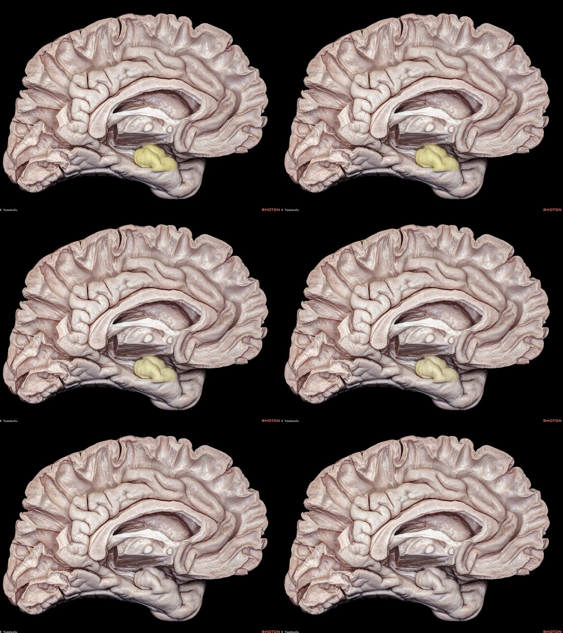
▼这是 钩回,该脑回呈一钩形向前延伸。
海马旁回和钩回在侧方包被海马。海马的功能与记忆相关。
This gyrus forms a hook like expansion anteriorly, called the uncus. The parahippocampus and uncus lie laterally and envelop the hippocampus. The hippocampus plays a role in memory function.
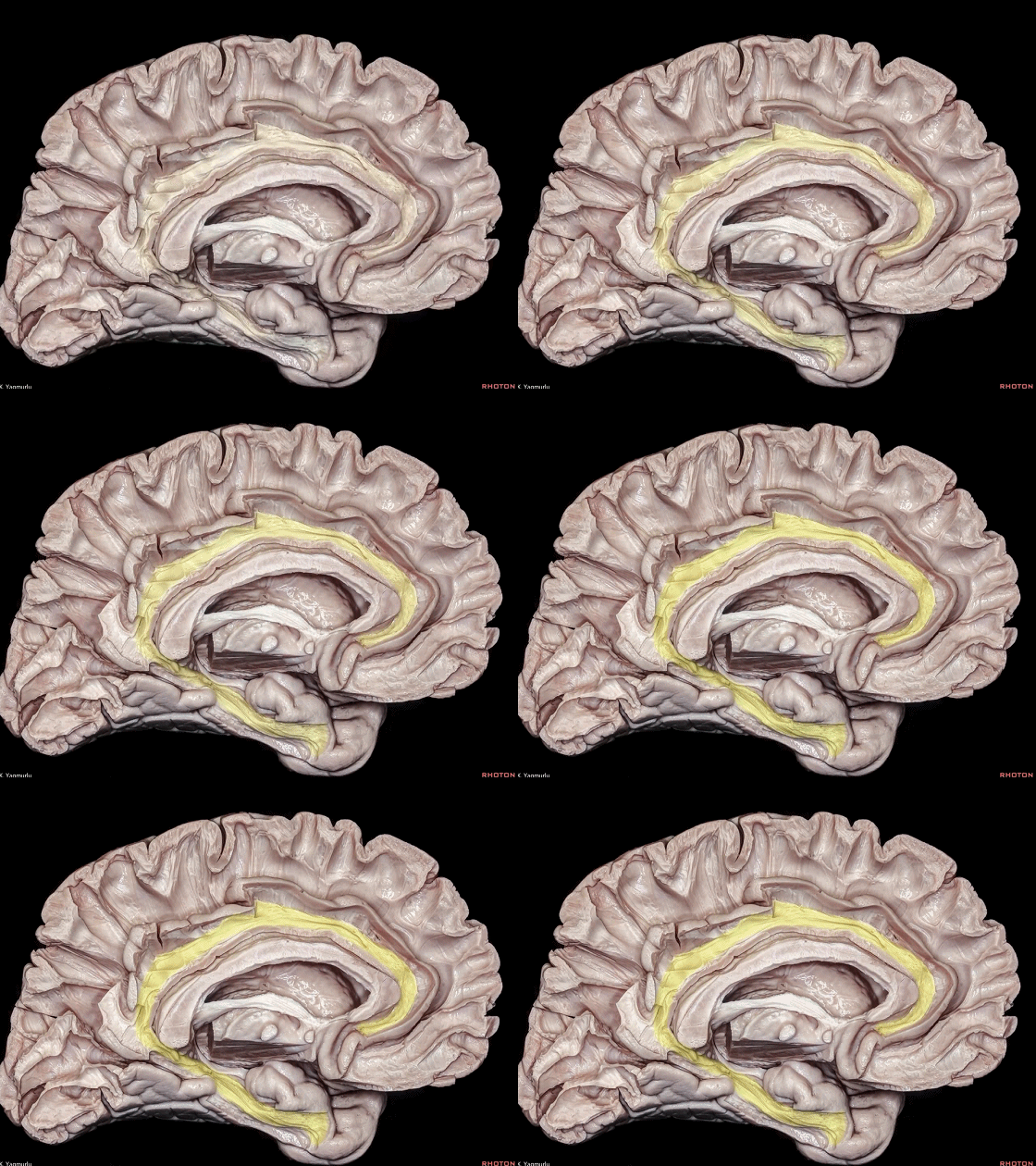
▼扣带(下图)属于长联络纤维通路。
The cingulum is a long association fiber pathway.
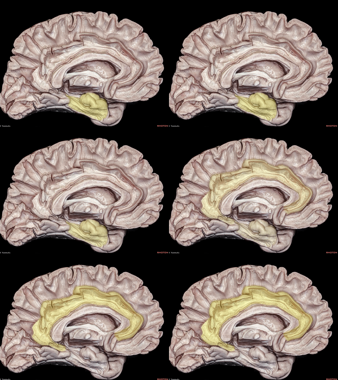
▼扣带 连接 海马旁回(下图)和扣带回(下图)。
The cingulum interconnects the parahipocampal and cingulate gyri.
▼这是 内侧观。
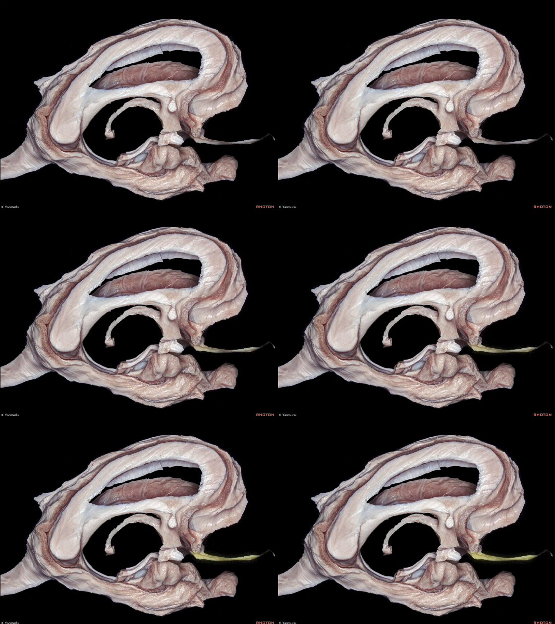
边缘系统的皮层下结构,主要包括 嗅觉系统、隔区、下丘脑、杏仁核。
下图示 嗅觉系统。
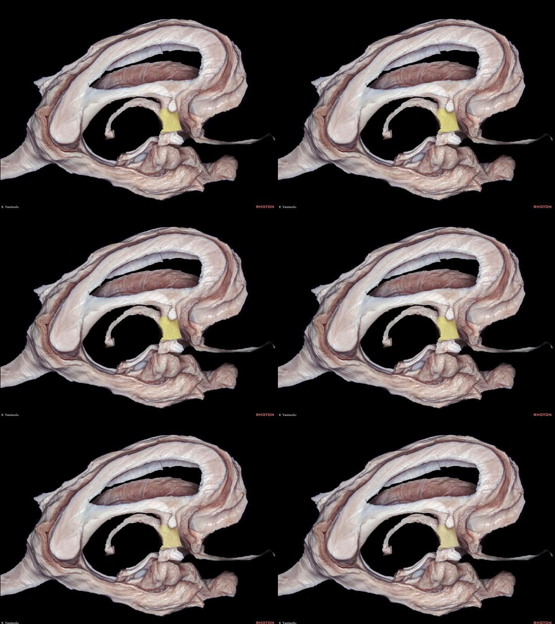
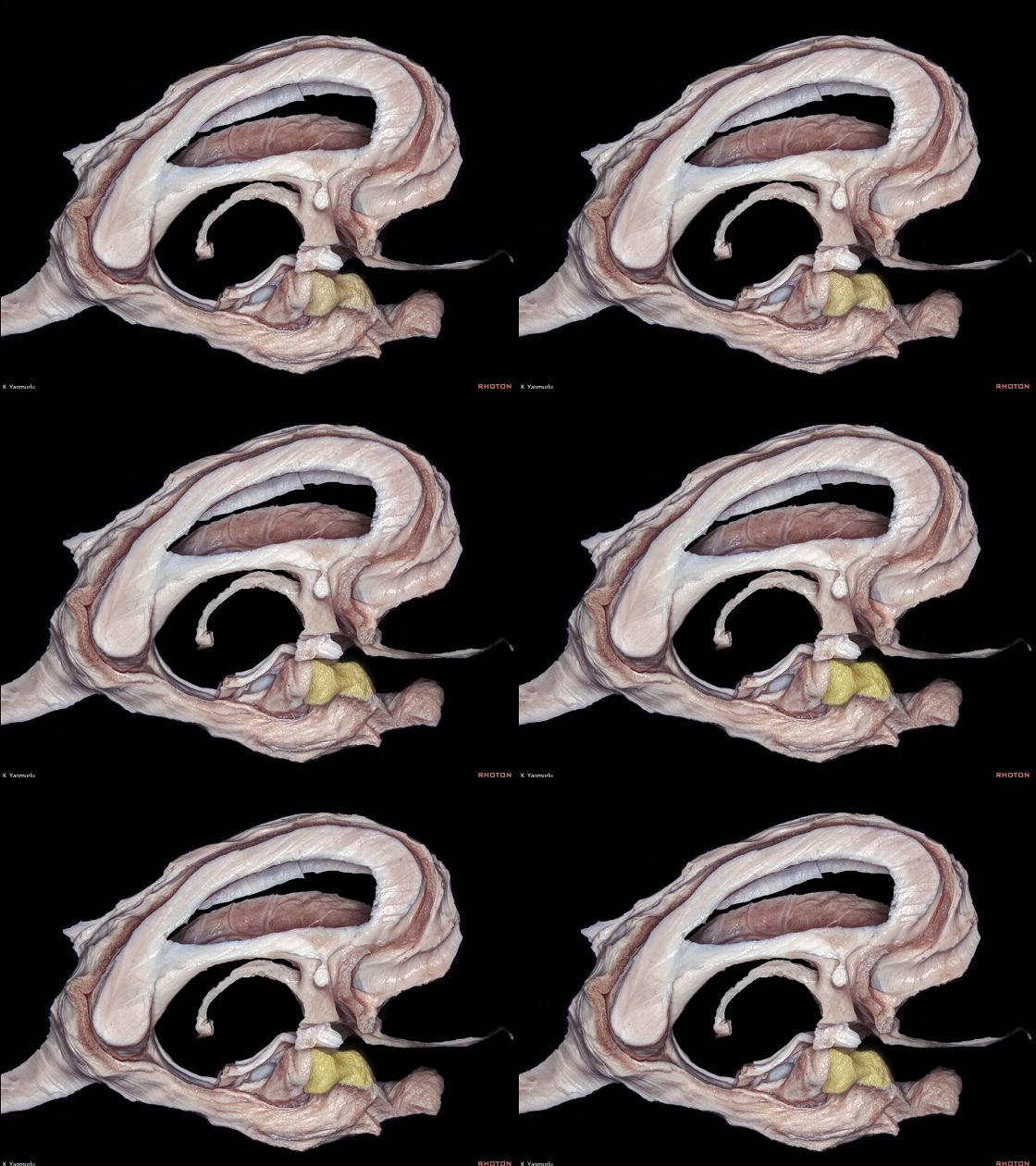
Medial view. The subcortical area of the limbic system, basically, includes the olfactory system, septal region, hypothalamus and amygdala.
▼这是 隔区。

▼ 这是 下丘脑。
▼ 这是 杏仁核。
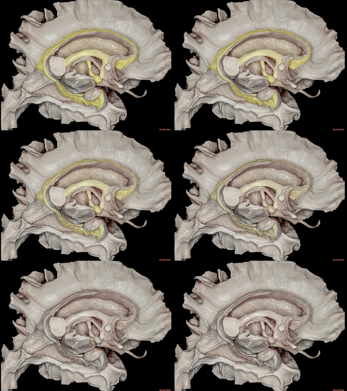
▼最著名的沟通边缘皮质和皮质下区域的边缘通路,即Papez环路(下图)。可作为情绪表达的神经基础。
The best known limbic pathway that interconnects the limbic cortical and subcortical areas, is Papez circuit.
▼具体说来,纤维从 海马结构(下图)发出,经过 穹窿(下图) 到达 乳头体(下图)。
To review, fibers leave the hippocampal formation and proceed through the fornix to reach the mamillary body.
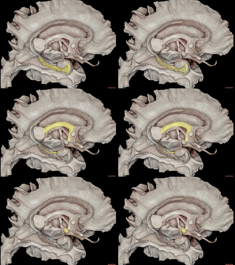
▼从乳头体(下图)发出一条新的通路,上行至 丘脑前核(下图),即 乳头丘脑束。
From here, a new pathway, the mammillothalamic tract, ascends up to the anterior thalamic nucleus.
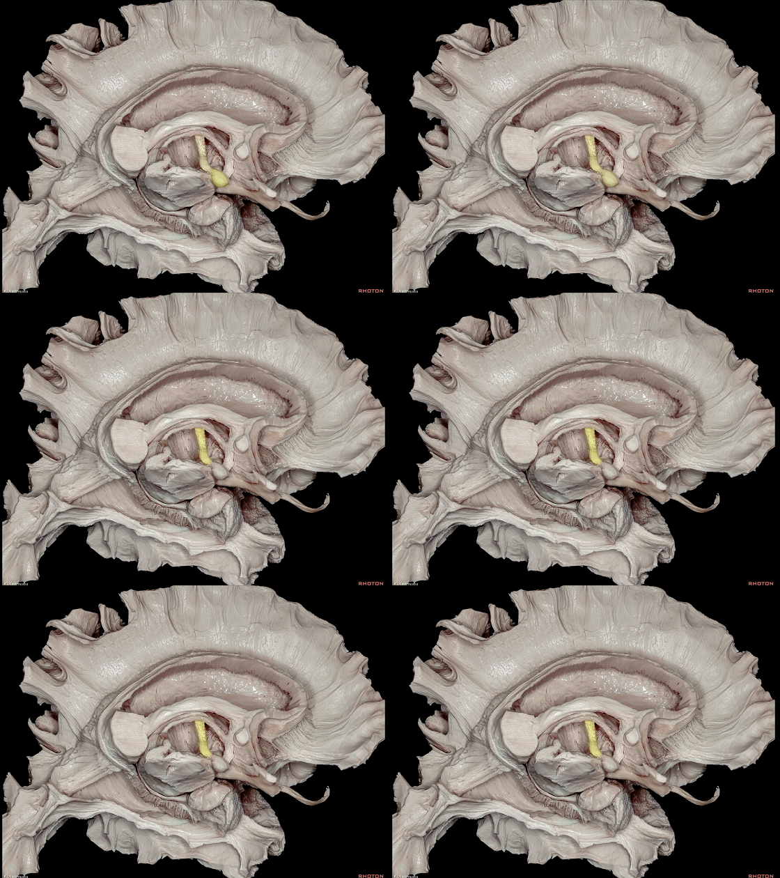
▼丘脑前核(下图)发出纤维继续行至扣带回(下图),其通路构成丘脑扣带纤维。
The information in the anterior thalamic nucleus continues to the cingulate gyrus by means of the thalamocingulate fibers.
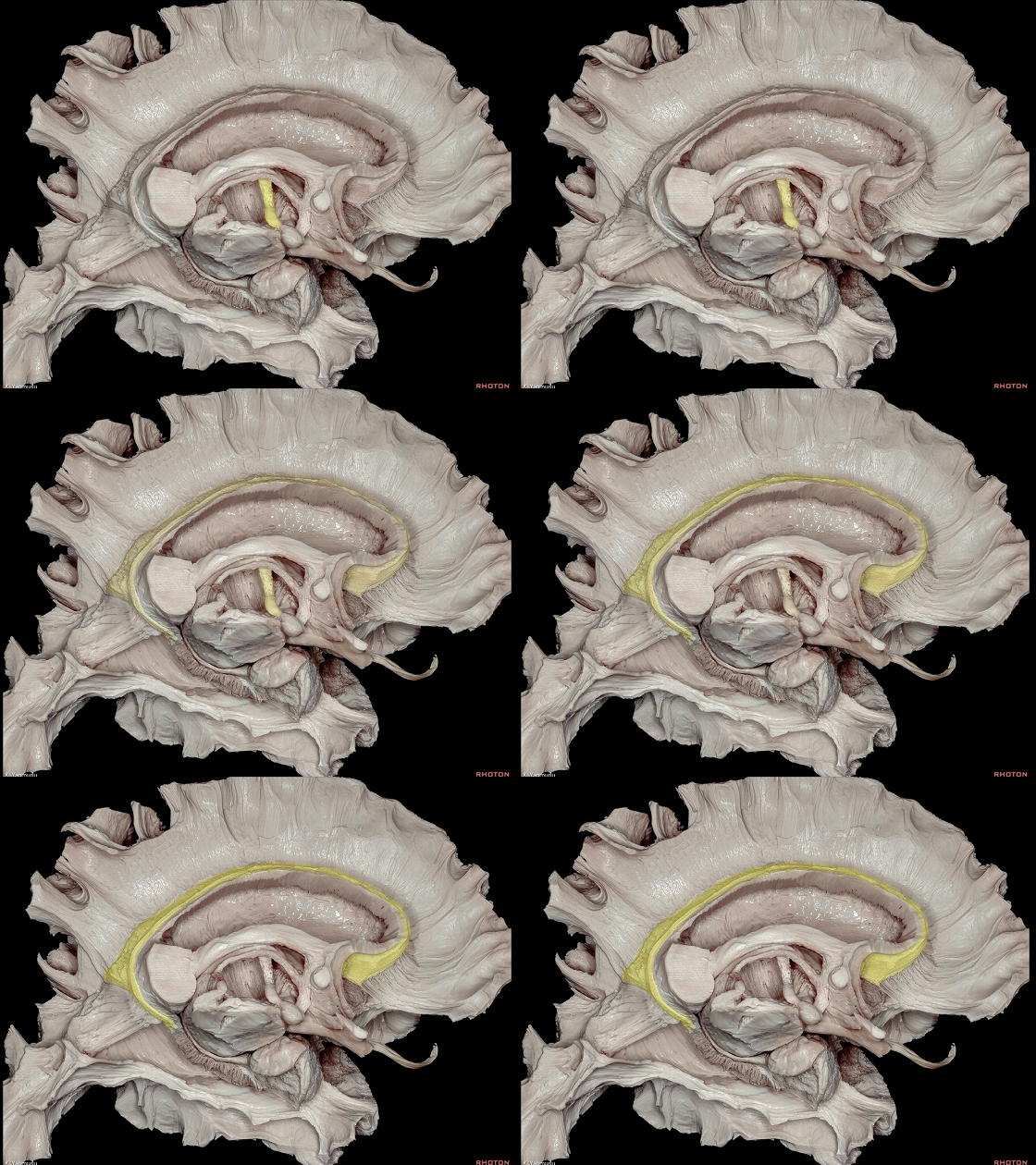
▼丘脑扣带纤维行经 内囊(下图)。
the thalamocingulate fibers pass through the internal capsule.
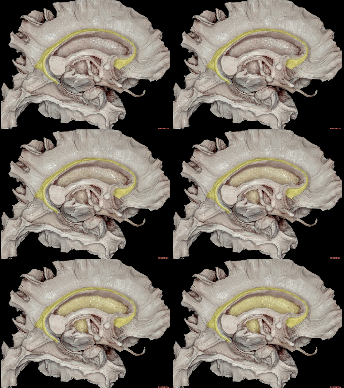
▼扣带回(下图)再将信号经由扣带传递至 海马旁回(下图)。海马旁回再投射至海马体。
From the cingulate gyrus, the information carries to the parahippocampal gyrus by the cingulum. The parahippocampal gyrus projects to the hippocampal formation.
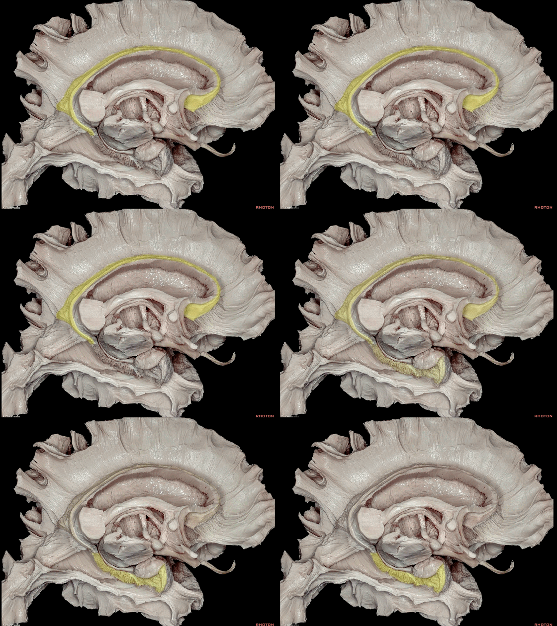
▼以上就是沟通边缘皮质和皮质下区域的边缘通路,即 Papez环路(海马环路)。
目前研究认为,Papez环路是一个重要的与学习、记忆密切相关的脑结构。
Hence, the circuit is formed.


