


本文为Rhoton解剖视频中《Navigating the Orbit》这一章节,主要讲解了眼眶的骨性结构、眶内结构、各种眼眶入路等内容。视频时间较长,笔者将部分内容按顺序重新编排,共截取270张图片。
笔者水平所限,错误之处请批评指正!
(因文章篇幅较长,脑医汇特将文章分为上、中、下三篇,欢迎大家阅读,分享!)

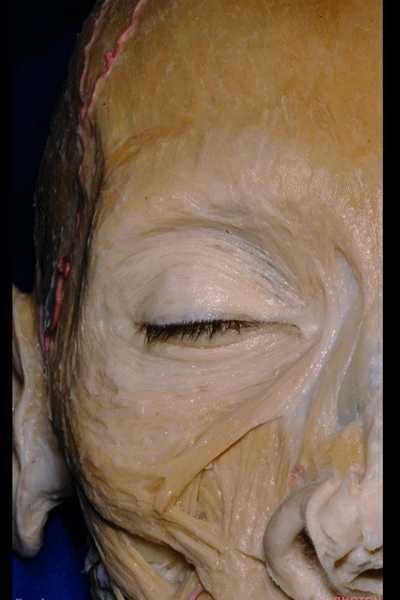
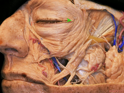
▼现在我们从前方来看眼眶。这是眼轮匝肌。
We wanna just look at the orbit a little bit from anteriorly. Here we see the orbicularis oculi.
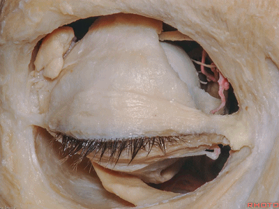
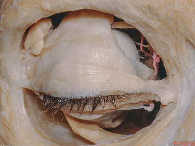
▼在眼眶前部,上方,与上睑提肌相结合的是上睑板
And, looking into the anterior orbit,above here, incorporated into the levator is the superior tarsal plate of the eye,
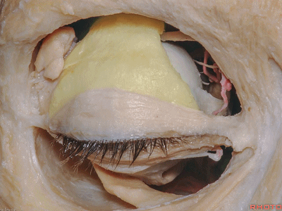
▼这是内侧和外侧眦韧带。
medial and lateral canthal ligament.
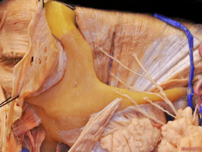
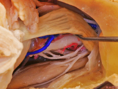
▼可见走行于上直肌下方的上斜肌肌腱。
We see diving in under the superior rectus, the tendon of the superior oblique.
▼下方可见附着于上颌骨的下斜肌
And below we see the attached to the maxilla, the inferior oblique
▼下斜肌 走行于下直肌下方,至上斜肌附近附着于眼球的外侧部。
the inferior oblique that passes below the inferior rectus to attach near the superior oblique along the lateral part of the globe.
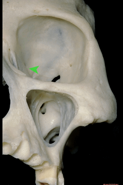
▼深入观察眶顶,可见上斜肌肌腱 附着于此处(箭头处)的额骨。该处的额骨上有一小凹陷。
As you look into the orbital roof,we see the tendon of the superior oblique anchored here to frontal bone.There's a little dimple in the bone at that point.
▼这是滑车上神经
The supratrochlear

▼这是眶上神经
superior orbital nerve

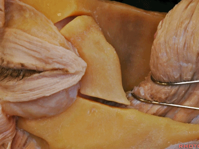
▼下图示上睑提肌、上斜肌、内直肌和外直肌。
the levator, superior oblique,and medial and lateral rectus muscles.
▼抬起上睑提肌,可见上直肌
Here we've elevated the levator, we see the superior rectus,
▼下图示 上斜肌肌腱
the tendon of the superior oblique,
▼下斜肌行于下直肌下方
the inferior oblique passing below the inferior rectus
▼这是外直肌 和 内直肌。
the lateral and medial rectus muscles.
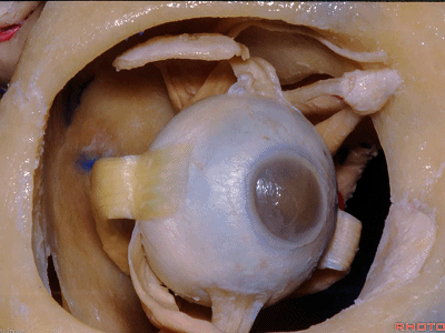
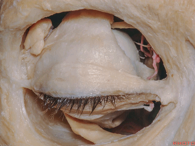
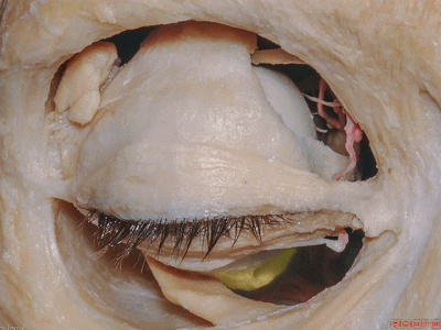
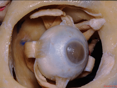
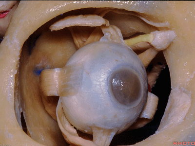
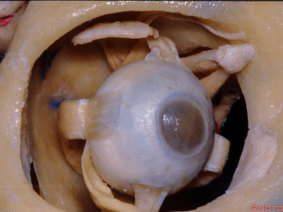
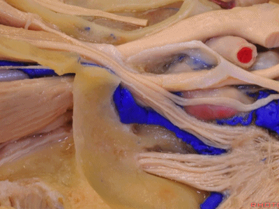
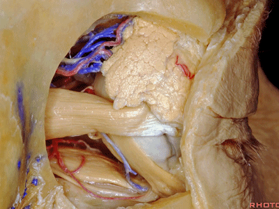
▼从前方进入眼眶时,附着于眶缘的是眶隔,其分隔后方的眼球和眶脂肪与前方的巩膜和角膜。
And, when you enter the orbit from anteriorly, attached along the orbital rim is the orbital septum, that separates the globe and the orbital fat from the sclera and cornea anteriorly.
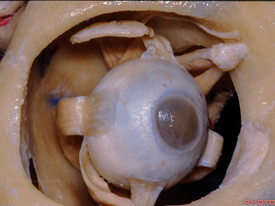
▼下图示巩膜和角膜。
sclera and cornea anteriorly.

![]()
▼到达眶顶、眶外侧壁或两者的入路,可通过这一小骨辦完成
And for the approaches to roof and lateral wall and both involved, you can use this small bone flap

▼这在之前已介绍过,详见文章《眶颧入路---Rhoton解剖视频学习笔记系列》。这一入路可到达眼眶,或可进入外侧裂。
we've shown before. That gives access to the orbit, or along the Sylvian fissure.
▼对于巨大病变,可行单骨片眶颧开颅,或双骨片,或三骨片眶颧入路暴露该区域。
For larger lesions you can do one-piece orbitozygomatic craniotomy, or two-piece,or three-piece approaches to this area.
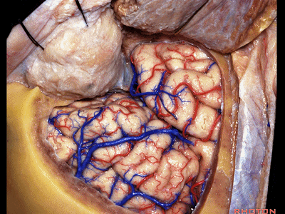
▼或者也可作一小骨瓣打开眶顶和外侧壁,显露硬膜和眶骨膜。
Or you can use very small craniotomies of the superior and lateral wall that give access to dura and the periorbita.


因此可以选择上述不同的入路作不同的骨瓣。
So you can tailor these approaches from above using a variety of flaps.
▼但对于眶外侧入路,需注意面神经的颞支,其支配额肌以及眼轮匝肌。
but lateral orbitotomy, you always in doing it wanna be very careful of these branches of the facial nerve that go to frontalis and the orbicularis oculi.
▼但也可经所谓的外眦切开术。我认为现今这个入路更受欢迎,从这里(箭头处)稍向后暴露出眶外侧壁,从而来进行眶外侧切开术。
But you can do it through a lateral sort of canthotomy. I think a more favoured one today is to come through this area, and then turn back a little bit on the lateral orbital wall for a lateral orbitotomy.
▼暴露额骨、颧骨。
And here we see frontal,zygomatic bone.
▼然后行眶外侧切开术,沿着此处的颧骨,去除眶外侧缘和部分眶外侧壁。可进一步向后扩展至眶尖。
you can do the lateral orbitotomy here along the zygomatic bone, and lift up the lateral orbital rim and some of the lateral orbital wall. You can extend this back to lateral to the orbital apex.
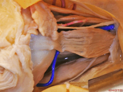
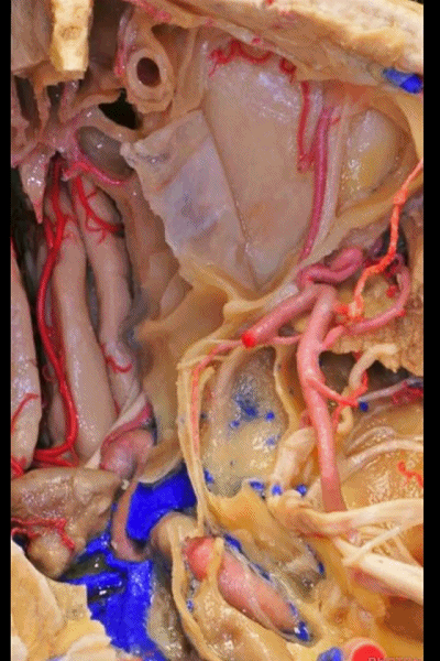
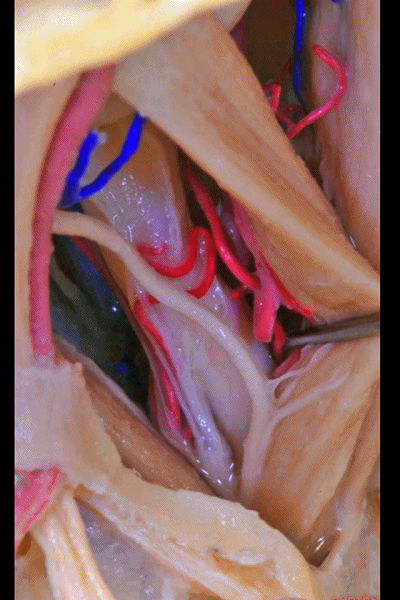
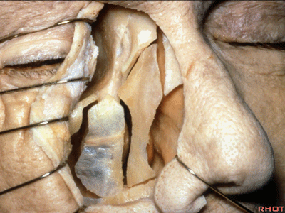
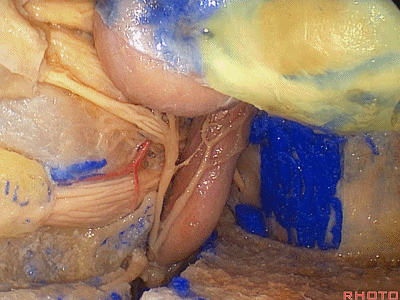
▼现在从外侧向内暴露,可见外直肌、下直肌
Here looking in from laterally we see the lateral rectus,inferior rectus

▼这支纤长的神经向前,发自动眼神经下支,支配下斜肌。
and then this really long nerve that's going forward from the inferior division to the Inferior oblique, that's...forward.

▼上方的是泪腺神经
Lacrimal nerve above.
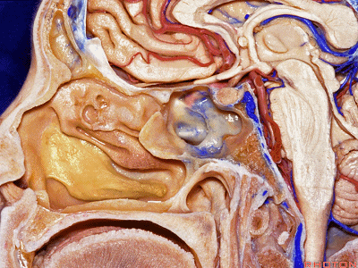
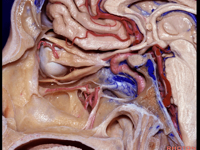
▼如今,对于海绵窦前部(箭头处)的暴露,也可通过该入路进行。
And, one of the things that's happening today is that for getting into anterior part of cavernous sinus,the bone in this area has been removed.

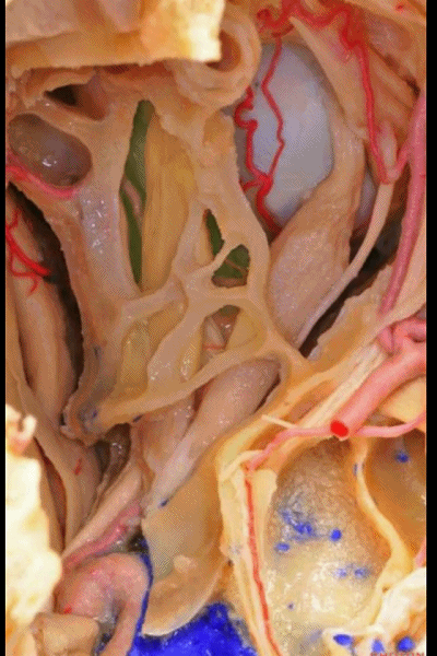
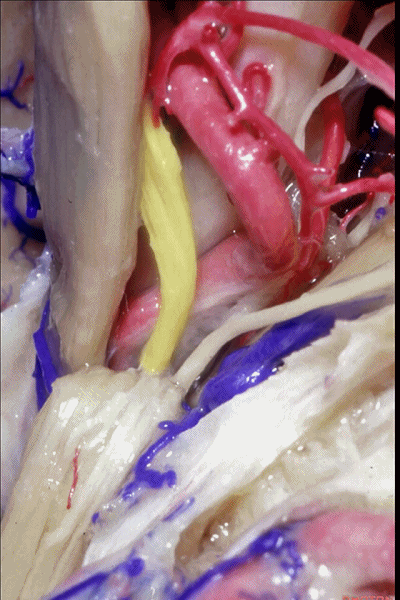
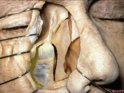
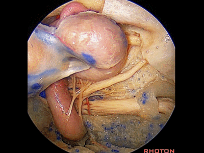
▼现在抬起外直肌,可见视网膜中央动脉,位于视神经下方。
here we just lift up the lateral rectus and we see the central retinal artery here below the nerve.
▼我们可进一步向后方切除眶上裂的外侧缘,即可进入海绵窦的前部,即通过上述入路向后扩展。
And, you can elevate backwards and remove the lateral edge of the superior orbital fissure, and then get into the anterior part of cavernous sinus through this approach, and work back through this area.
▼我们可从侧方打开总腱环,即可暴露从海绵窦前部一直向前至眶内。
You can divide the annular tendon laterally, and have access to anterior cavernous sinus all the way forward into the orbit.
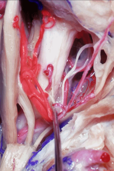
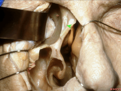
▼再简单看看眶外侧入路,去除眶外侧壁,可见泪腺。
Just another quick lateral orbitotomy here, and lateral orbital wall remove,lacrimal gland,
▼这是外直肌
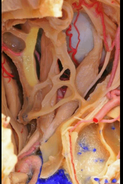
▼随后将外直肌抬起。这里再次暴露出视神经的下外方为视网膜中央动脉,这在术中需时刻注意保护。损伤该血管即会导致失明。
and then the lateral rectus elevated. And again in this area, inferolateral below the optic nerve,you have the central retinal artery that is always to be preserved if possible.If lost, it leads to a blind eye.
▼我们还可以通过上颌窦打开眶底
Then you can access the orbit through the maxillary sinus through the floor here,
▼或打开上颌窦后壁进入翼腭窝,可见翼管
or you can get in through the possible wall to the pterygopalatine fossa,
▼或向内进入蝶窦和筛窦。这是眼眶的筛板壁,恰位于上颌骨上方。
or work medially into the sphenoid sinus and ethmoids. The ethmoidal wall of the orbit is here just above the maxilla.
▼因此,我们可利用该区域,经眶底入眶。
So that, you can get in through this area, come through the floor and have great access to the floor of the orbit.

▼我们可进入上颌窦,从前方将其打开
And you can come into the maxillary sinus here from anteriorly,

▼随后从下方经眶底进入眶内。这常用于Graves病的眶部减压。
and then work below, through the floor of the orbit.This is often used for orbital decompression in Graves.
▼这里可见筛窦气房 在眶下方延伸的范围。这里显露出了所有筛窦气房。因此我们也可利用筛窦,进行经筛窦的眼眶入路,通过眶内侧壁,以及此处的眶底进入眶内。
And here you see how far the ethmoid air cells extend below the orbit.These are all ethmoid air cells that have been drilled out.So the route through the ethmoid,you can come in through the nose through the ethmoid and access medial wall of orbit,as well as floor in this area.
▼也可经筛窦进入上颌窦,并向前暴露所有这片区域,直至下斜肌。
And then you can get into maxillary sinus and have access to all of this area forward to the inferior oblique muscle.
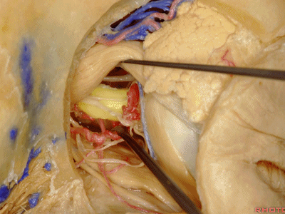
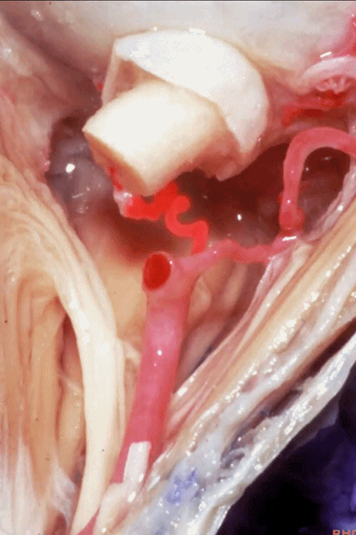
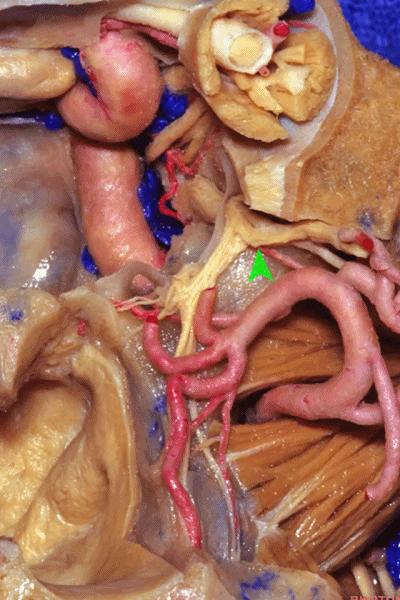
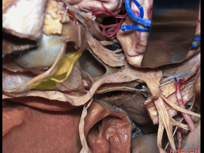
▼我们来看下方入路,这里看到的又是视网膜中央动脉, 从下方进入视神经。
And just a view from below but we see again the central retinal artery entering the optic nerve from below.
▼在视神经下方,动眼神经下支的一部分在视神经下方行至内直肌。
And this is a view from below the nerve, part of inferior division going under the optic nerve to the medial rectus.
▼这是一例少见的眼动脉,其向内走行于视神经下方。
And this is one of these uncommon ophthalmic arteries that passes medially below the optic nerve.

▼将眼动脉牵向后方,这是视网膜中央动脉
Here we pull that ophthalmic artery posteriorly, and you see the central retinal artery
▼视网膜中央动脉进入视神经的下表面。
entering the lower surface of the optic nerve.
![]()
▼通过内侧上颌骨切开术进入眶内的入路有多种术式。
可以仅仅在此处切开皮肤,向后沿着上颌骨额突、泪骨、筛骨眶板(下图)到达眶内侧壁。
And then there're a variety of medial maxillotomy approaches that you can access the orbit. You can just do an incision here, and work back along the frontal process of the maxilla, along the lacrimal bone back to the orbital plate to access medial orbit.

▼也可作局限的内侧上颌骨切开术,在中鼻甲下方进入鼻腔,然后经由筛窦气房进入眶内侧壁。
You can do a small medial maxillotomy and get into the nasal cavity under the middle turbinate,and work through the ethmoid air cells into the medial orbit,
▼这就是向内侧,从中鼻甲下方,经筛窦入眶。
you have medially then,you can get under the middle turbinate,work through the ethmoids.

▼也可从前方打开上颌窦,经筛窦和上颌窦顶,进入眶内侧。
Or you can get into the anterior wall of the maxillary sinus, work through the ethmoids, and the roof of the maxillary sinus to the medial orbit.
▼这是经上颌窦入眶。

▼或仅仅作一小切口(下图箭头),通过内眦切开,向后至眶尖。
Or just a small incision, a sort of medial canthotomy approach back to the orbital apex.
▼但是 经由上颌骨的入路,通过筛窦、外侧蝶窦、以及鼻腔,可暴露所有这片区域。
But...using these routes through maxilla,through the ethmoids,through the lateral sphenoid,through the nasal cavity,you can access all of this area.
▼这是眶尖,附近为总腱环。另外,利用鼻窦从内向外的入路也可到达所有上述区域。
Here we see the orbital apex, adjacent the annular tendon. But, coming through the sinuses medially you have access to all of this area.

![]()
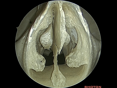
▼来看看鼻腔,这是下鼻甲、中鼻甲、上鼻甲。
And just the view as you work through the nasal cavity, inferior, middle, superior turbinate.
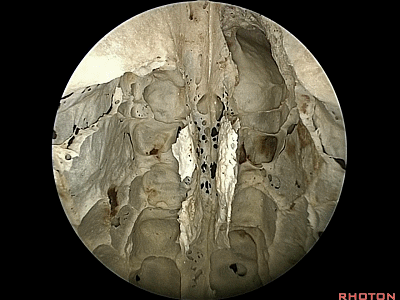
▼切除中鼻甲和上鼻甲,打开鼻腔外侧壁的筛窦气房。即可显露筛骨纸板,即为眶的内侧壁
We remove middle and superior turbinate and open the ethmoid air cells in the later wall of the nasal cavity.And you're up to the lamina papyracea here, the ethmoidal medial wall of the orbit,
▼打开即可见内侧的眶骨膜。
and here the periorbita medially.
▼因此利用这些经鼻腔入路,通过筛窦、上颌窦顶,可以对眶底和眶外侧壁实现广泛的暴露。
And using the approaches through nasal cavity, through ethmoid, roof of maxillary sinus,you have fairly wide access to the floor and lateral wall of the orbit
▼这是从中鼻甲下方进入
So that you get in under the middle turbinate,
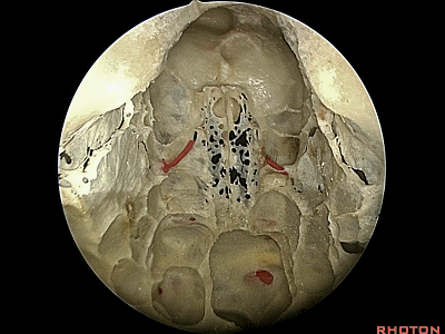
▼我们可向外侧,通过中鼻甲,打开筛窦(下图)。
So you can work lateral to the middle turbinate,through the ethmoids.
▼在开放筛窦后,即可暴露所有眶板区域,直至由额骨构成的筛房顶壁(下图)的下方。通过此处还可进入前颅窝。
And when you work through the ethmoids,well, you expose all of this orbital plate below the roof of the ethmoids here that's formed by frontal bone.That leads to the anterior fossa.
▼因此我们仅仅从中鼻甲下方,即可暴露整个眶内侧壁。下图示眶内侧壁
But just getting in under the middle turbinate, you have access to all of this medial wall of the orbit.
▼去除筛窦气房,即可暴露眶尖附近的眶内侧壁。
and remove ethmoid air cells, and it delivers you up here to medial wall of orbit near the orbital apex.
▼还可打开部分海绵窦,其位于眶尖后方。
You can access some of cavernous sinus here behind the orbital apex.

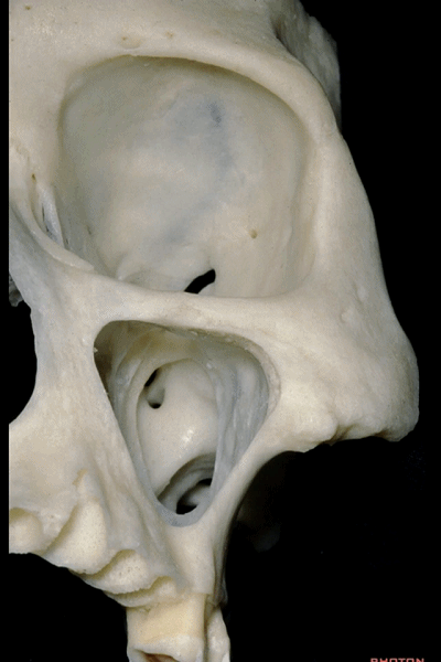
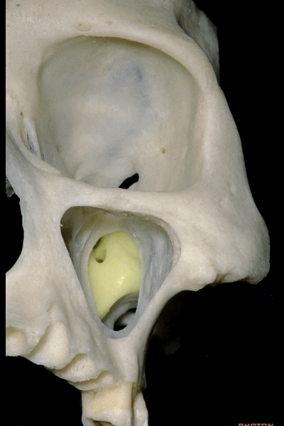
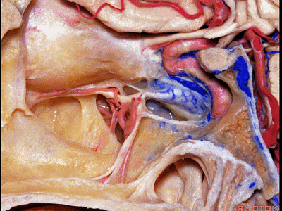
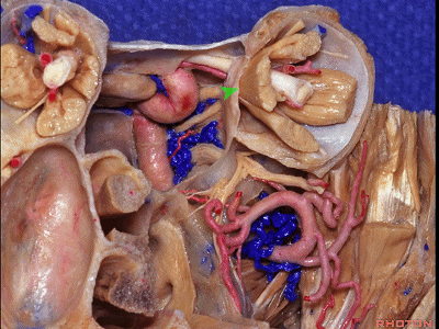
▼蝶骨小翼的内侧部,有视神经管通过,包绕于视神经及眶上裂的结构,即为总腱环,眼球各直肌起源于此。
And it's good as you think of the osseous anatomy to think of the related neural and vascular structures. So that, here at the medial part of the lesser wing, we have the optic canal, and then, here surrounding the optic nerve and superior orbital fissure,we have the annular tendon from which the rectus muscles arise.

▼视神经管在大多数情况下,位于蝶窦的上外侧,但在少数情况下,筛窦可向后延伸,此时的视神经管可位于筛窦的后部。
And the optic canal can either be...well, most commonly, in the superior lateral part of the sphenoid sinus, but at times, the ethmoid can extend posteriorly,and you'll find the optic canal in the posterior part of the ethmoid sinus.
▼此处,走行于眶底的是上颌神经,在眶下裂的内侧部。
Below here, along the floor of the orbit, we have V2.And below the medial part of the inferior orbital fissure,
▼有开口与翼腭窝相沟通,此处包含翼管的前端、圆孔,内走行有上颌神经,其沿着眶底走行。
opening into the fissure is the pterygopalatine fossa, where we see the anterior end of the vidian canal, and foramen rotundum transmitting V2, that runs along the floor of the orbit. So as we look at osseous anatomy, we always want to have that view of the related neurovascular anatomy.
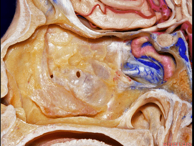
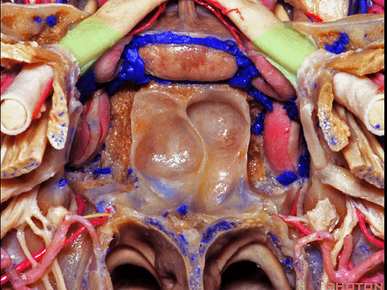
▼通过此入路,还可暴露蝶鞍(下图)
And working through that area, you can access sella,

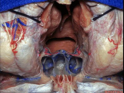
▼继续暴露右侧的海绵窦前内侧壁、动眼神经、眼神经、外展神经、上颌神经,
anteromedial wall of cavernous sinus, 3rd nerve,V1,6th nerve, V2,...right side,
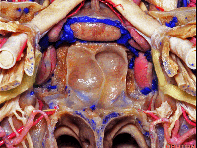
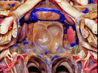
▼这是左侧所见。向前经眶板(箭头处)
same anatomy on the left side.And then, once you work forward through the orbital plate,
▼即可经眶内侧壁,循着上述神经血管至眼眶内侧部。
well, you're into the medial orbital wall and can follow all of these structures into the medial orbit.

▼有时,当病变极其复杂时,我们可进行低位上颌骨切开术
And then sometimes the pathology gets very complicated, and you can use a lower maxillotomy

▼从牙槽突向上至眶底。
from the alveolar process, up to the floor of the orbit.

▼同时联合额颞开颅,则可显露眶底、眶外侧壁、海绵窦、眼神经、上颌神经、下颌神经。这基本上可暴露所有前外侧颅底的复杂病变。
And combine that then with a frontotemporal craniotomy that gives you access to floor, lateral wall of orbit, cavernous sinus, and V1, V2, V3. That can basically expose all of the anterolateral skull base for very complicated lesions.
▼最后,从上方,我们可作一双额骨瓣开颅,来暴露复杂病变
And from above, you can use bifrontal craniotomy to access complicated lesions that may

▼其范围包括双侧眼眶的内侧壁
involve both medial wall of both orbits,

▼或向后扩展至蝶窦。
or extend back into the sphenoid sinus.
▼用该入路我可以切除累及所有这些鼻窦、眶内侧壁的脊索瘤。
And I've removed chordomas involving all of this area of sinus, medial wall of orbit.
▼另外还可进行硬膜下操作。
以上就是眼眶的解剖及相关入路的概述。
And you have intracranial access.That's an overview of the different walls of the orbit and the intraorbital anatomy.

我们学习解剖的目的是通过我们最完美的手术技术使每一例患者得到最佳的救治。
We've studied all of this anatomy to make what is a delicate, fateful, awesome experience for our patients more accurate, gentle and safe.
