本文为Rhoton解剖视频中《Navigating the Orbit》这一章节,主要讲解了眼眶的骨性结构、眶内结构、各种眼眶入路等内容。视频时间较长,笔者将部分内容按顺序重新编排,共截取270张图片。
笔者水平所限,错误之处请批评指正!
(因文章篇幅较长,脑医汇特将文章分为上、中、下三篇,欢迎大家阅读,分享!)

▼再来看周围的结构,下图可见额窦
So that as you look at the surrounding structures, you wanna see the frontal sinus medially
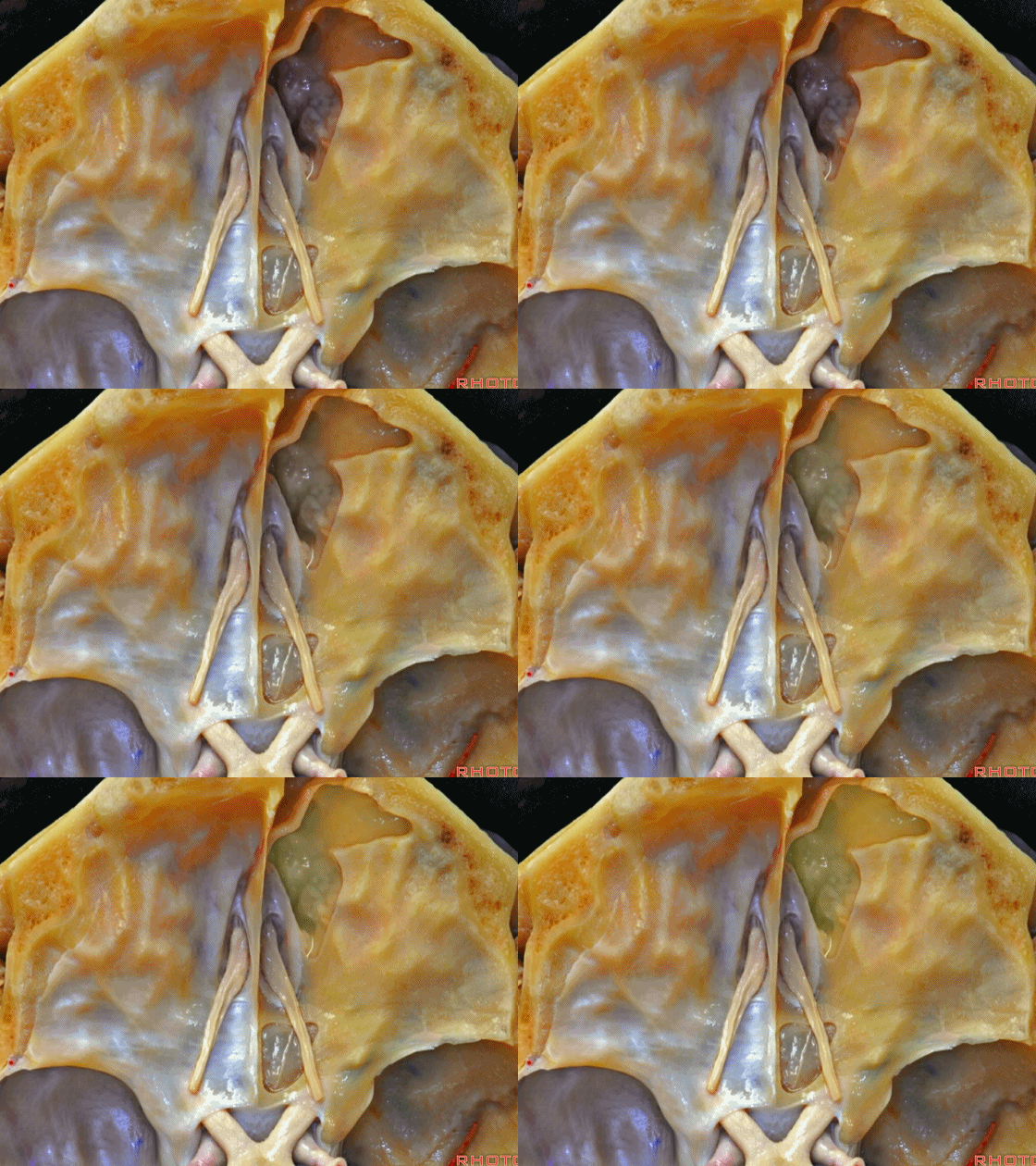
▼下图示筛窦、蝶窦。
the ethmoid, and then the sphenoid.
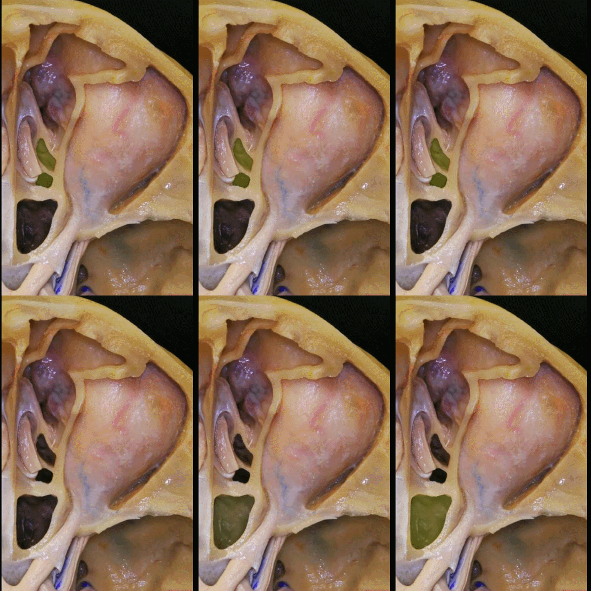
▼去除眶顶即暴露眶骨膜。其内部填满了脂肪。对于穿行于脂肪内的结构我们需要掌握,但同样需要了解一点,即在眶内进行手术时,需要尽可能保留这些脂肪,因为去除脂肪,将使得眶内的肌肉和神经形成疤痕化,从而影响眶内结构的功能。
眼眶手术,神经外科对其作出的一项革新是自动牵开器的应用,这可在术中保留解剖层面的同时保护眶内脂肪而维持重要结构的功能。
And then if you remove the orbital roof you see the periorbita, and inside of this it's filled with fat.And we wanna understand the structures passing through that fat,and also understand that when we're doing orbital surgery,you wanna preserve that fat as much as possible,because if you lose that fat,then the muscles and nerve become scar together and the orbit doesn't function very well. So for working in the orbit,one of the innovations of brain surgery which are self-retaining retractors become very important in maintaining those surgical planes for surgery while preserving the fat that makes the orbit function as well as it does.
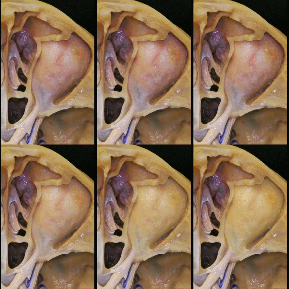
▼这是眶上裂
Here we see the superior orbital fissure,
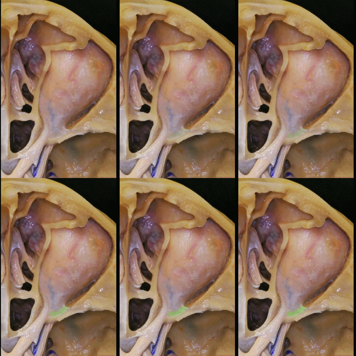
▼这是视神经管
the optic canal
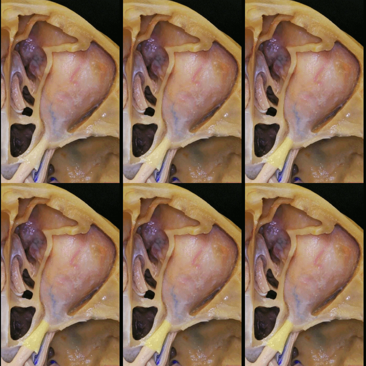
▼眶内侧壁的鼻窦可为进入眶内的通路
the sinuses along the medial wall that are routes to the orbit,
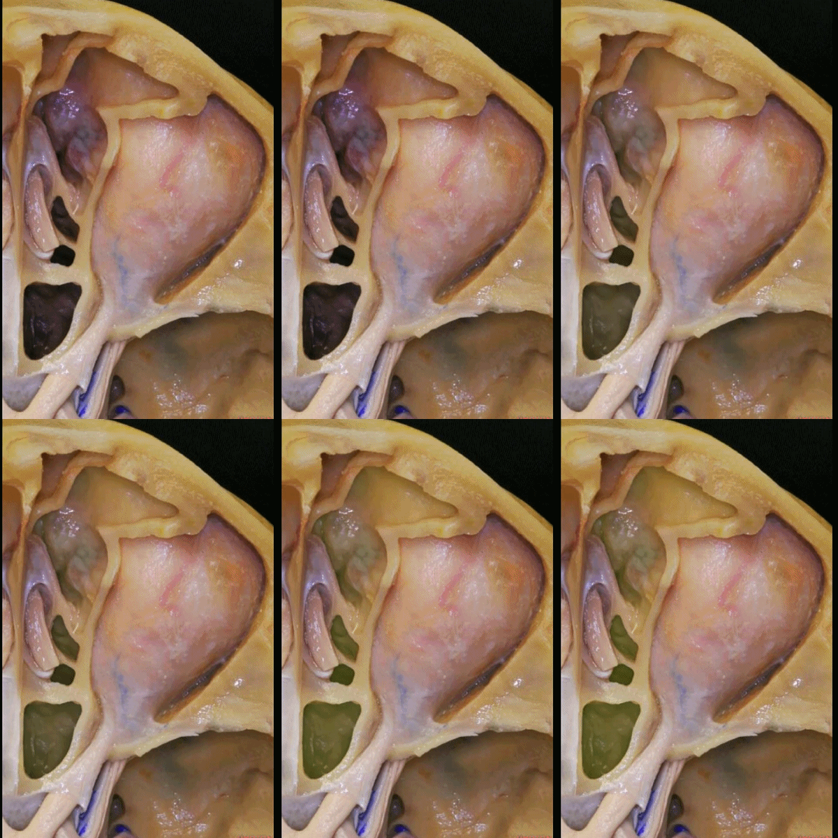
▼而眶外侧壁,可为眶外侧入路提供路径。
or the lateral orbital wall that provides the route for the lateral orbitotomy.
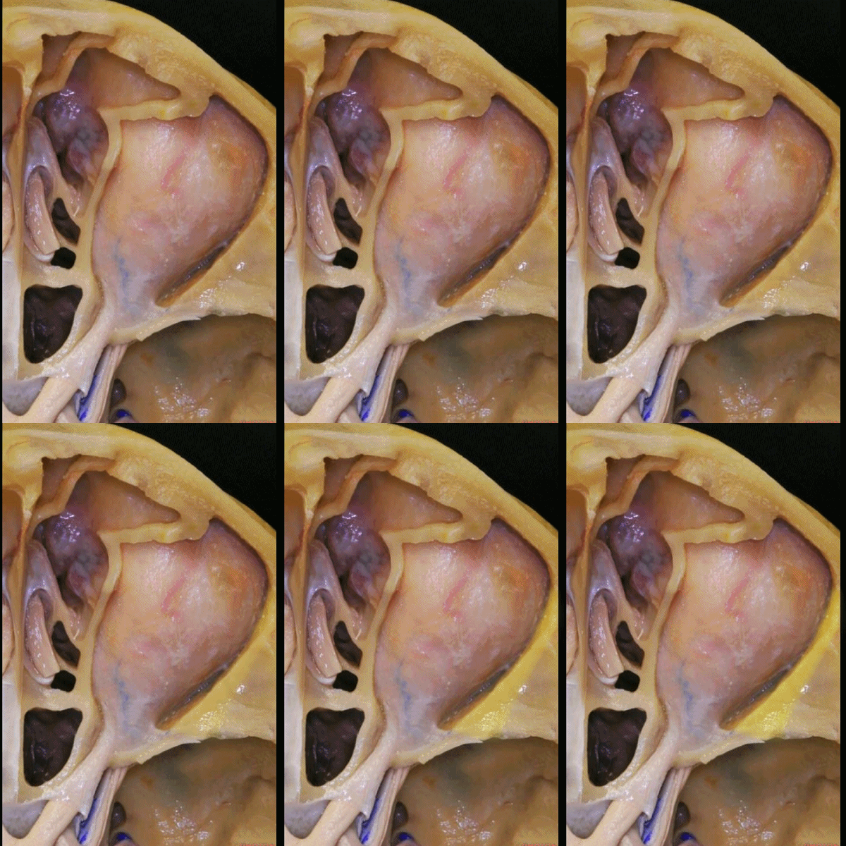
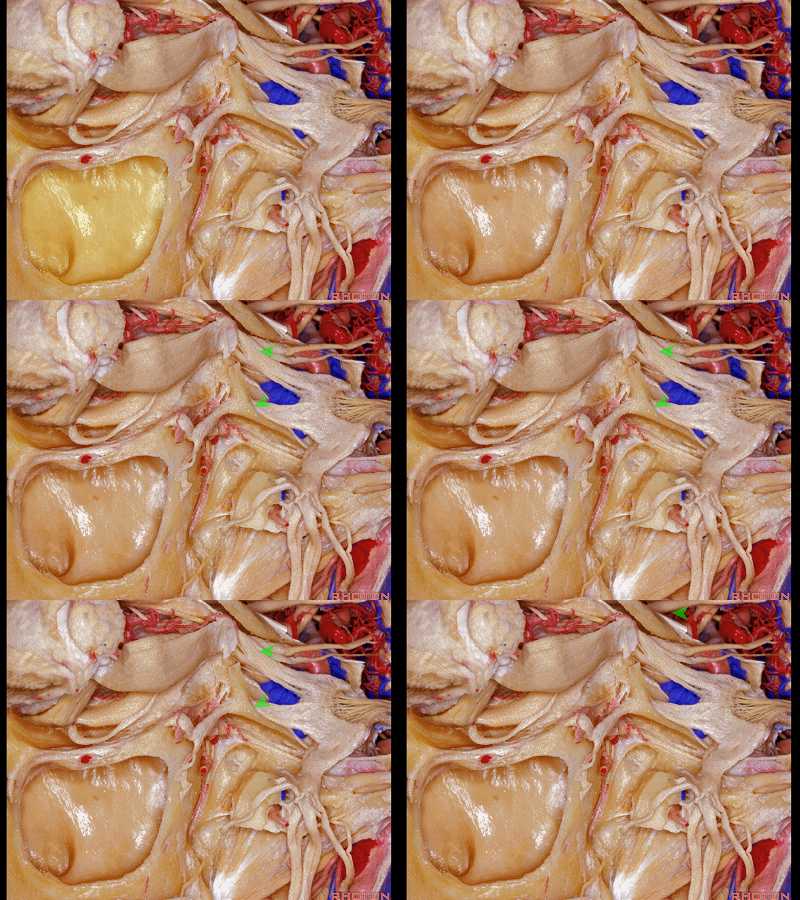
▼在处理视神经附近时,我们需要了解眶内有一些结构,沿着外侧壁入眶,但随后即越过视神经上方走向内侧,从而阻碍我们暴露视神经(下图)
And we want to understand as we work toward the optic nerve,that there are a group of structures in the orbit that enter the orbit on the lateral wall of the orbit but then cross medially above the optic nerve and tend to block access to the nerve
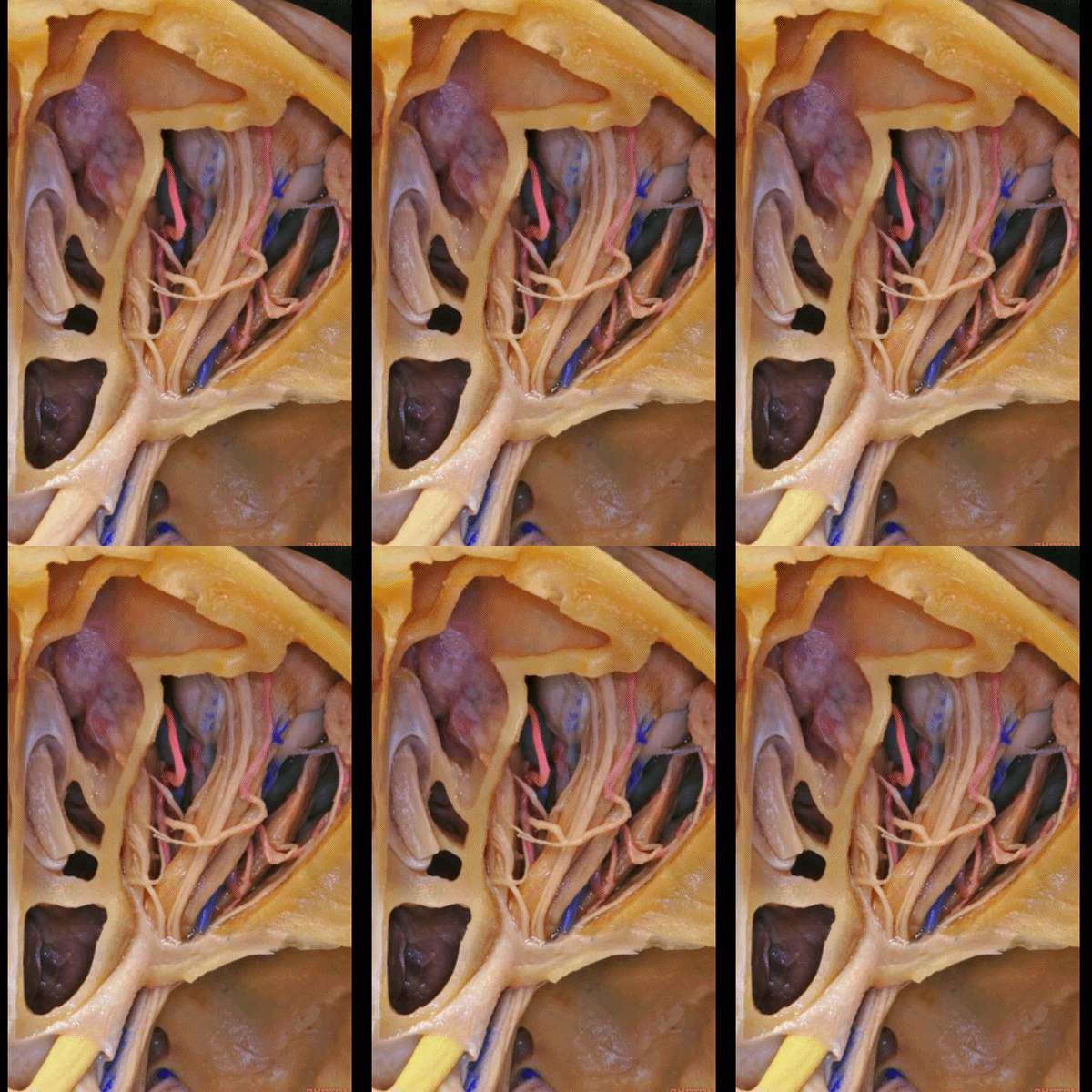
▼其中之一即为滑车神经。
one of these is the trochlear nerve.
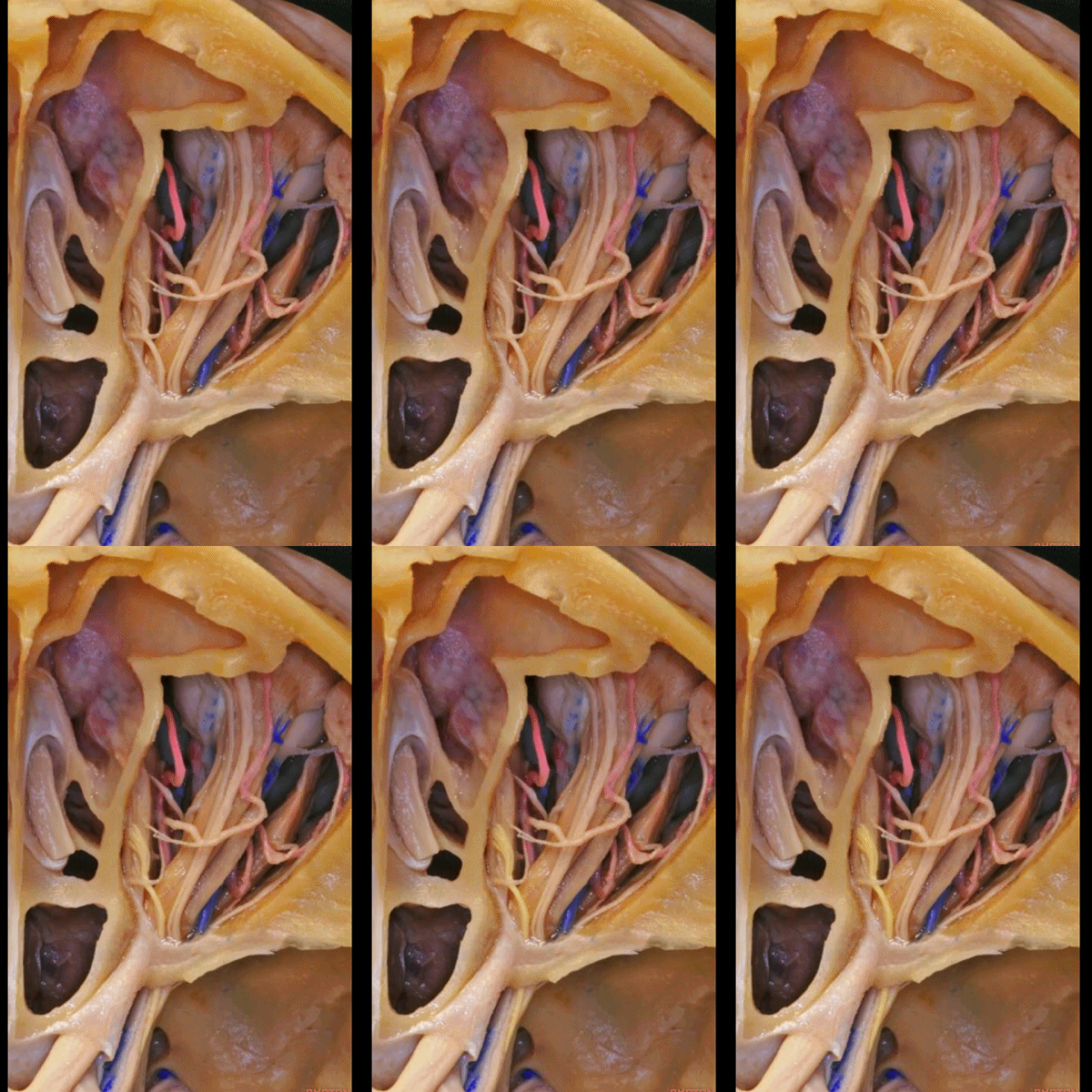
▼切开总腱环的位置可位于上斜肌和上睑提肌之间。但这一切口需注意保护滑车神经。
You can divide the annular tendon here between superior oblique and levator. But if you make an incision, you wanna preserve trochlear nerve.
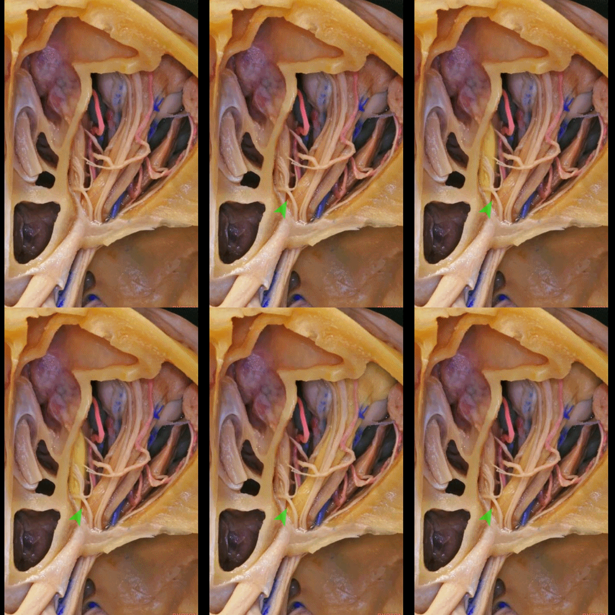
▼同样地,额神经以及其分支眶上神经和滑车上神经 在眶内的走行也是从外向内。
Also, frontal nerve with its supraorbital and supratrochlear branches passes from lateral to medial here in the orbit.
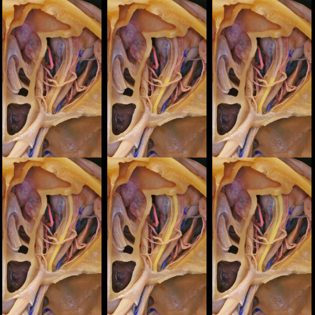
▼另外还有数个从外向内横跨于眶内的结构。其中一个是眼上静脉,从上方越过视神经。
And we'll talk about a number of other structures that cross from lateral to medial. Another one is the superior ophthalmic vein that crosses above the optic nerve
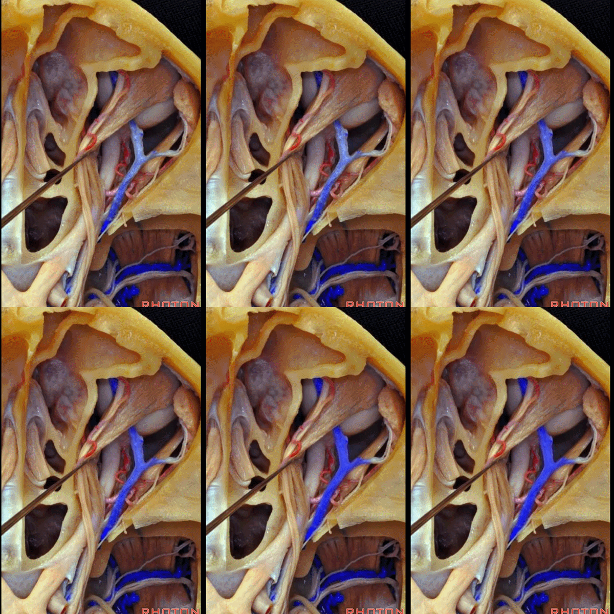
▼眼动脉同样从上方越过视神经。
as does the ophthalmic artery.
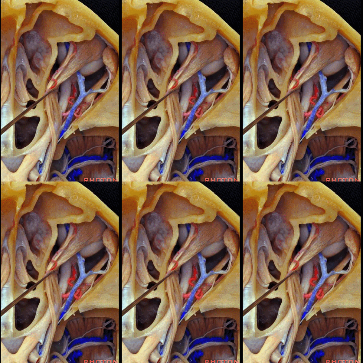
▼再来看眶尖。我们从上直肌和外直肌之间分离,即从外侧打开眶尖。
And here we're back at the orbital apex.We've divided between the superior and lateral rectus here.You can open the orbital apex from laterally.
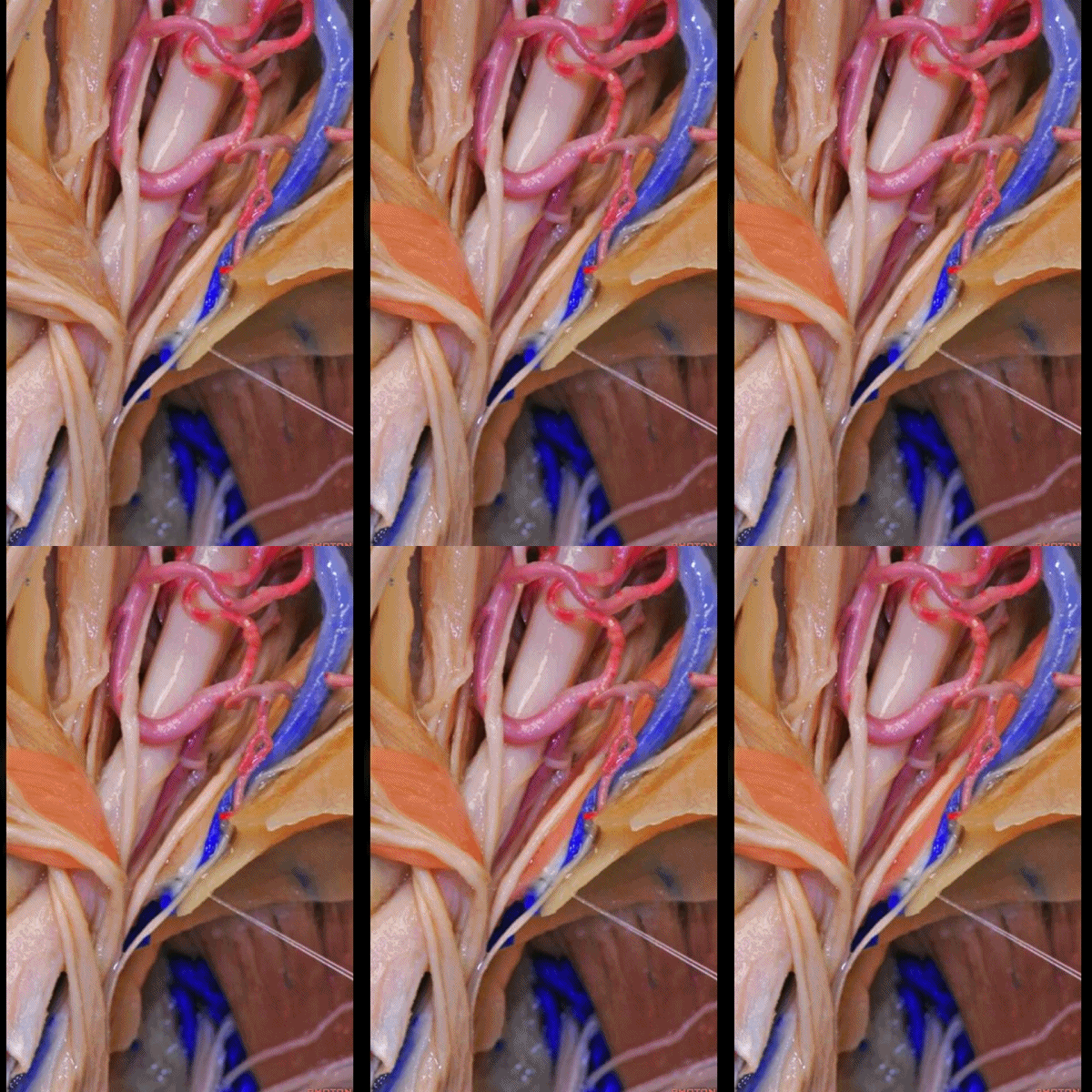
▼这是发自眼神经的额神经,于总腱环外进入眼眶。
Here we see the frontal branch of V1 pass to the orbit outside the annular tendon.
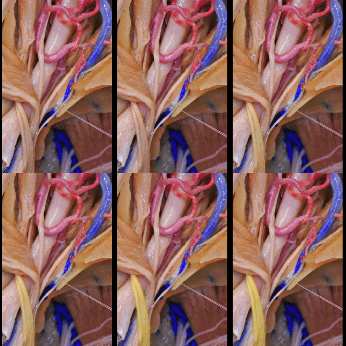
▼但同样为眼神经分支的鼻睫神经(下图),则行于总腱环内,它也是横跨于视神经上方的众多结构之一。
But the nasociliary nerve, the branch of V1,passes through the annular tendon,and it's a number of...one of the structures that passes above the optic nerve.
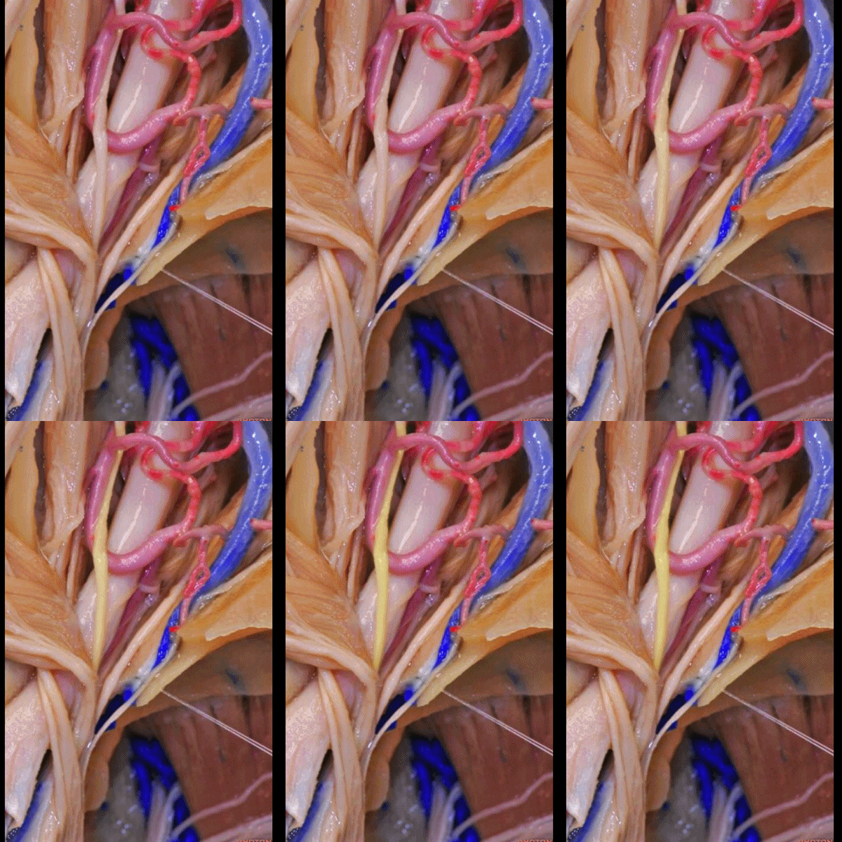
▼这是外展神经,其穿行于总腱环内,支配外直肌。
We see the 6th nerve pass through the annular tendon to the lateral rectus.
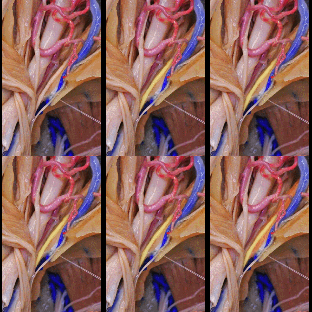
▼这是泪腺神经,其向上行至泪腺区域。
And here's the lacrimal nerve, that passes up to the area of lacrimal gland.
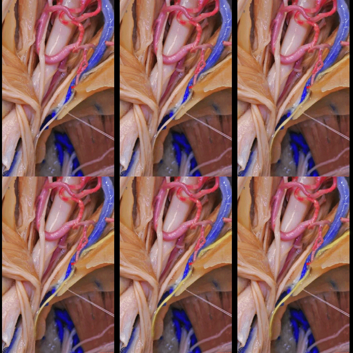
▼这是眼动脉,在90%的病例中,其从外向内走行于视神经上方,但10-15%的情况下,其走行于视神经下方。
Here's the ophthalmic artery, which in 90% of cases will pass above the optic nerve from lateral to medial,but in about 10-15% of cases you'll find the ophthalmic artery below the nerve.
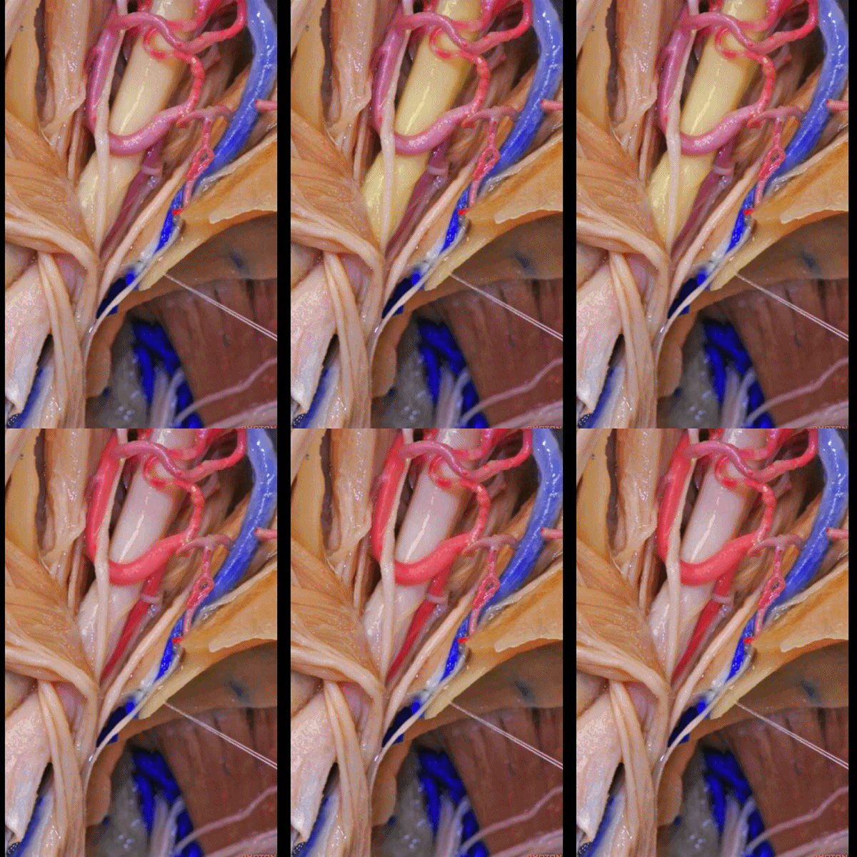
▼我们切开总腱环,将外直肌翻向外侧。可见外展神经进入外直肌
Just another picture we've divided the annular tendon between the lateral rectus that we've folded laterally here.We see the abducens nerve entering it,
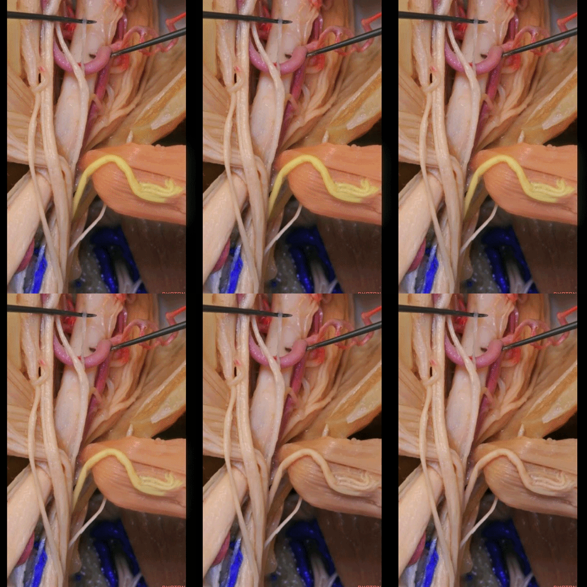
▼鼻睫神经(下图)在上方跨越视神经
the nasociliary crossing above here the optic nerve,
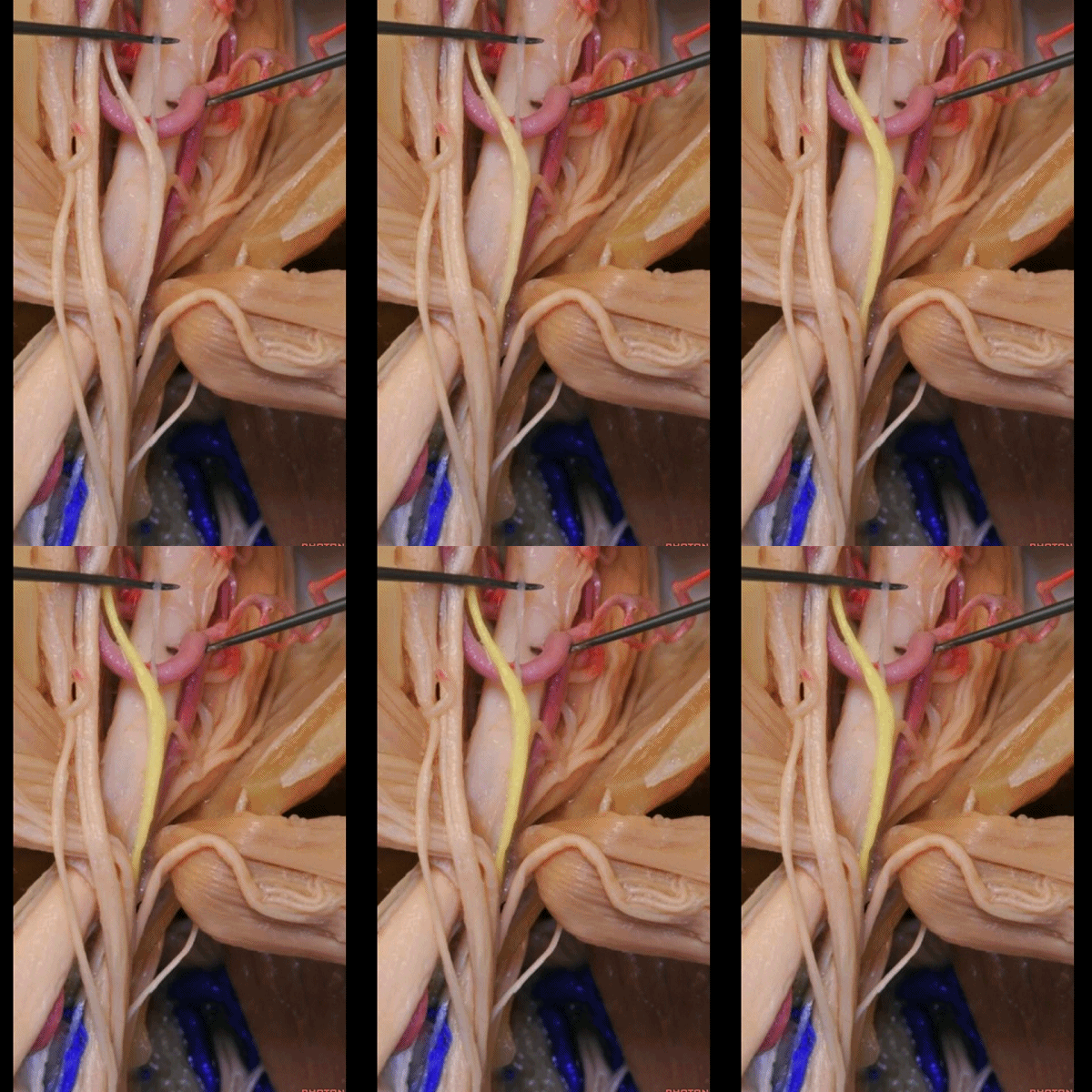
▼同样的还有额神经(下图)和滑车神经。
as does the frontal branch and the trochlear nerve here.
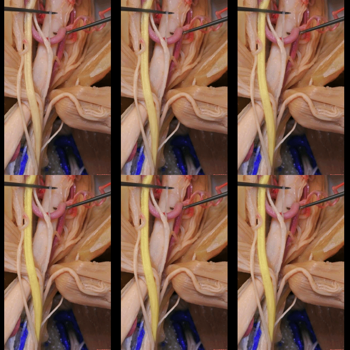
▼下图示滑车神经
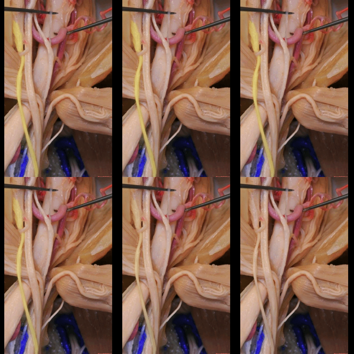
▼这是睫状长神经的一支,内含行至眼球的交感纤维。
Here we see one of the long ciliary branches that carries sympathetics to the globe.
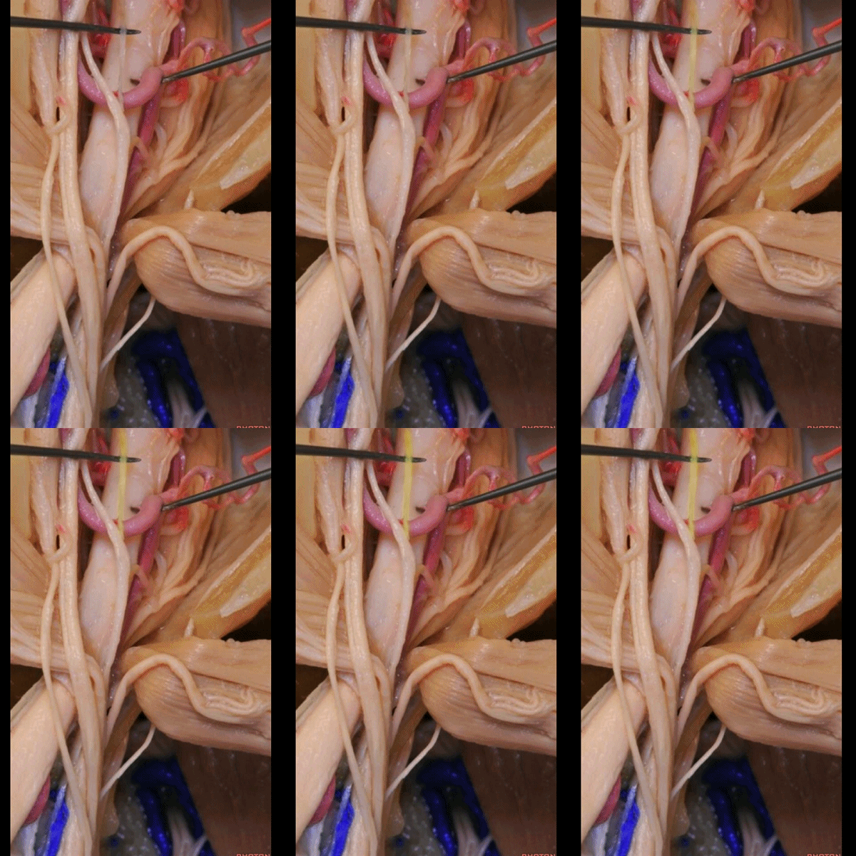
▼这是动眼神经下支,内含进入睫状神经节的副交感节前运动纤维。
But here we see the inferior division of the oculomotor nerve that gives rise to the motor, parasympathetic root of the ciliary ganglion in this area.
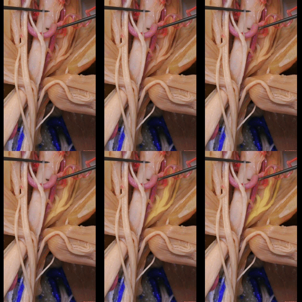
▼相关内容稍后再会讨论,但最先需要掌握的就是上述骨性结构及眶内复杂的解剖。
And we'll talk a little bit more about that, but you wanna understand the osseous wall as well as the complex anatomy inside the orbit.

![]()
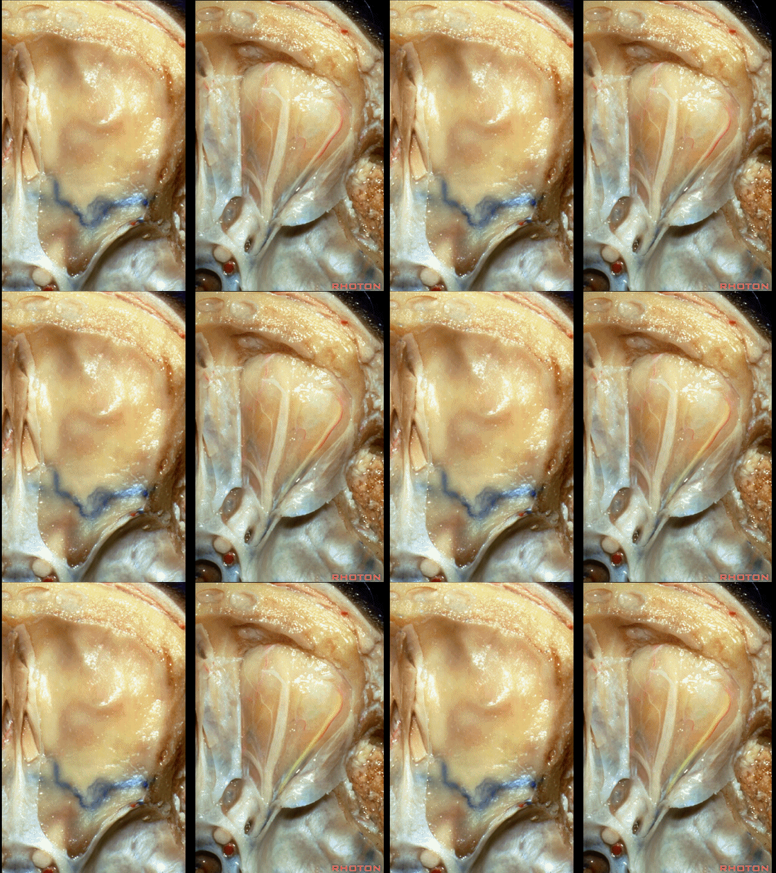
▼接下来我们分层打开眶顶,打开眶骨膜
Just when we take off the orbital roof,and open the periorbita,
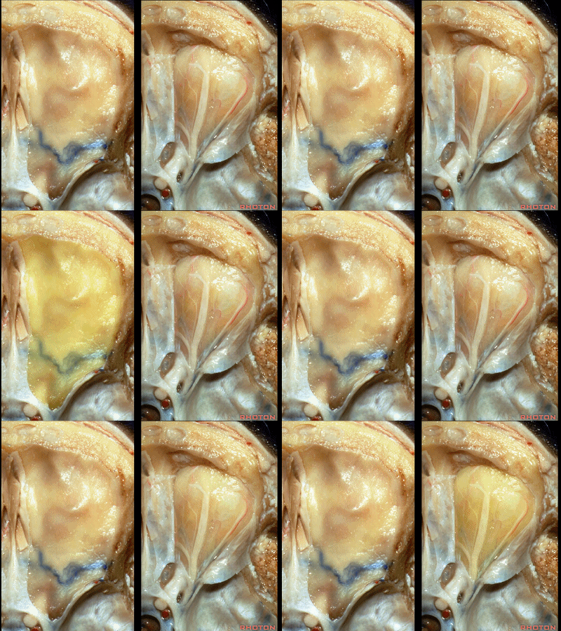
▼首先暴露的神经为支配上斜肌的滑车神经
the first nerves that we see are trochlear nerve to superior oblique,
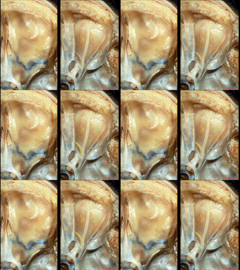
▼还有发出滑车上神经及眶上神经的额神经
the frontal branch gives rise to supratrochlear and supraorbital,
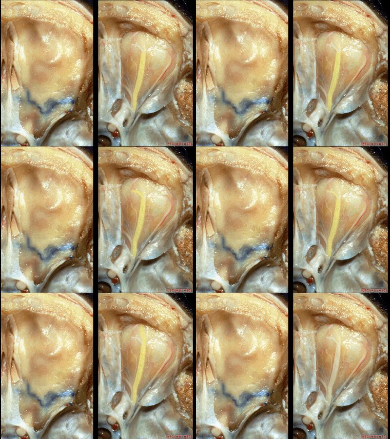
▼外侧的泪腺神经
and then laterally the lacrimal nerve.
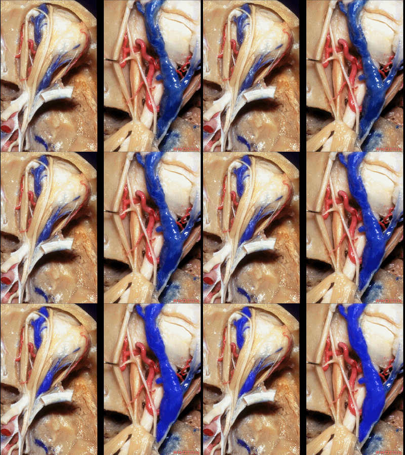
▼进一步深入,则可见眼上静脉,其越过视神经上方。
And then as we go deeper we see the superior ophthalmic vein that passes above the optic nerve.
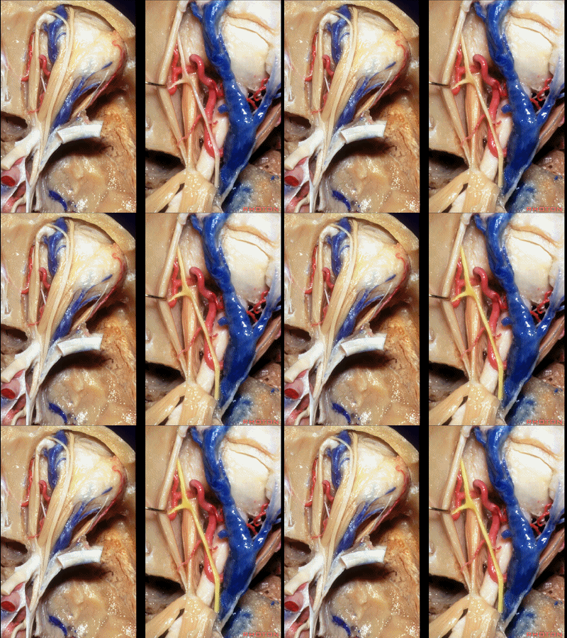
▼在其附近,其他横跨视神经上方的结构包括鼻睫神经(下图),以及眼动脉。
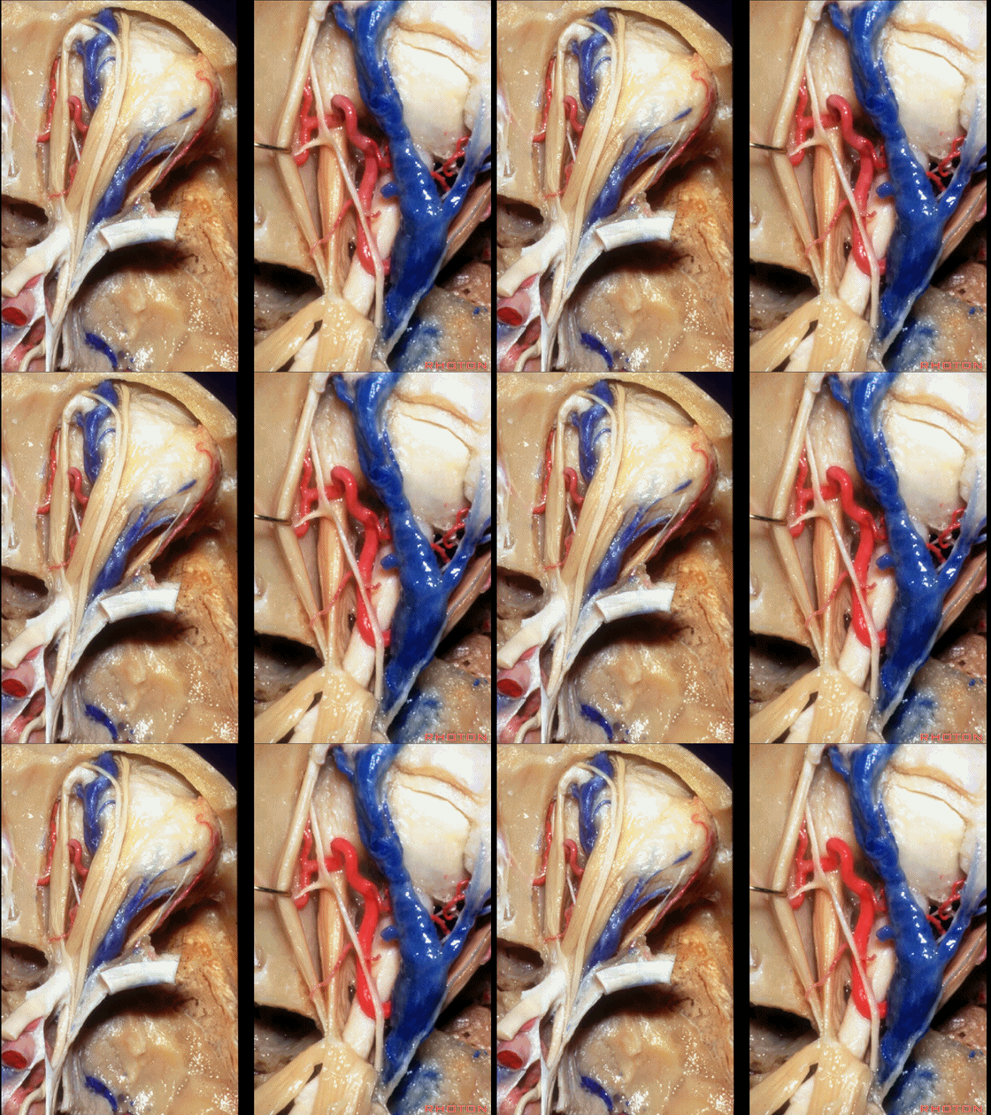
And then adjacent it, other structures that pass above the nerve, the nasociliary nerve,and the ophthalmic artery.
▼下图示眼动脉
▼这支走行于视神经下方的是 动眼神经下支的分支
Here we see another nerve that passes below the optic nerve. What is that nerve? That's the branch of the inferior division inferior division of 3rd nerve
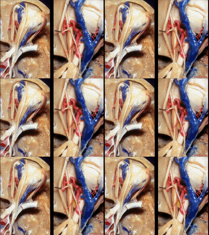
▼这里暴露的是支配内直肌的动眼神经下支。其走行于视神经下方。
Here we see this branch to the medial rectus again that passes below the optic nerve to reach the medial rectus, the branch of the inferior division.
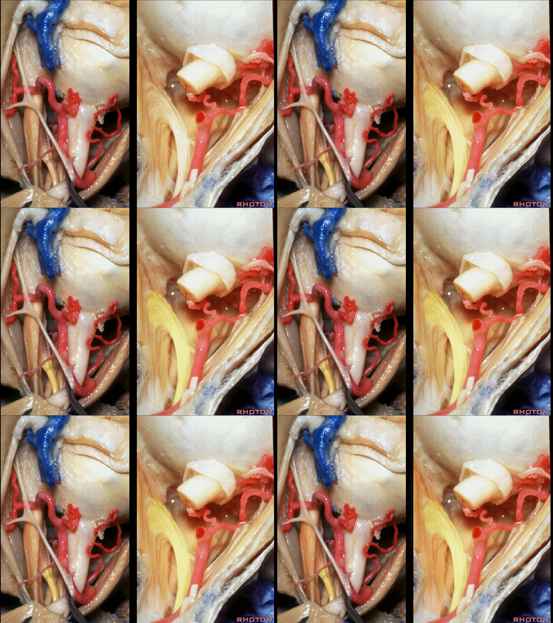
▼动眼神经下支 支配 内直肌(下图)
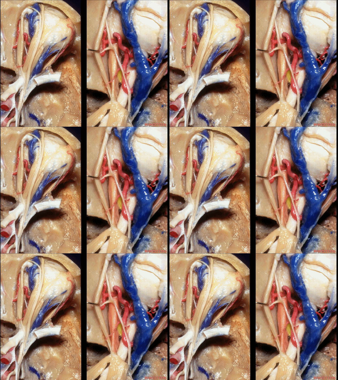
▼眶尖外侧部最重要的一个解剖,即为发源于眼动脉的视网膜中央动脉。
One of the important bits of anatomy, very critical to dealing here at the lateral part of the orbital apex is the origin of the central retinal artery.
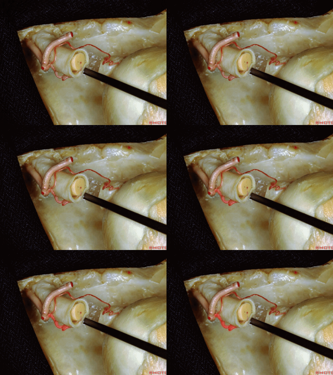
▼它通常是第一个发自眼动脉的分支,并在动脉跨越视神经之前即发出。
And it's commonly the first branch of the ophthalmic artery before it crosses above the nerve.
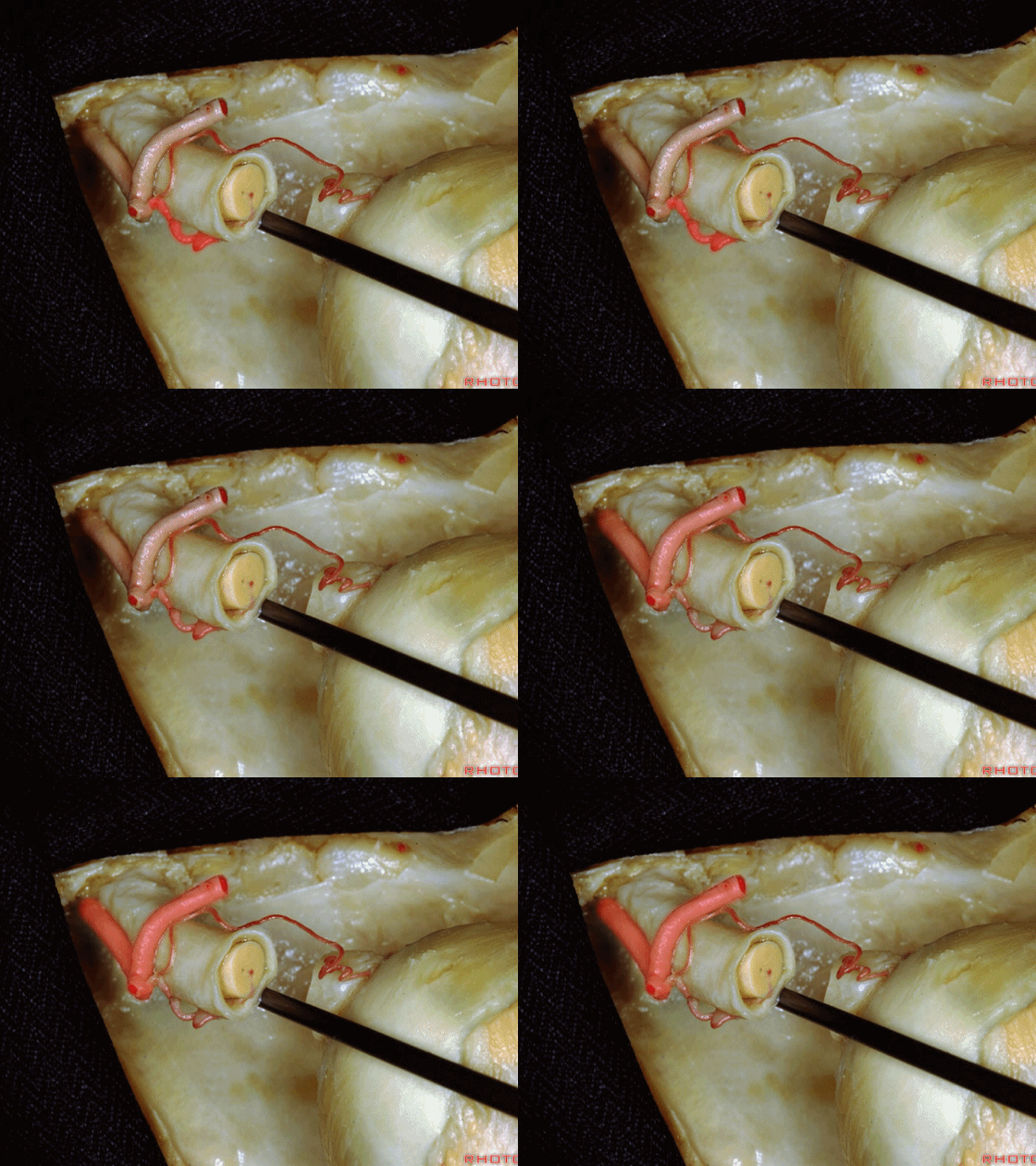
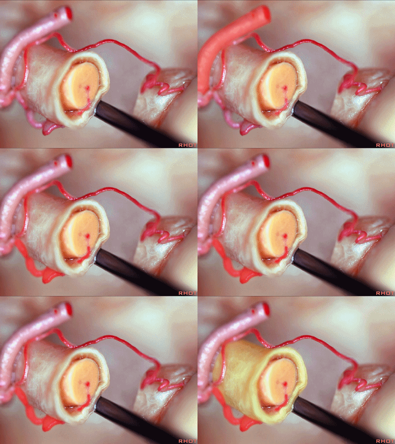
▼视网膜中央动脉 穿入视神经鞘并向前行至眼球,进入视神经中央。
And here just a picture of the central retinal artery. The artery enters through the sheath and then passes forward to the globe. and the center of the optic nerve.
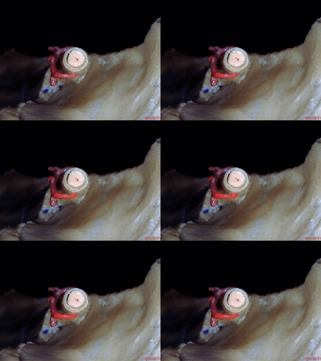

▼微小的出血、轻微的电凝,即可损坏该细小动脉,若损伤了这支小动脉,患者将失去该侧视力。
And just losing that tiny little branch, just get a little bleeding, a little bipolar, and a tiny artery and it's lost and the patient can end up with a blind eye.

▼因此需要特别注意保护视网膜中央动脉,特别在眶尖外侧部、视神经外下侧进行操作时。
So it's very important to think about saving the central retinal artery in here below and lateral to the optic nerve along the lateral part of the orbital apex.

▼在处理眼眶及视神经管区域时
As you approach the orbit, and working from the optic canal
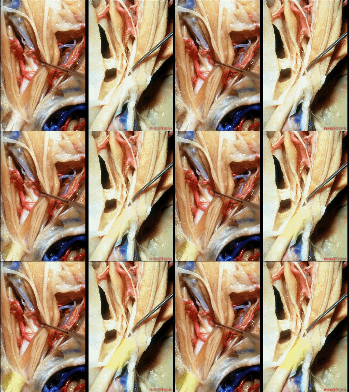
▼可在总腱环内侧,于上睑提肌和上斜肌之间分离。
you can divide the annular tendon mediallybetween the levator and superior oblique.
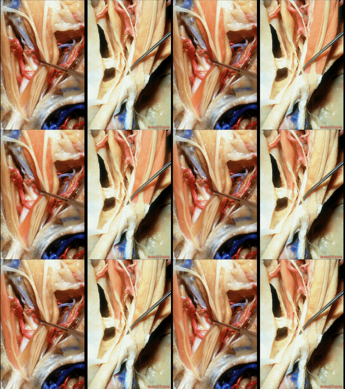
▼但需注意保护滑车神经,这里可见总腱环已打开,而滑车神经也得以保留。
But you wanna preserve the trochlear nerve, and here it's been divided with the trochlear nerve preserved.
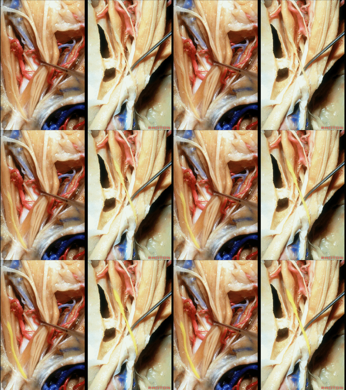
▼也可在总腱环外侧进行分离,以处理累及眶上裂的病变。但在上睑提肌和外直肌之间分离总腱环外侧时,需将眼上静脉留在外侧。
You can also divide the annular tendon laterally if you're working with a lesion coming through the superior orbital fissure. But to divide the annular tendon laterally between levator and lateral rectus you wanna let the superior ophthalmic vein go laterally.
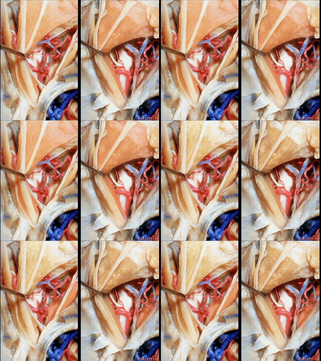
▼眼上静脉较为粗大,下图示眼上静脉。
It's quite a large vein,
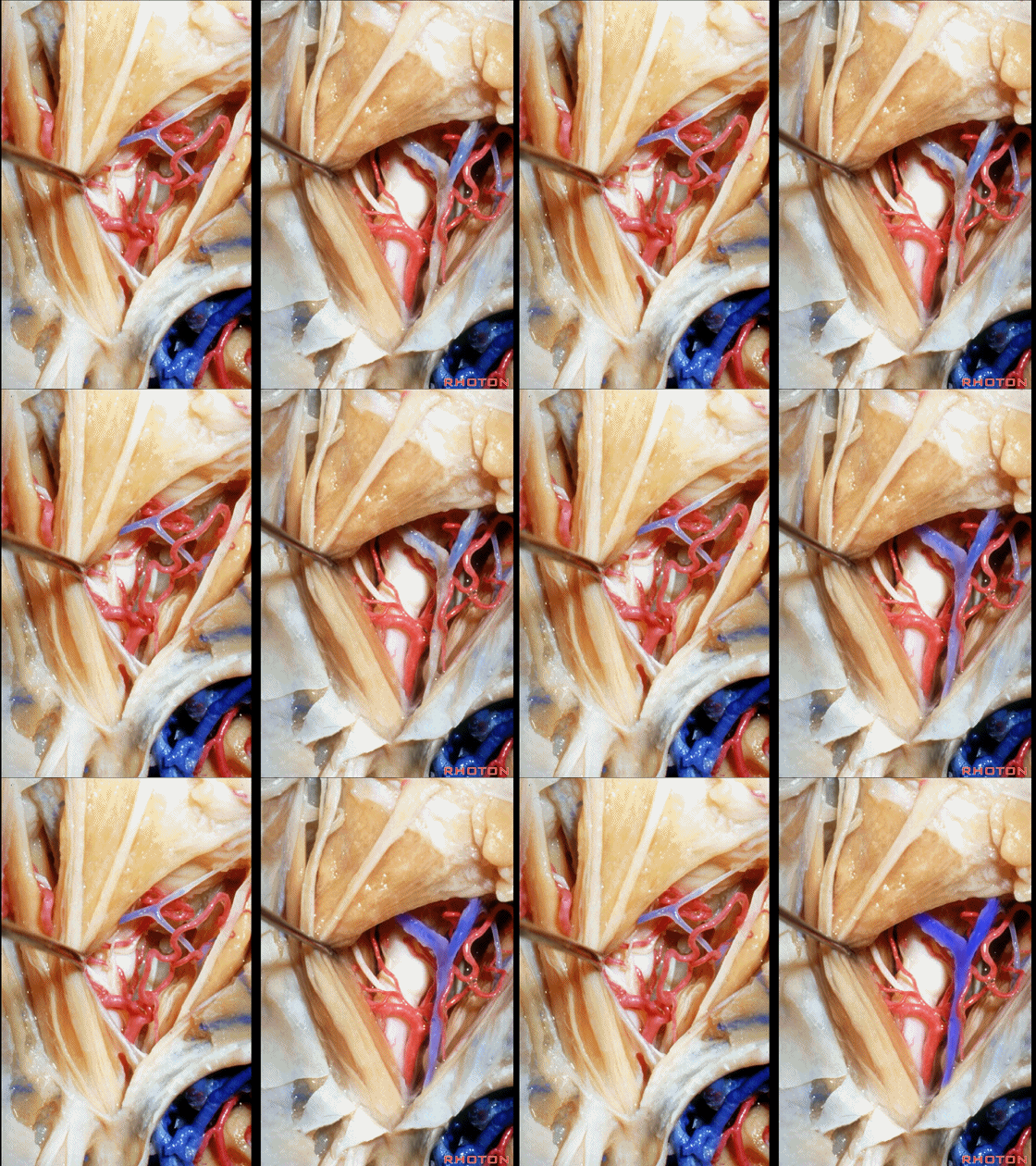
▼若将眼上静脉与上睑提肌一起推向内侧,则会阻挡对于眶尖部视神经外侧的暴露。
and if you pull it medially with the levator, it blocks access to the orbital apex on the lateral side of the optic nerve.
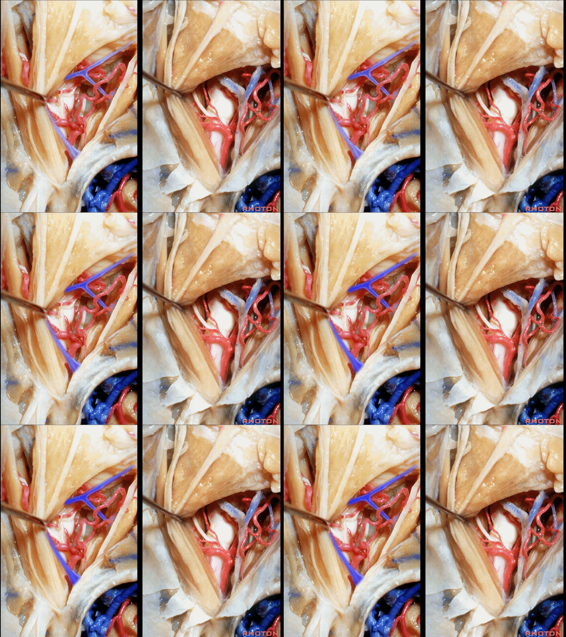
▼因此,恰当的方式是将眼上静脉牵向外侧,将上睑提肌牵向内侧。
So it's better to let that vein go laterally, and let the levator without the vein be retracted medially.
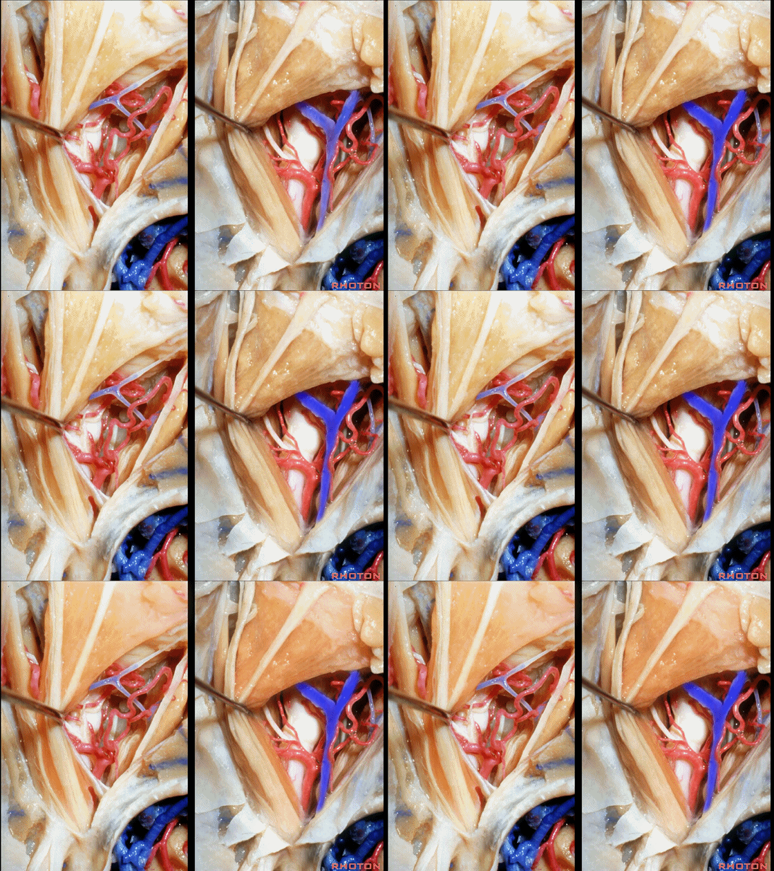
▼一旦从上睑提肌和由外展神经支配的外直肌之间打开,即可见动眼神经分成上支和下支。下图示上睑提肌(左侧)
Here once you open between the levator and here 6th nerve going to lateral rectus, you see the division of the oculomotor into superior and inferior division.
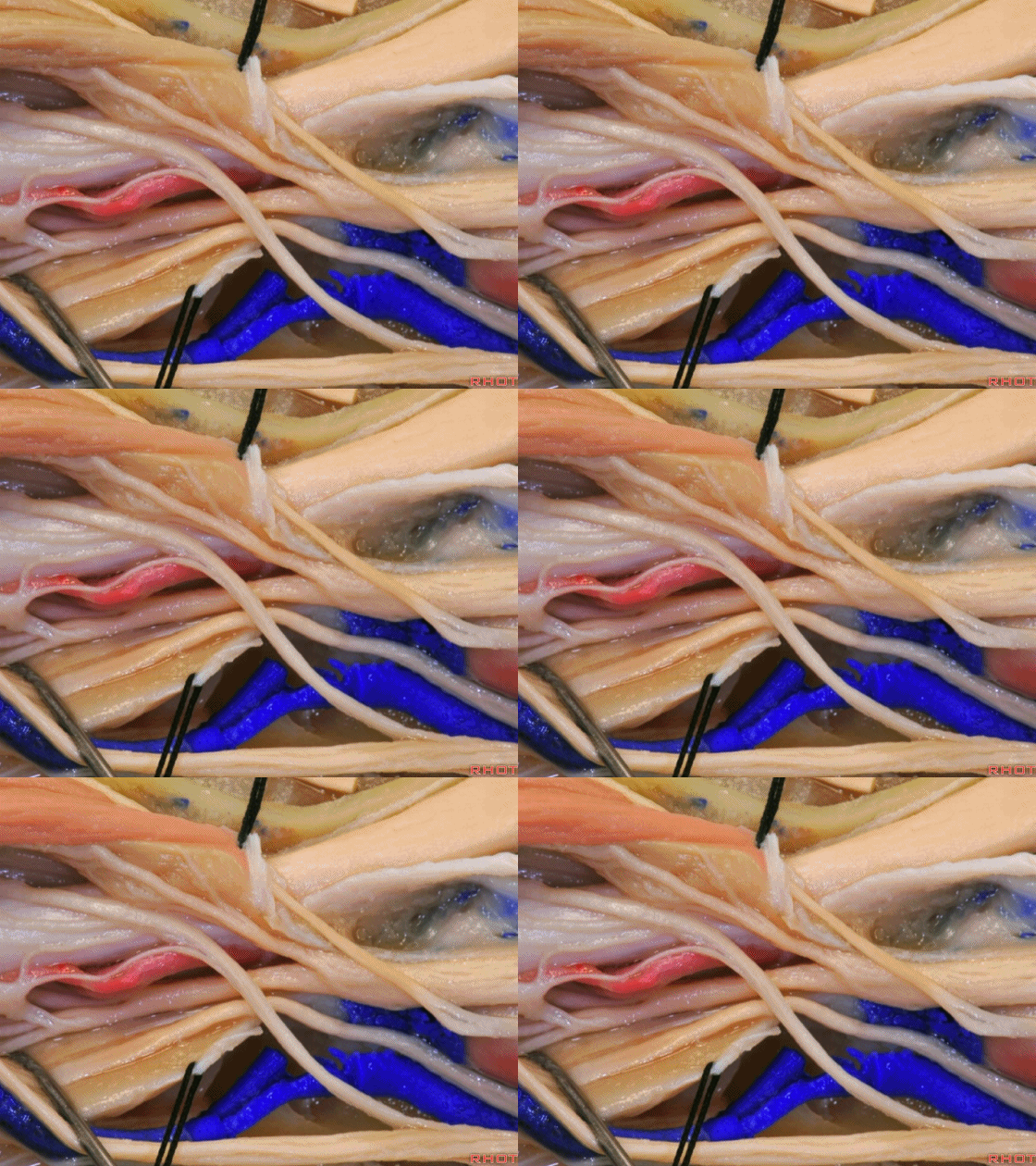
▼下图示外展神经
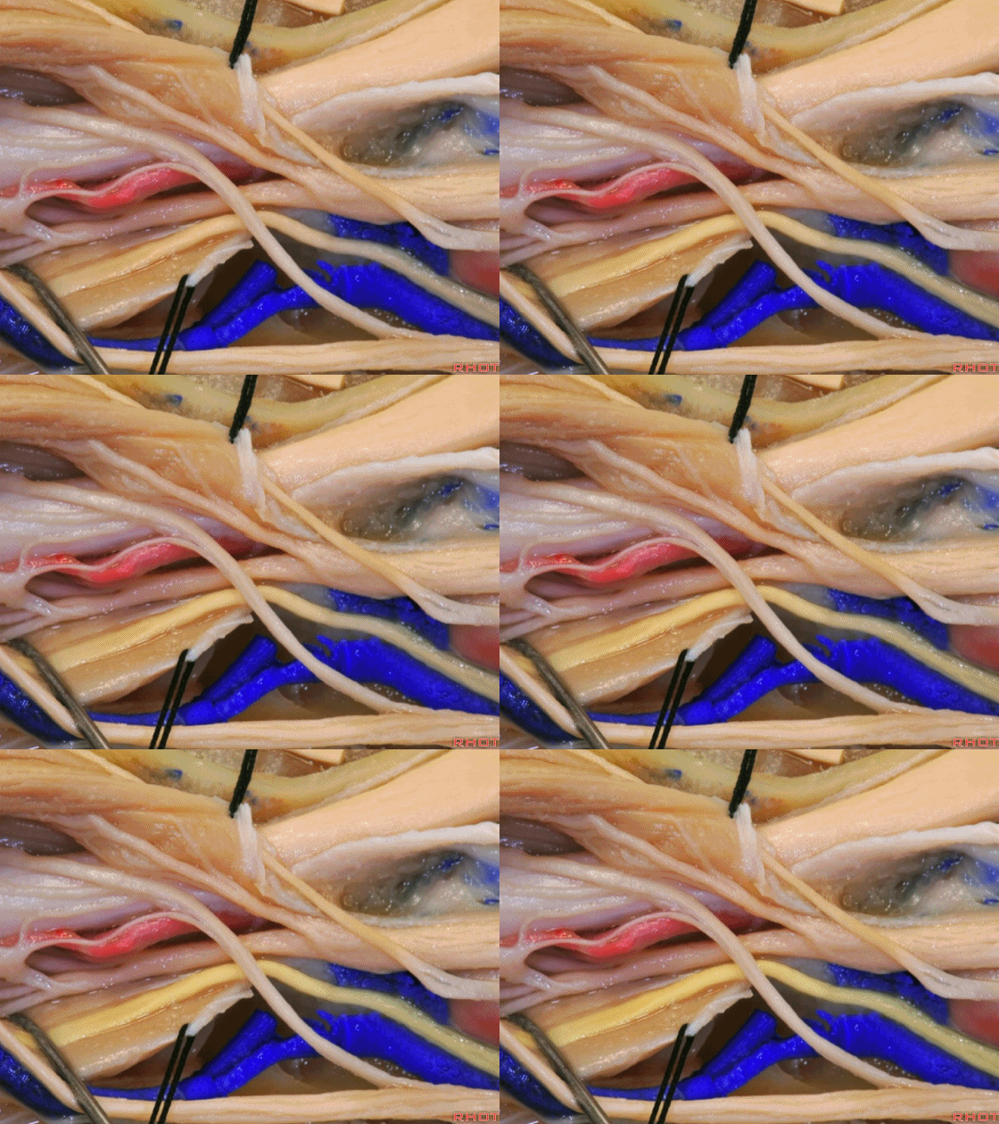
▼下图示动眼神经 分为上支、下支
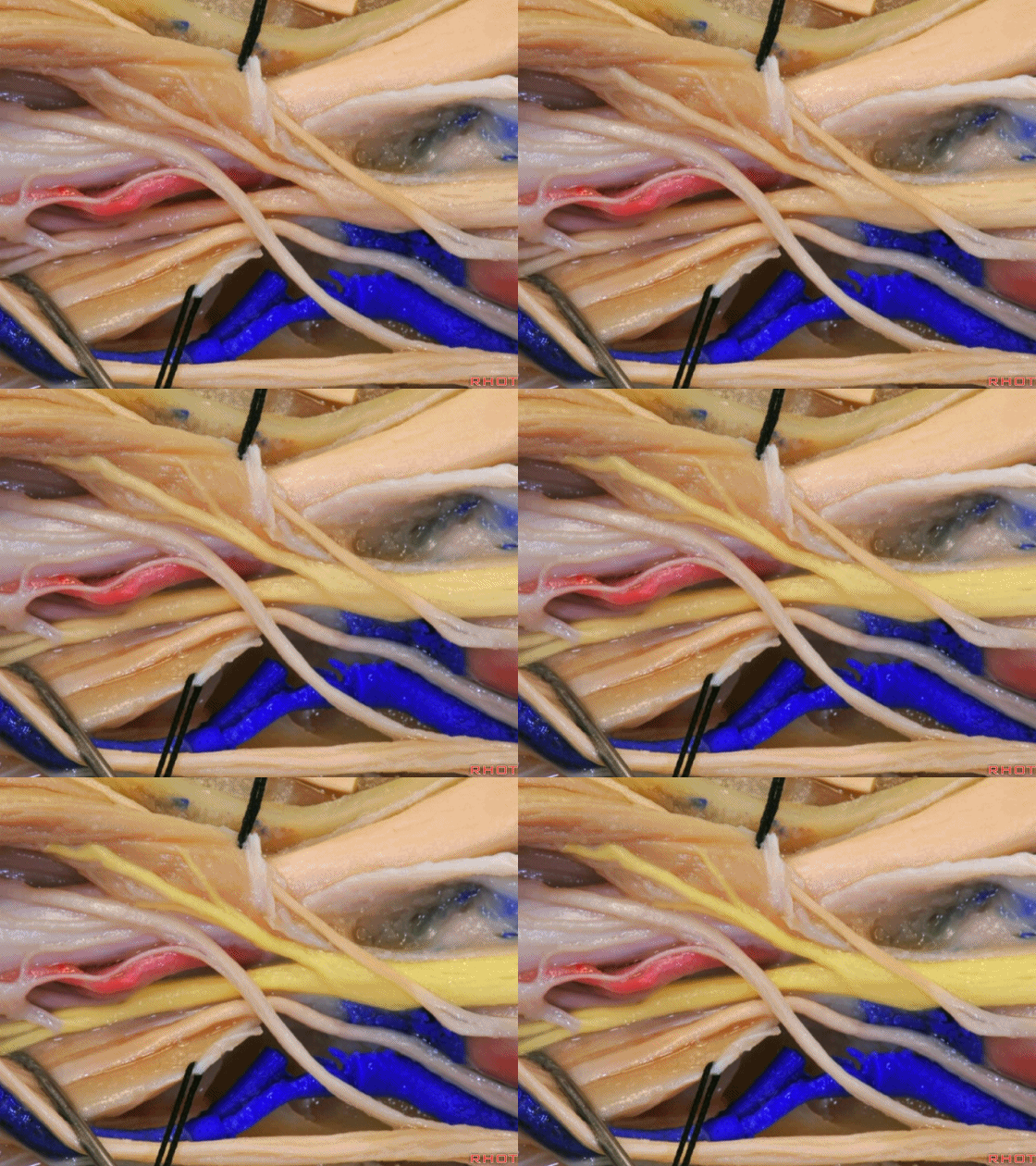
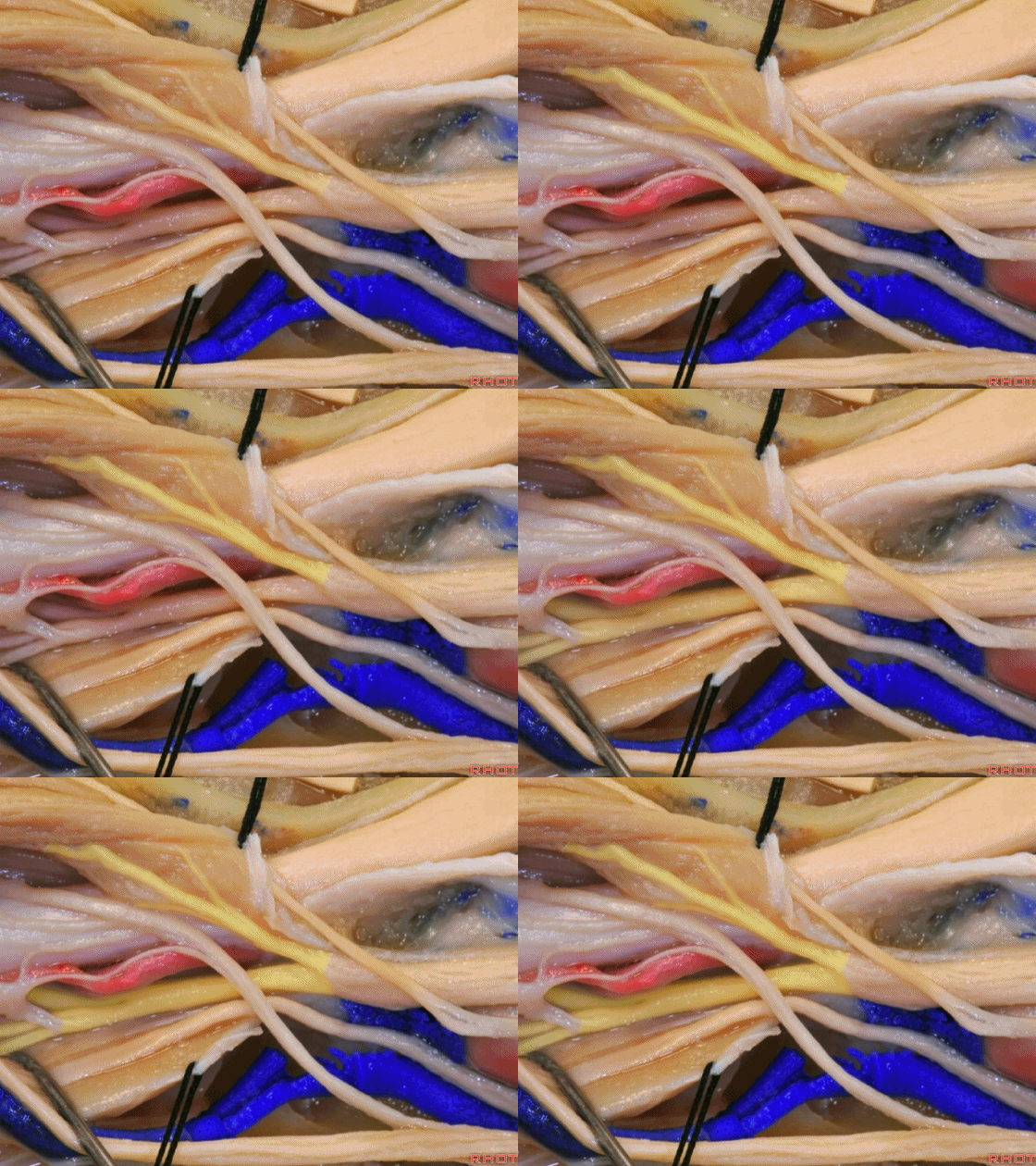
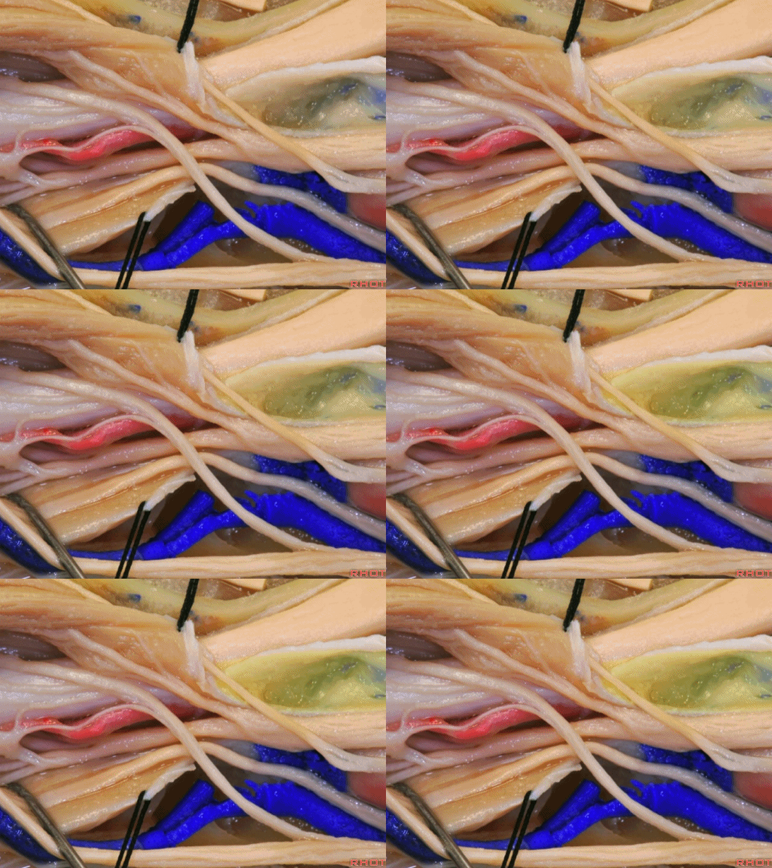
▼动眼神经分叉处位于前床突下方。下图示前床突(已磨除)
Those occur below the area of the anterior clinoid.
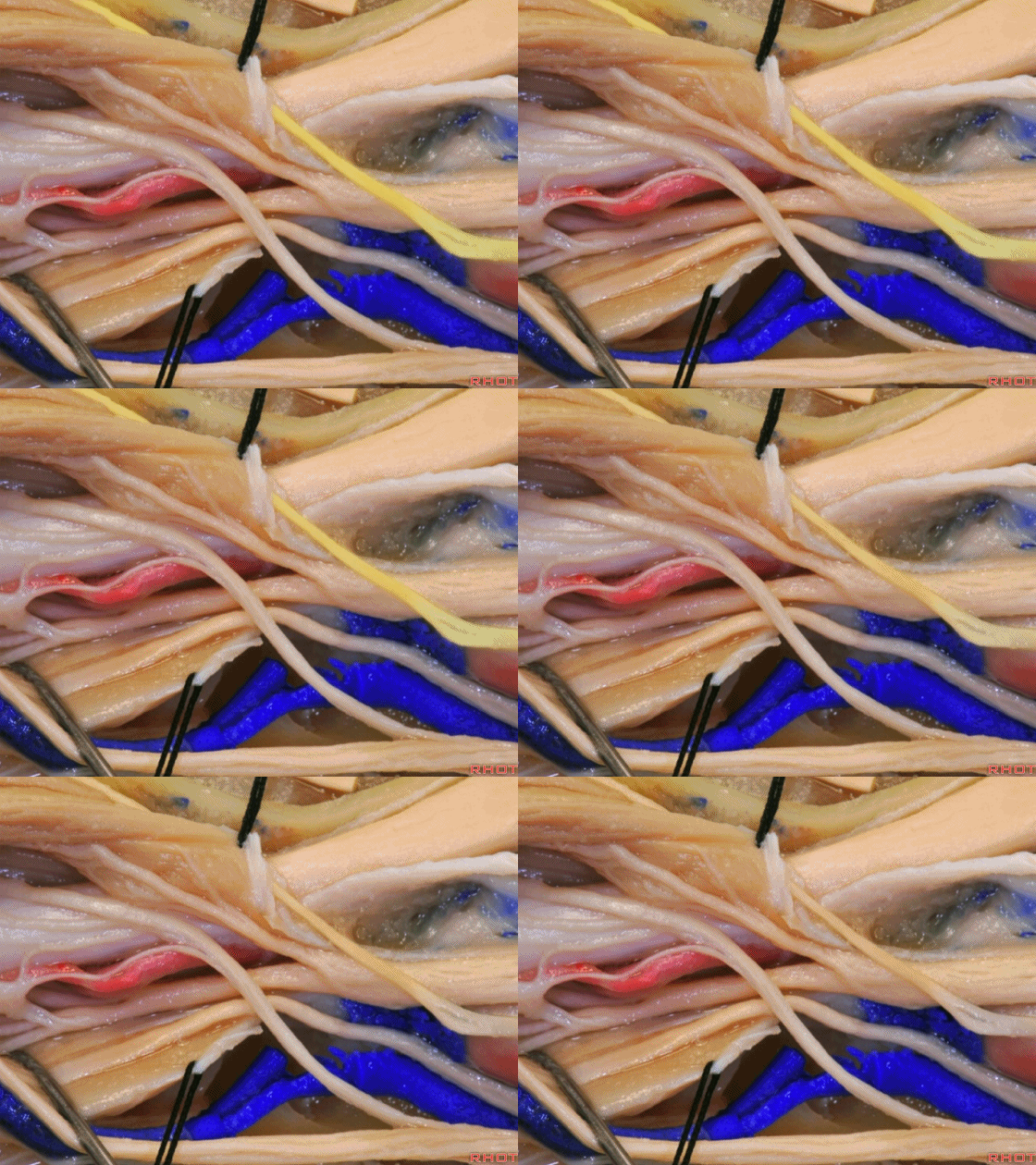
▼下图示向内走行的滑车神经。
the 4th nerve is passing medially.
▼这是鼻睫神经,穿过总腱环行于视神经之上。
And here we see nasociliary nerve pass through the annular tendon above the optic nerve.
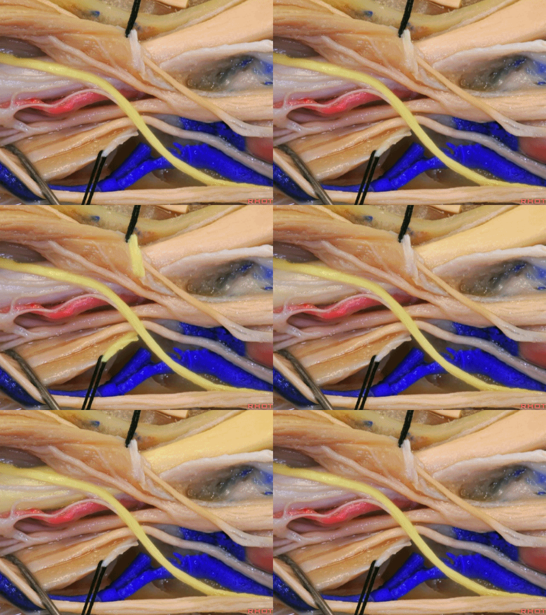
▼这是睫状神经节。
here we see ciliary ganglion.
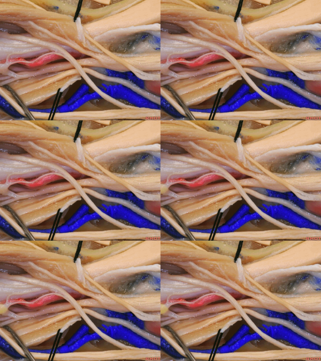
▼这是一支感觉神经,支配角膜的感觉。它并不在睫状神经节内形成突触。
Can anyone tell me what nerve that is? That's the sensory root, that's the nerve that carries corneal sensation.It doesn't synapse in the ciliary ganglion.
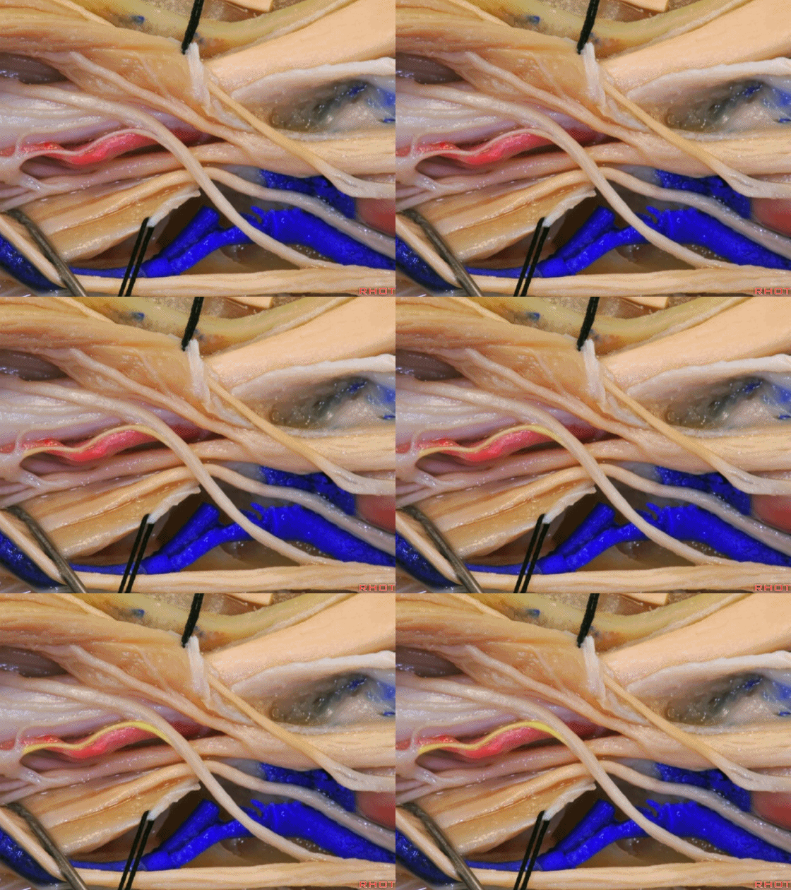
▼这是动眼神经下支。其内发出副交感根,在睫状神经节内换元控制瞳孔的舒缩。
But here's inferior division of III. This is the parasympathetic root, the motor root of the ciliary ganglion that controls the pupil.
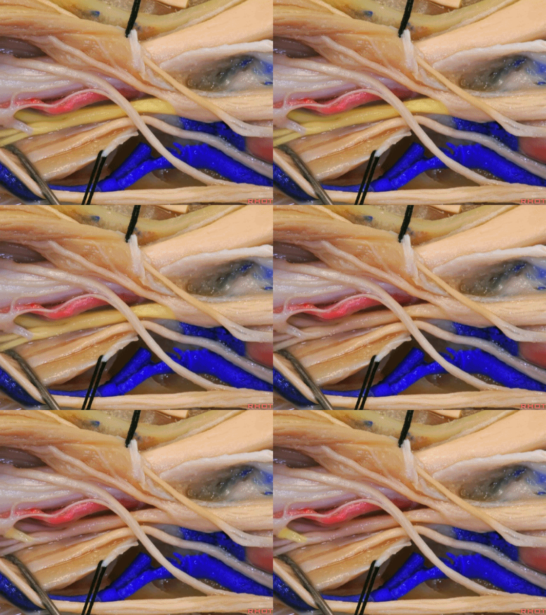
![]()
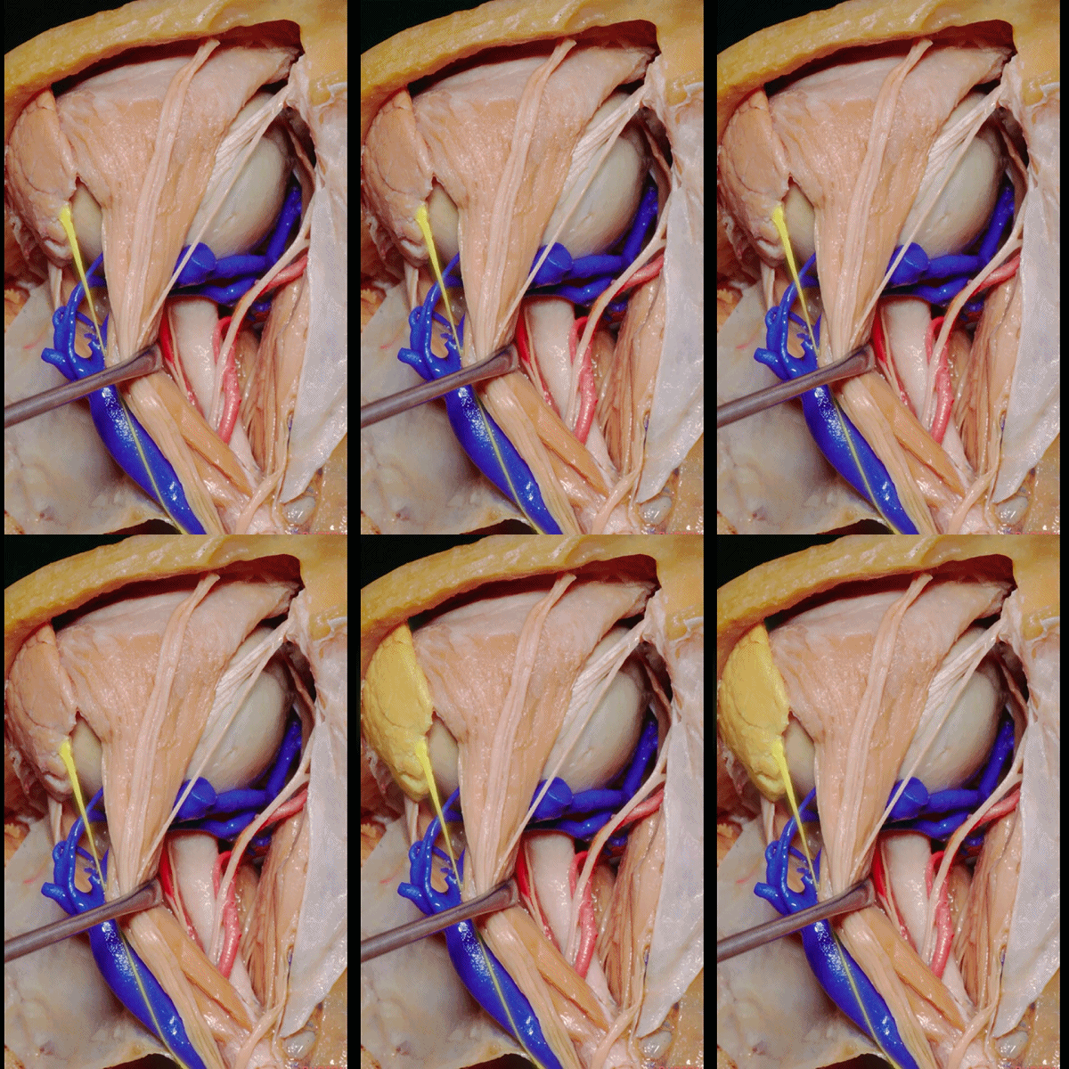
▼再来看看通过眶内侧壁的入路。这是上斜肌,及其骨性附着处
Here's just the approach along the medial orbital wall. Here, the trochlea of the superior oblique muscle
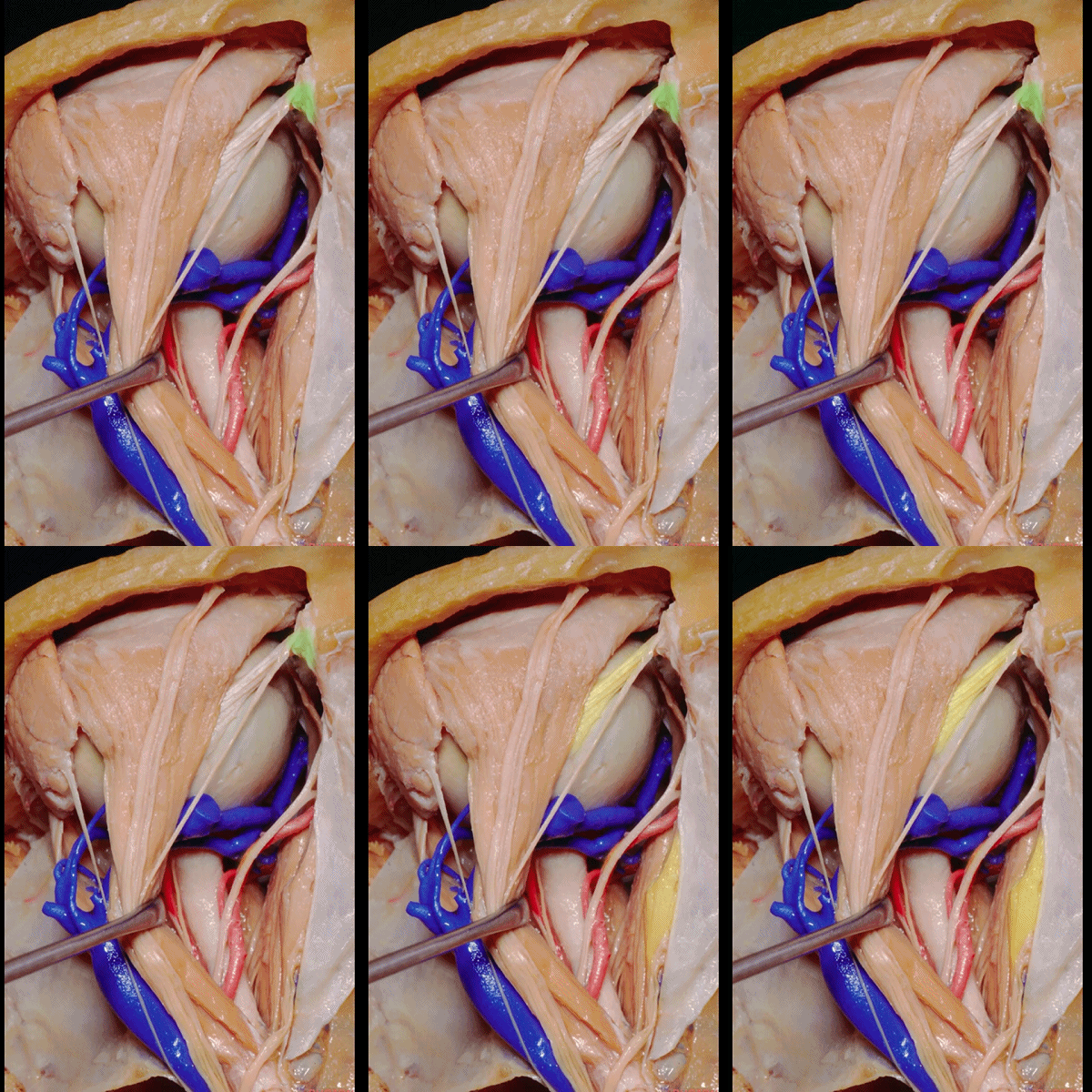
▼上斜肌走行于上直肌下方,附着于眼球的外侧部(箭头处)。
diving in under the superior rectus and attaching to the lateral part of the globe.
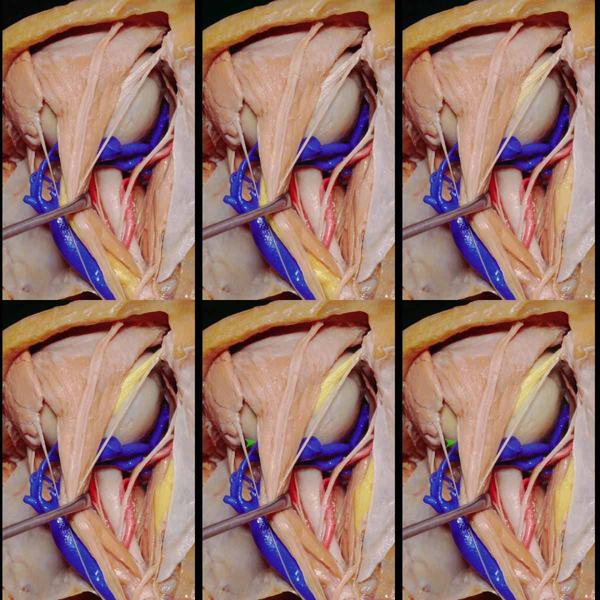
▼这是鼻睫神经
But here's nasociliary nerve,
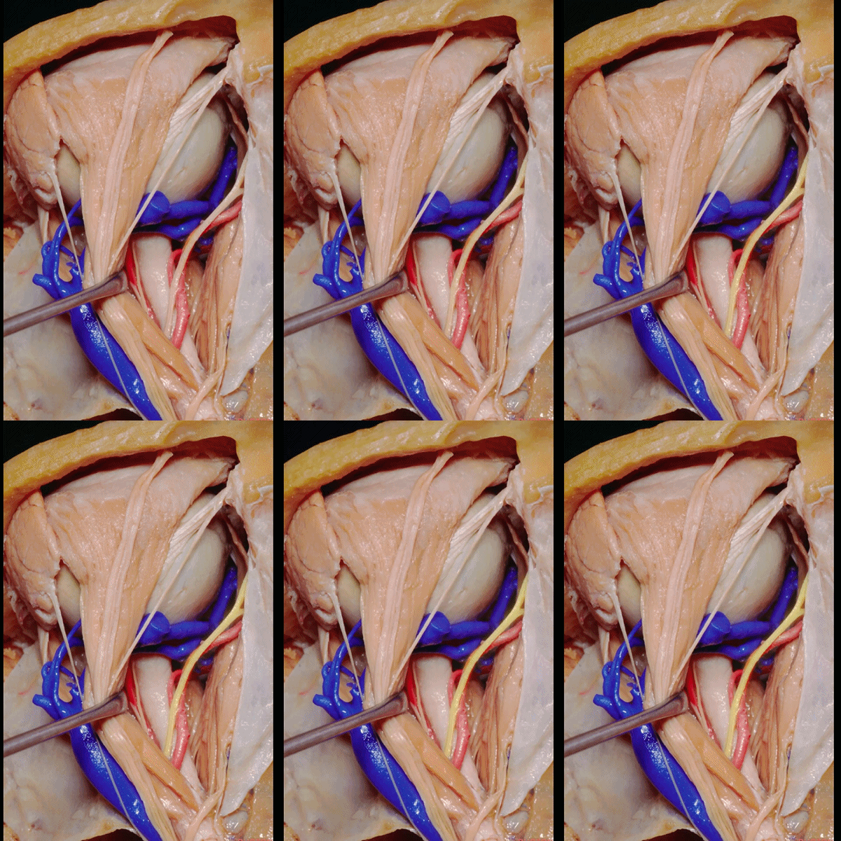
▼这是眼动脉
ophthalmic artery
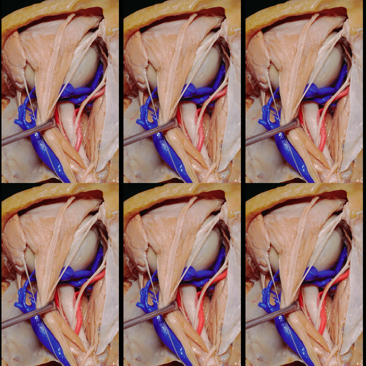
▼这是泪腺神经,支配泪腺,发自眼神经。
Here's lacrimal branch going to lacrimal gland that comes from V1.
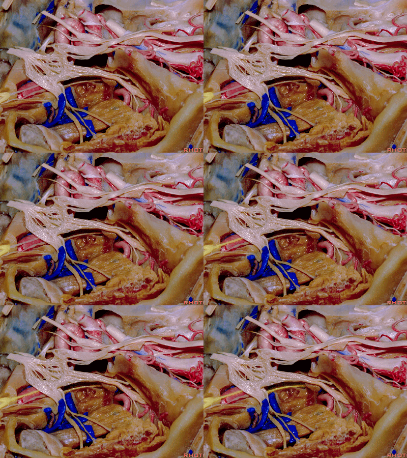
▼支配泪腺的副交感纤维是如何走行的?该通路起源于后颅窝的面神经、岩前大神经、翼管神经
But, lacrimation starts back in the facial nerve in the posterior fossa, greater petrosal,to vidian nerve,
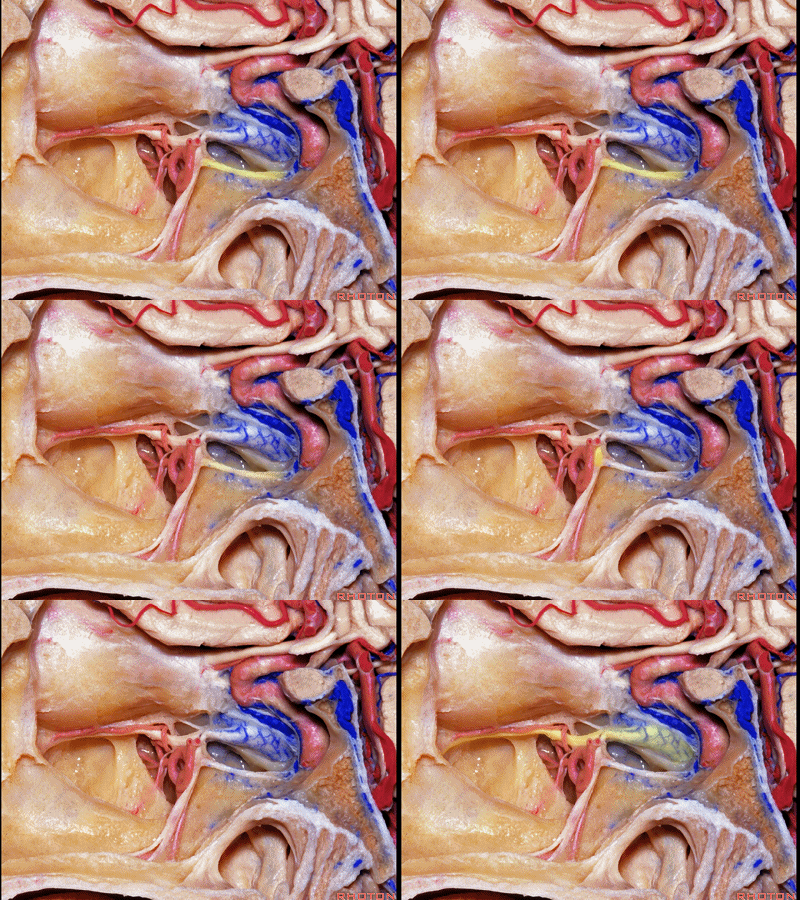
▼翼管神经行于翼管内,到达翼腭神经节,随后在眶底进入上颌神经。
How does parasympathetic innervation get to lacrimation? It gets there from vidian nerve that we saw coming through vidian canal to pterygopalatine ganglion. It gets into V2 along the floor of the orbit.
▼上颌神经发出颧神经,其支配眶外侧的部分皮肤感觉。
From V2...the V2 gives off the zygomatic nerve that innervates some of the skin around the lateral orbit.
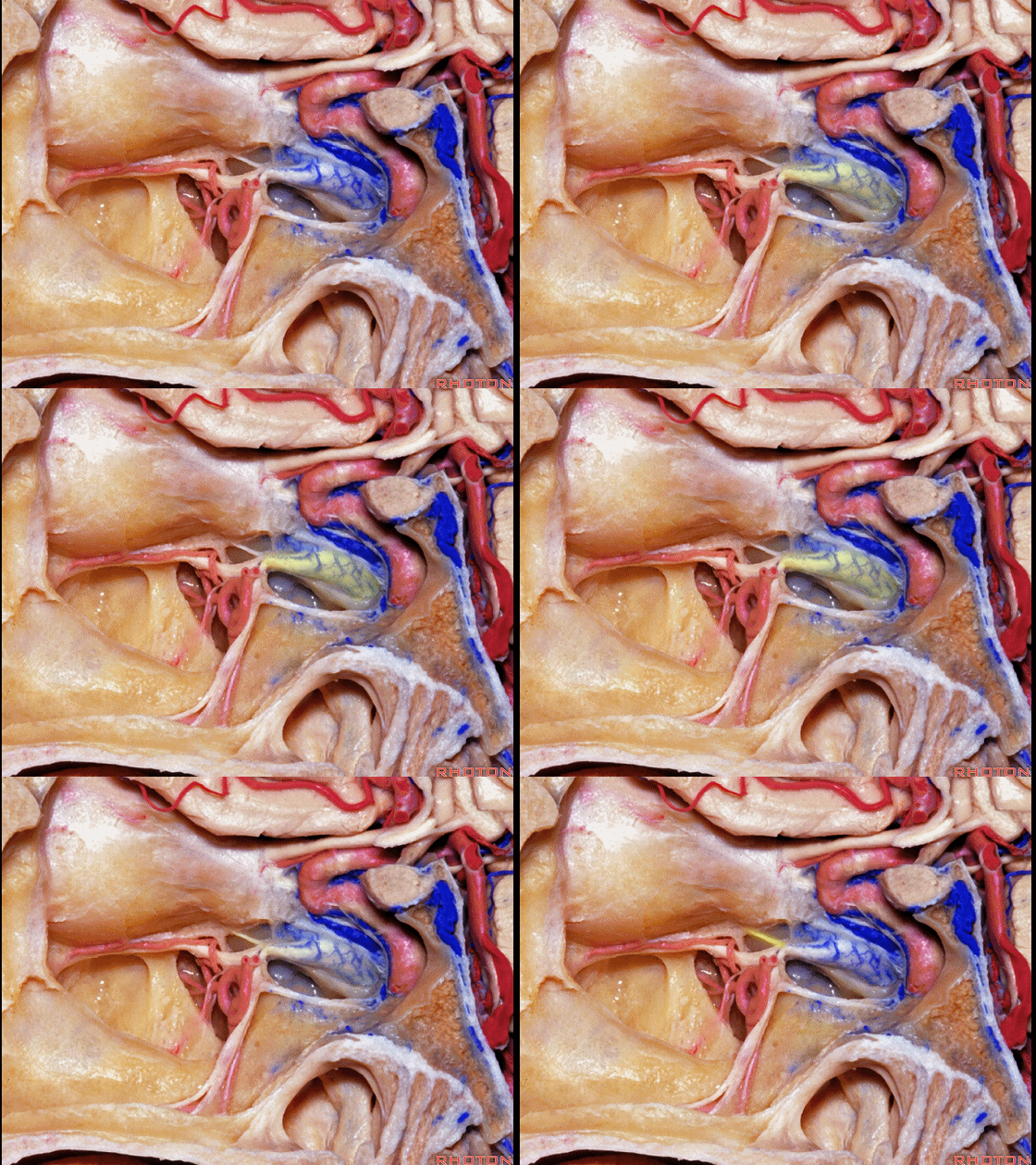
▼这些副交感分支即随着上颌神经的颧支进入眼神经的泪腺神经
those parasympathetic branches that get into the zygomatic branch of V2 jump to the lacrimal nerve
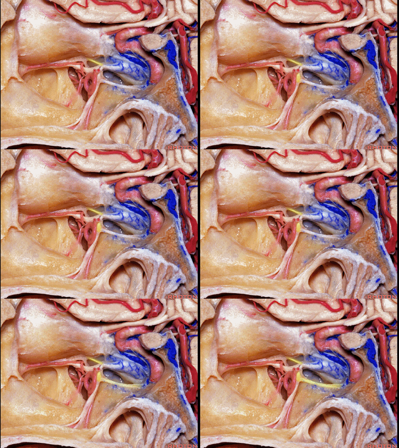
▼而泪腺神经即为到达泪腺的副交感最后通路。
the final pathway for lacrimation then comes through lacrimal nerve.
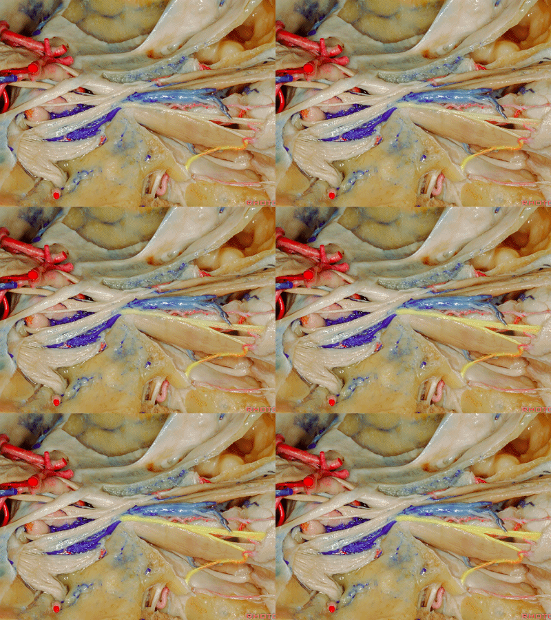
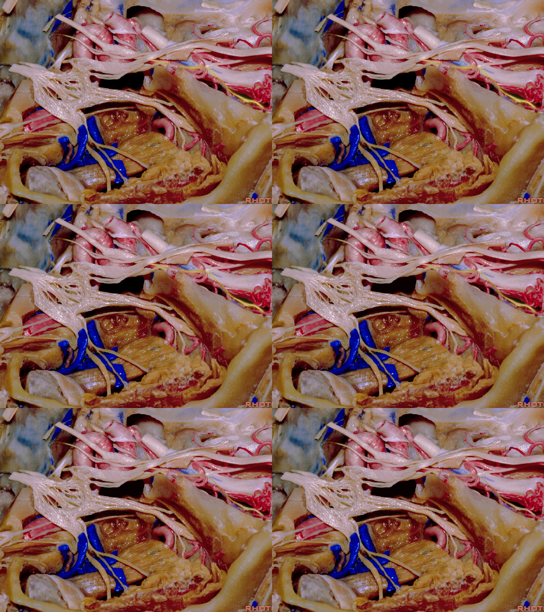
▼这是上述相关结构的概览,在颅中窝,眼神经的分支进入眶尖,上颌神经沿着眶底外侧走行,下颌神经进入颞下窝。
you wanna see this all in the context of the adjacent structures,middle fossa,branches of V1 entering the orbital apex, V2 running along the lateral orbital floor, and then V3 here entering infratemporal fossa.
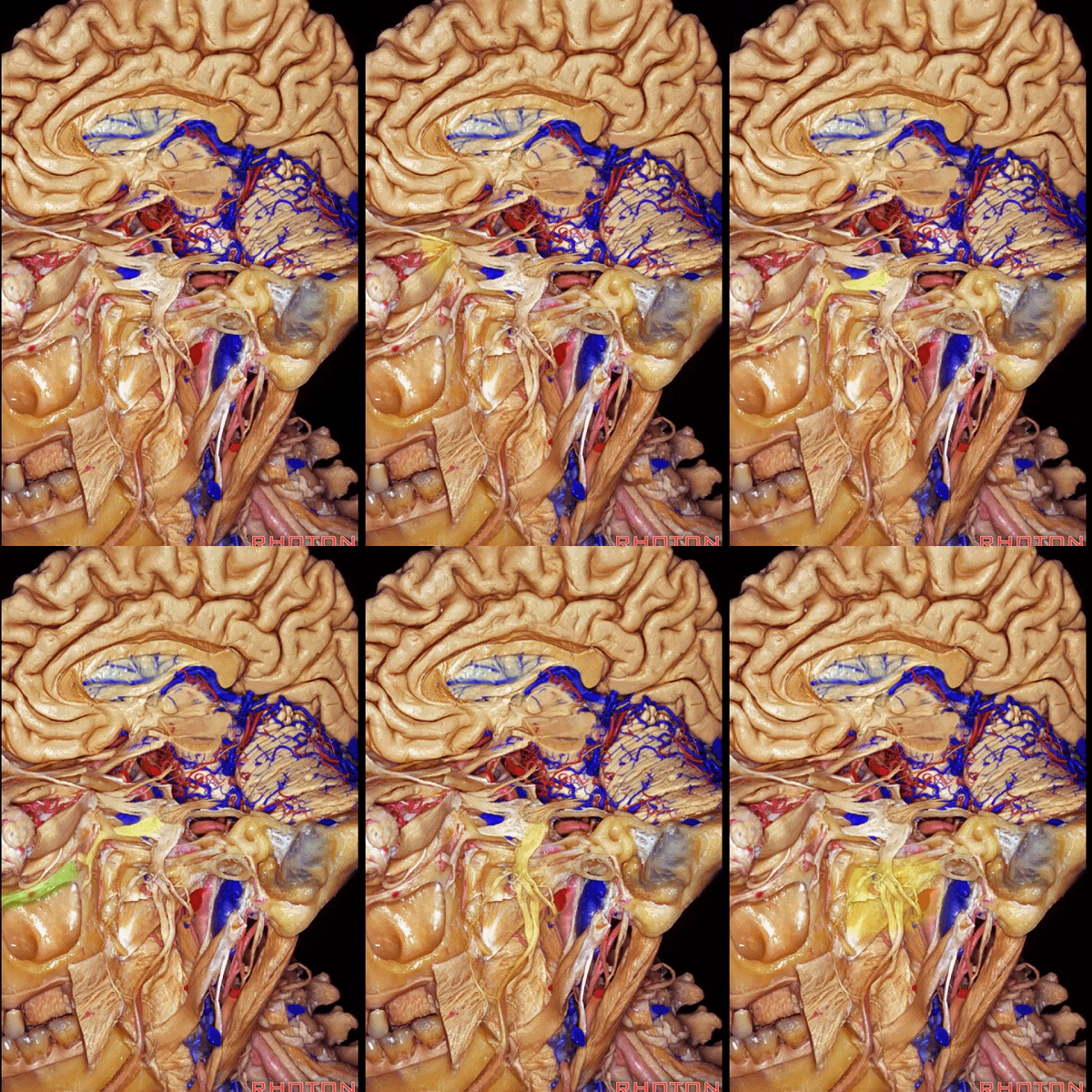
▼放大上述解剖结构,可见颅内病变侵入眶内的多条路径,包括从鼻窦、颅神经孔道、视神经管 进入眶内。我们需掌握所有这些复杂的解剖关系。
But as you blow up all of this anatomy where there...there're multiple routes that intracranial pathology can extend to the orbit out of the sinuses, or along the foramen of the cranial nerves, or along the optic canal,to the orbit. And you wanna understand all of these complex relationships.
![]()
▼再来快速复习一下后方的海绵窦,动眼神经行于前床突下方.
then we do just a quick review of cavernous sinus behind with 3rd nerve passing below the anterior clinoid,
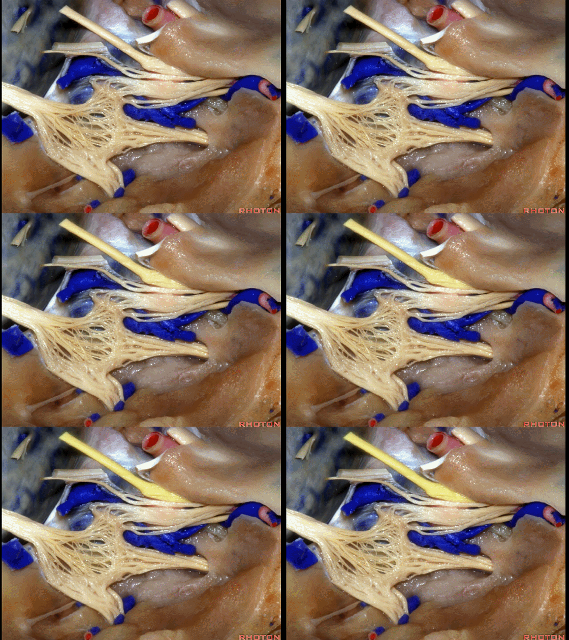
▼滑车神经、眼神经、外展神经于此处穿行于眶上裂
here, 4th nerve, V1,abducens nerve,here passing through the superior orbital fissure
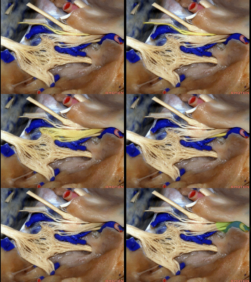
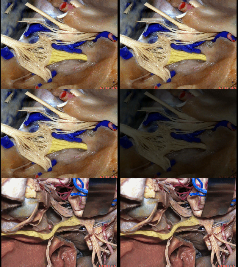
▼上颌神经于此处离开中颅窝,沿着眶底走形。
V2 exiting here middle fossa to run along the floor of the orbit.
▼我们移除了前床突。在前床突下方,可见滑车神经穿行至眶尖外侧部,向内走行支配上斜肌。
Here we've removed the clinoid. And just under the clinoid here,we see the 4th nerve passing from lateral part of orbital apex,to the medial part to innervate the superior oblique.
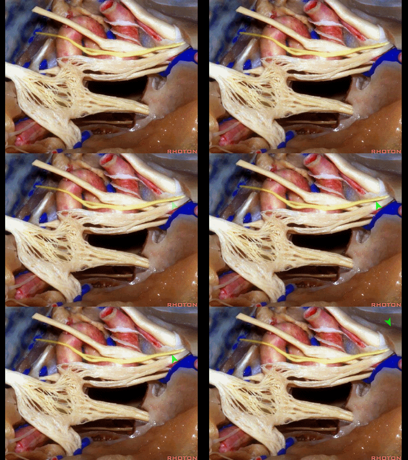
▼此处可见动眼神经分成上支和下支
And here we see the division of the 3rd nerve into superior and inferior division,
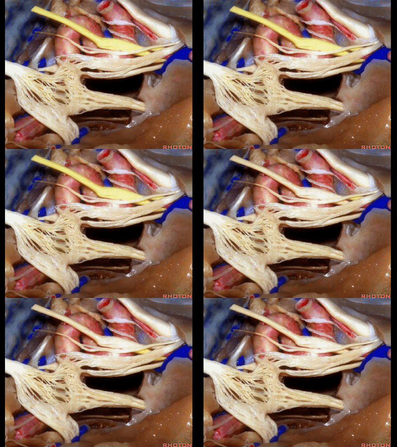
▼动眼神经分叉处恰位于前床突前部的下方。
this taking place below the anterior part of the anterior clinoid.
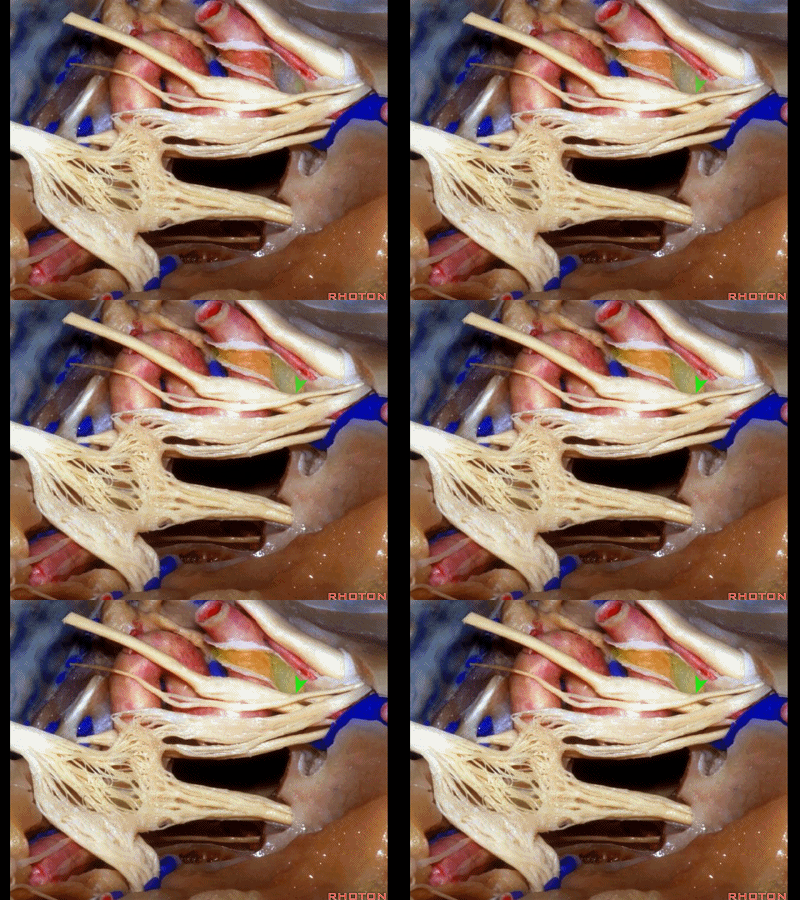
▼这是左侧眶尖,前床突已移除
Here we see another orbital apex, clinoid removed,
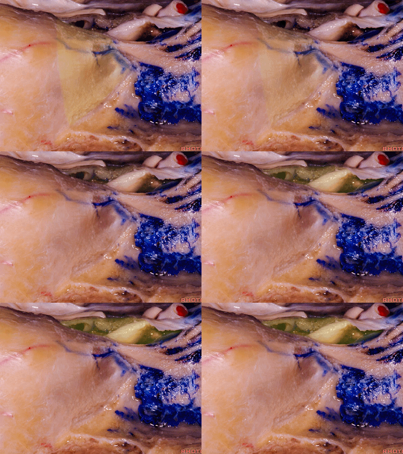
▼这是视神经管
optic canal.
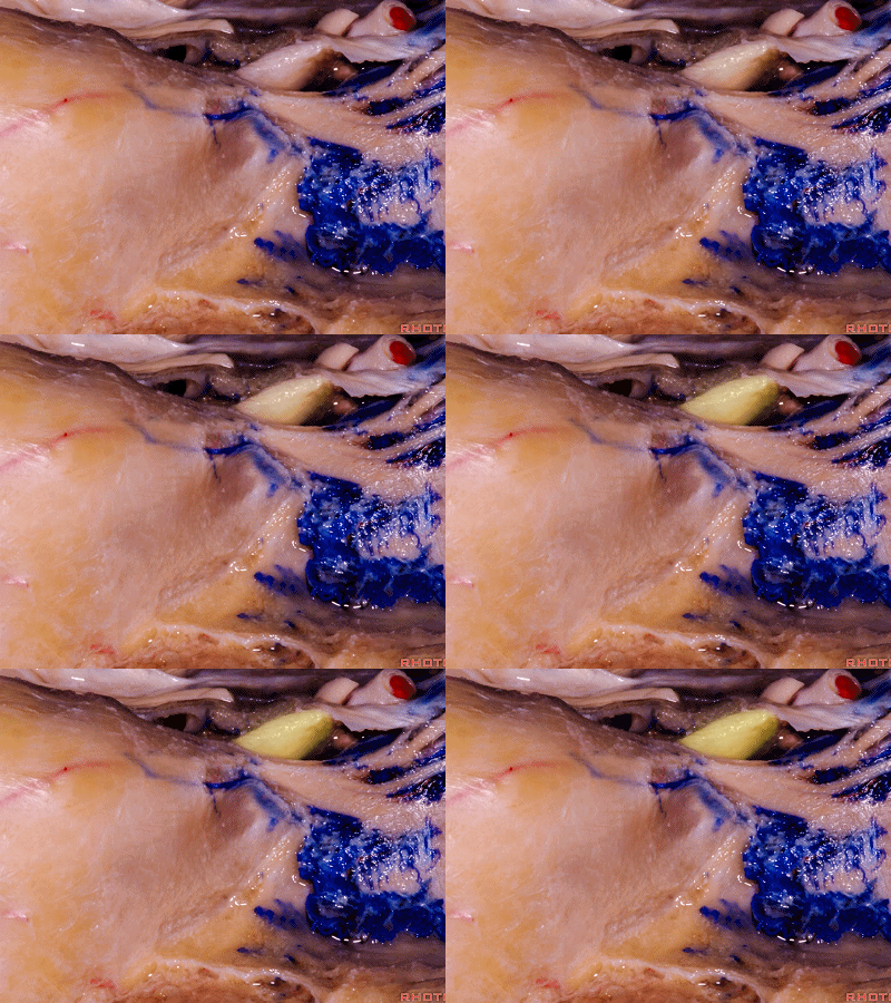
▼在视神经管外侧,可见这些结构穿行于动眼神经孔(属于总腱环的一部分),以及其他行于总腱环以外的结构。
And then, lateral to the optic canal, we see some of the structures passing through the oculomotor foramen of the annular tendon,and other structures passing outside.
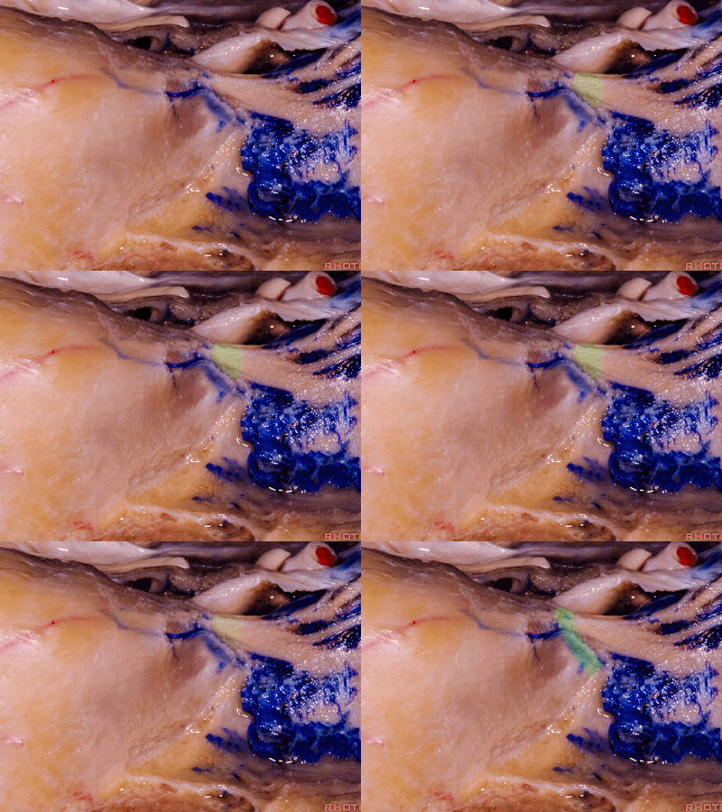
▼这里,我们移除前床突,以及眶上裂的外侧壁。
Here now, we see we've removed the clinoid,the lateral wall of the superior orbital fissure.
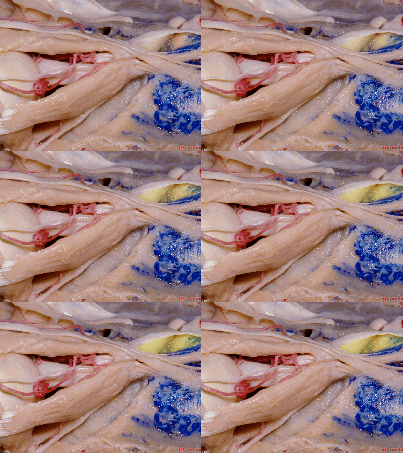
▼这是动眼神经孔
Here you see the oculomotor foramen
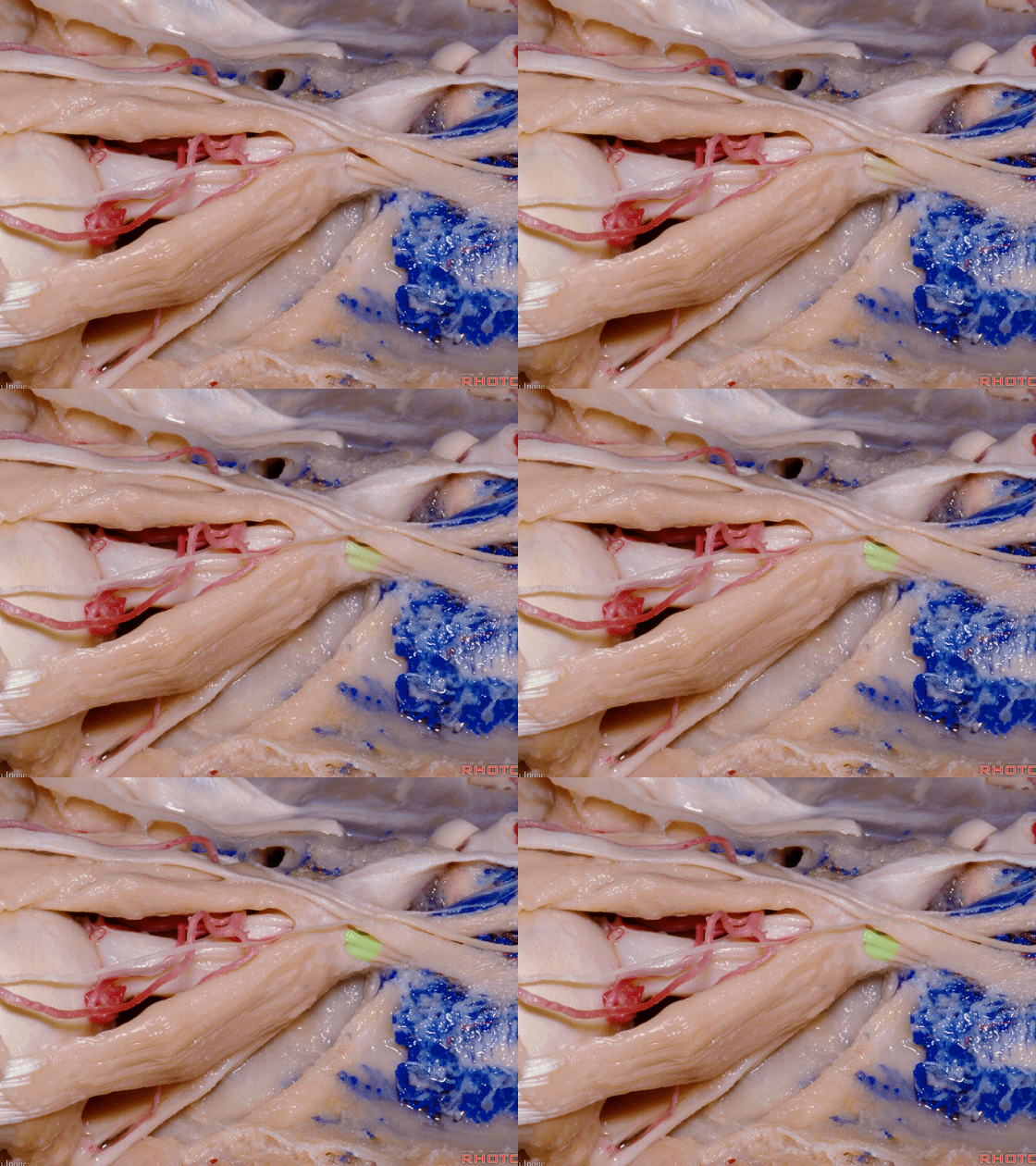
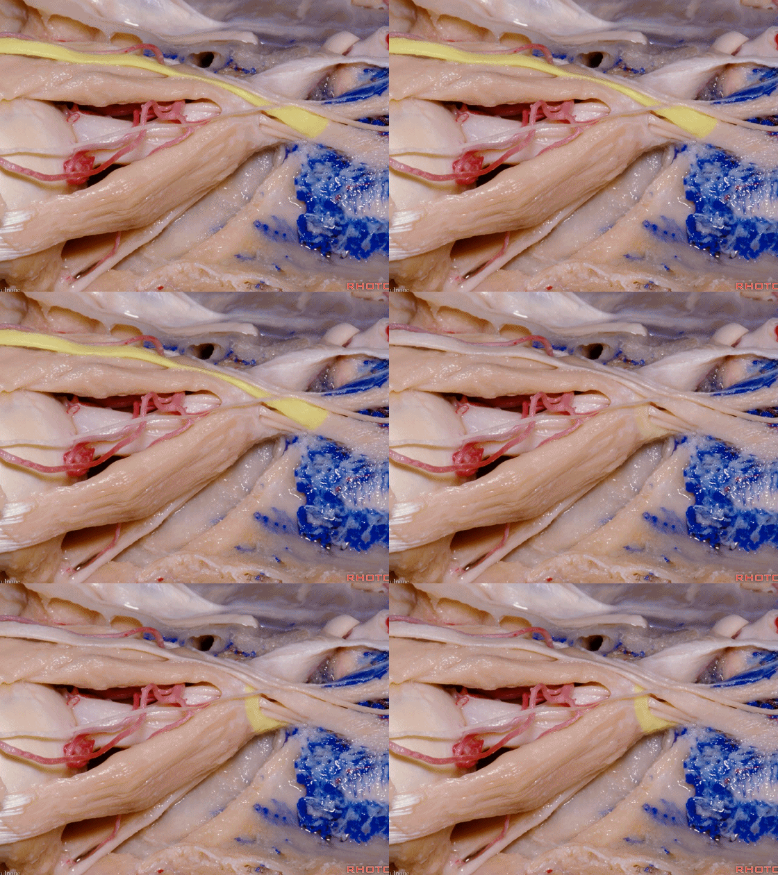
▼滑车神经于眶尖上方走行
the trochlear nerve passing above the orbital apex,
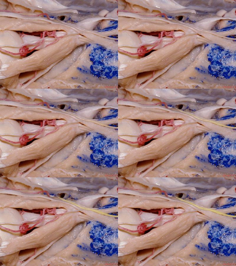
▼额神经行于总腱环以外。
frontal nerve passing outside the annular tendon.
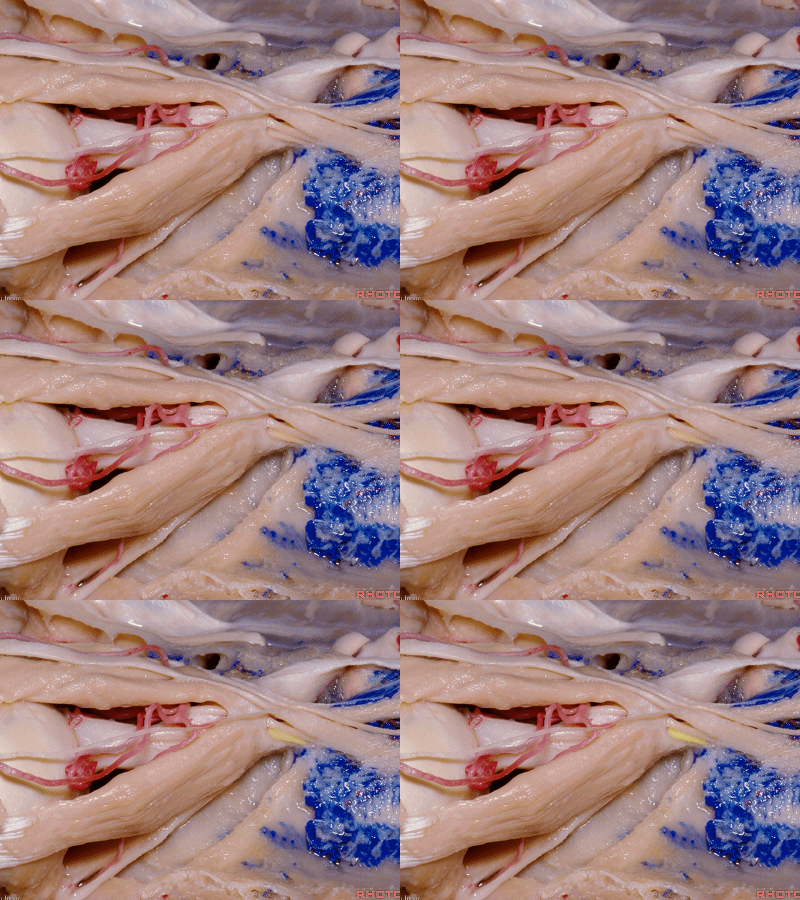
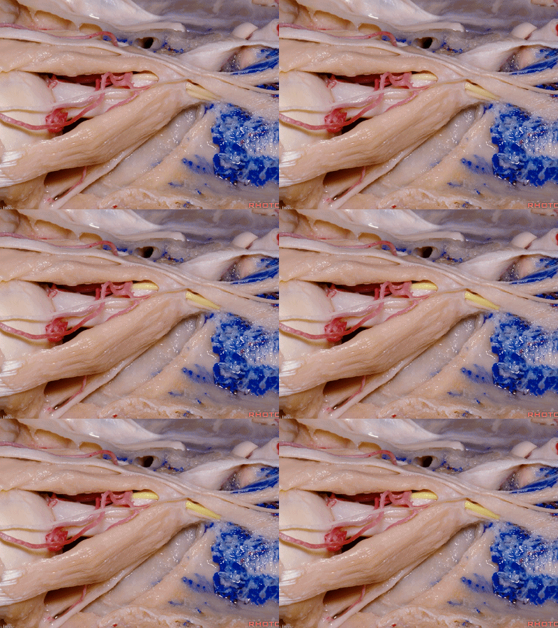
▼外展神经(下图)和鼻睫神经在眶尖处穿行于动眼神经孔的外侧缘。
But 6th nerve and nasociliary nerve passing through the lateral edge of the the oculomotor foramen here at the orbital apex.
▼下图示鼻睫神经
▼这里我们牵开额神经
Here we retract the frontal nerve.
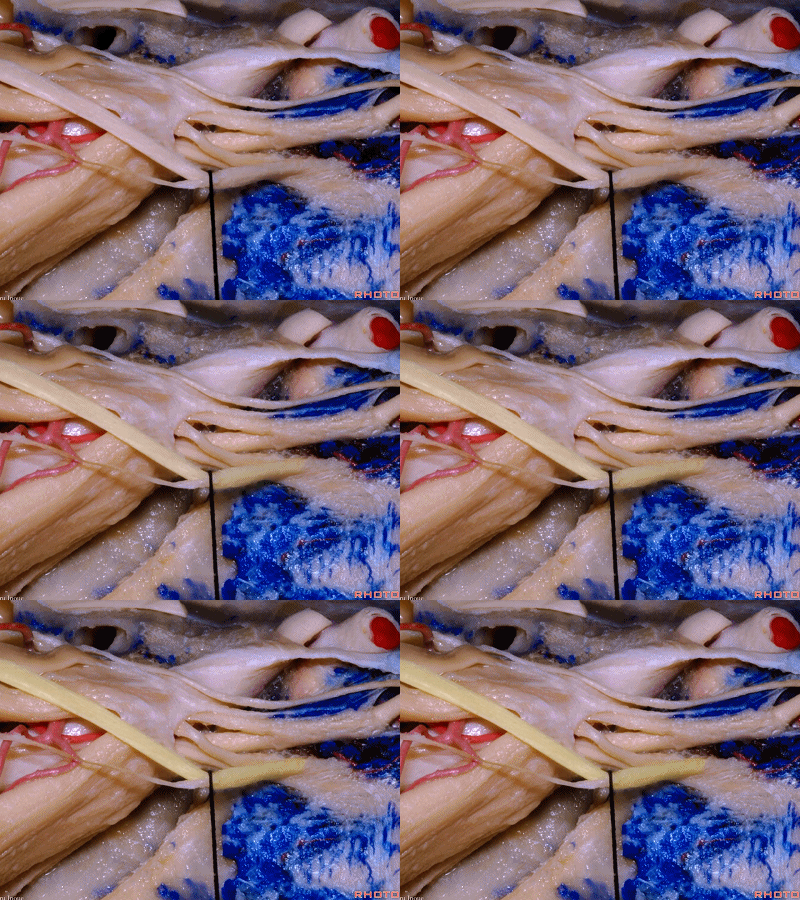
▼可见鼻睫神经
You see the nasociliary nerve
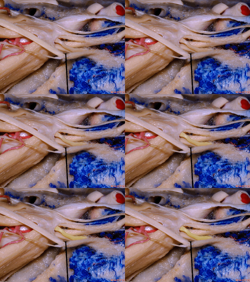
▼鼻睫神经下方为外展神经,其行至外直肌
and below it the 6th nerve going to the lateral rectus,
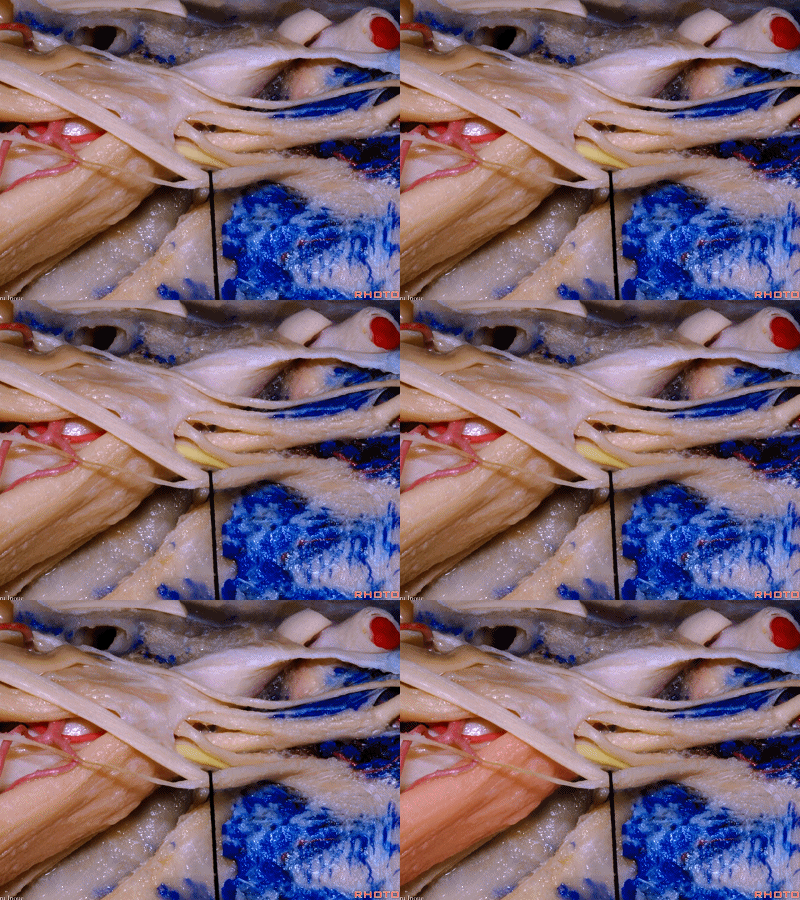
▼动眼神经在此处分成上支和下支
and here the division into superior and inferior divisions of 3rd nerve,
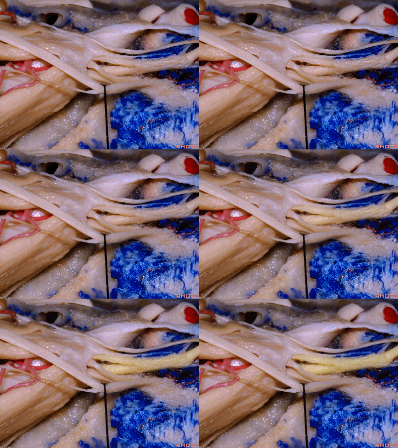
▼滑车神经向内侧走行于已被移除的前床突的前部下方。
and the trochlear nerve passing medially below the anterior part of the anterior clinoid that has been removed.
▼现在我们在总腱环的外侧进行分离
Here we've divided the annular tendon laterally here,
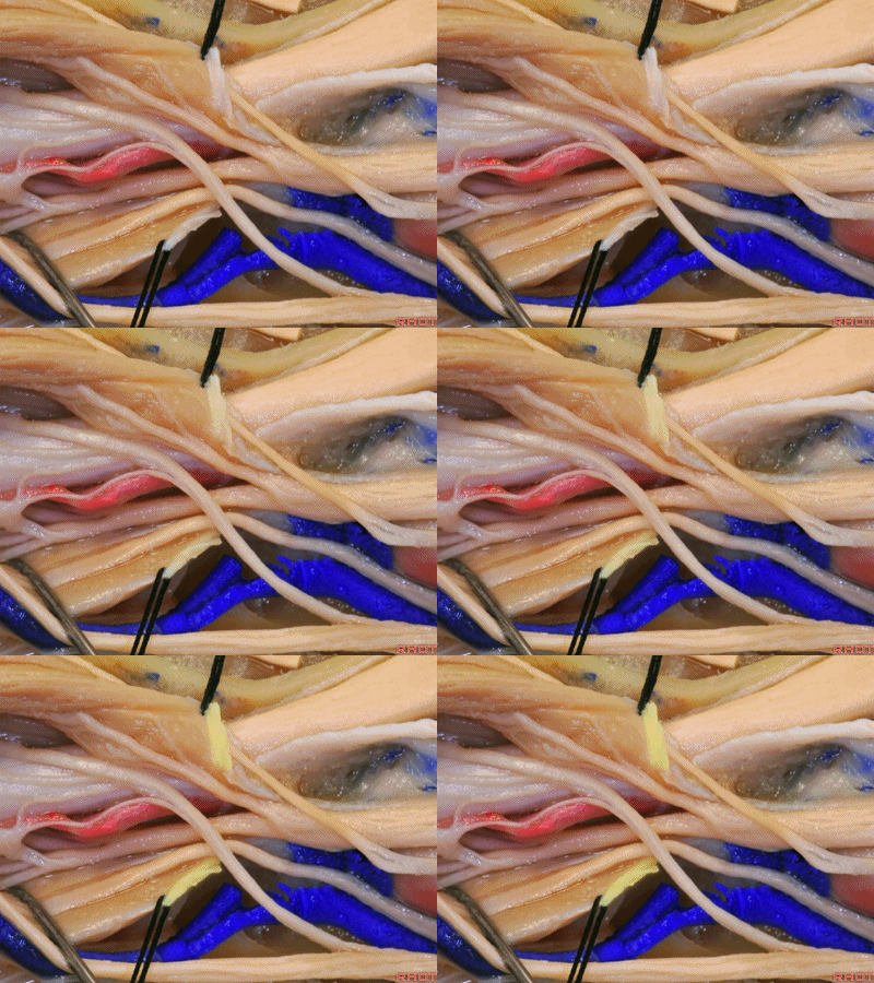
▼可见动眼神经上支进入上睑提肌、上直肌
and we see superior division of 3rd nerve going to levator, superior rectus,
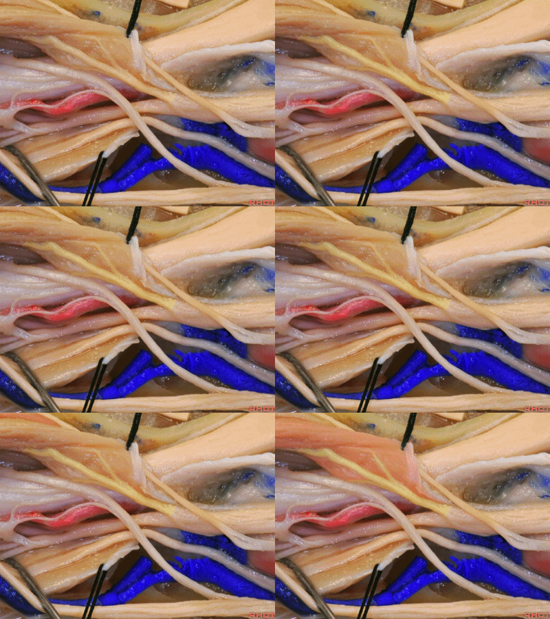
▼动眼神经下支进入下直肌、下斜肌
inferior division going to inferior rectus,inferior oblique,
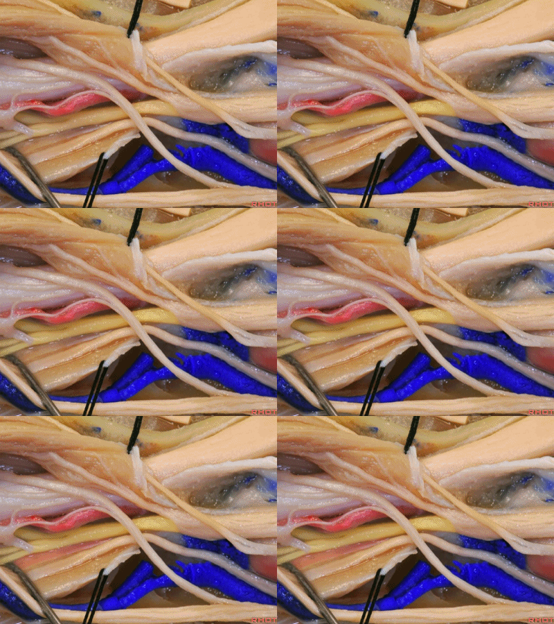
▼动眼神经下支发出副交感运动根进入睫状神经节。
giving rise also to the motor parasympathetic root of the ciliary ganglion.
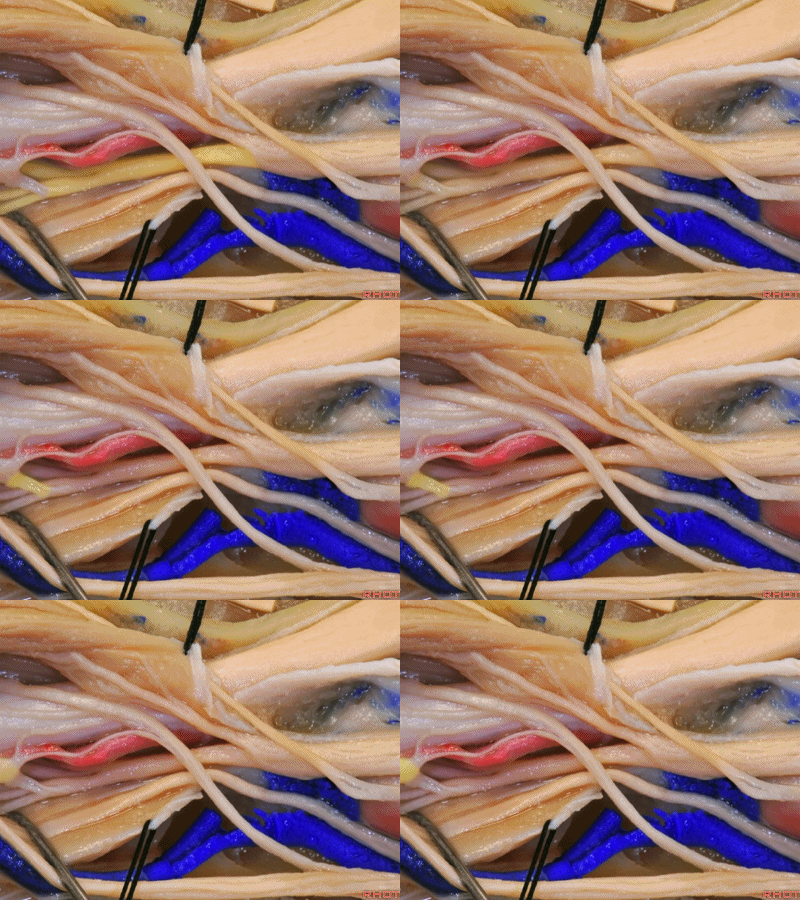
▼这是行经睫状神经节的感觉根,支配角膜的感觉,其发自眼神经。
And here we see the sensory root of the ciliary ganglion that conveys corneal sensation through V1.
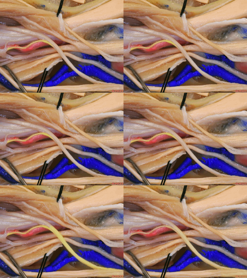
▼下图所示这些神经分支发自鼻睫神经(下图),负责眼球的交感功能。
And, some of the branches of nasociliary nerve that carry sympathetic function to the globe.
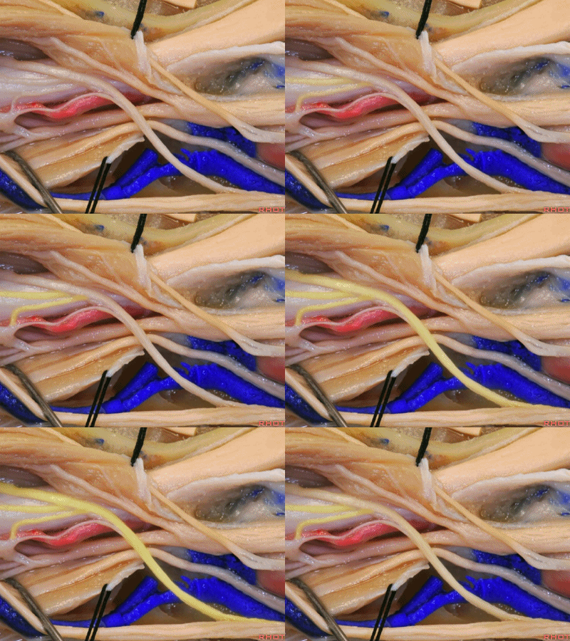
![]()


