本文为Rhoton解剖视频中《Navigating the Orbit》这一章节,主要讲解了眼眶的骨性结构、眶内结构、各种眼眶入路等内容。视频时间较长,笔者将部分内容按顺序重新编排,共截取270张图片。
笔者水平所限,错误之处请批评指正!
(因文章篇幅较长,脑医汇特将文章分为上、中、下三篇,欢迎大家阅读,分享!)

Navigating the orbit
▼我们今天的主题是眼眶,但也会涉及邻近的颅底。对于局限于眶内的病变,尤其前眶部,主要由眼科来处理。
We're talking about orbit today, but we include adjacent skull base because if a lesion is strictly within the orbit especially the anterior part, those lesions are dealt with predominantly by ophthalmology,
▼而如下情况则需要神经外科干预,例如病变侵犯了邻近颅底,或累及视神经管或眶上裂,或累及眶周骨质。因此,我们需要应对的不仅仅是眶部,还包括眶周的颅底。
but neurosurgery becomes involved if a lesion extends to the adjacent skull base,or extends through the optic canal or superior orbital fissure,or involves the bones surrounding the orbit. So,we're often asked not only to deal with the orbital part,but also we get called in because of the involvement of the adjacent skull base.
▼神经外科需处理的眶周病变常累及视神经管、眶上裂、毗邻的鼻窦壁包括额窦、筛窦以及蝶窦(下图)。病灶也可通过眶底侵及上颌窦
So that when we look at an orbit,neurosurgery tends to become involved if the lesion extends through the optic canal,or through the superior orbital fissure,or through the adjacent walls from either the sinuses, the frontal,ethmoid and sphenoid sinus.Lesions can also extend from below and involve the maxillary sinus as well as the orbit,
▼又如,在蝶骨嵴脑膜瘤中,眶外的病灶可累及眶外侧壁并侵入眶内。因此,眼眶及眶周病变均需要我们神经外科的干预。
or as in sphenoid ridge meningiomas, they can involve the lateral orbital wall and extend into the orbit. So, we often get involved when the adjacent areas as well as the orbit is involved.
▼眶内病变亦可累及颞窝、颞下窝(下图)
Lesions in the orbit can involve temporal fossa,infratemporal fossa,
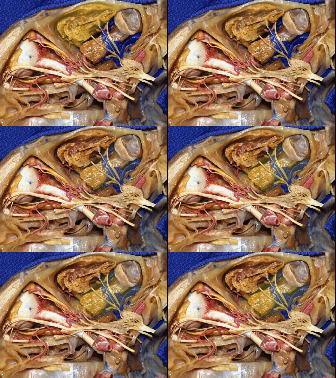
▼这是上颌神经(下图),从眶尖下方走行,进入翼腭窝,或经由眶下裂进入眶内。
or here we see V2, pass below the apex of the orbit, and extend into pterygopalatine fossa,or through the inferior orbital fissure into the orbit.
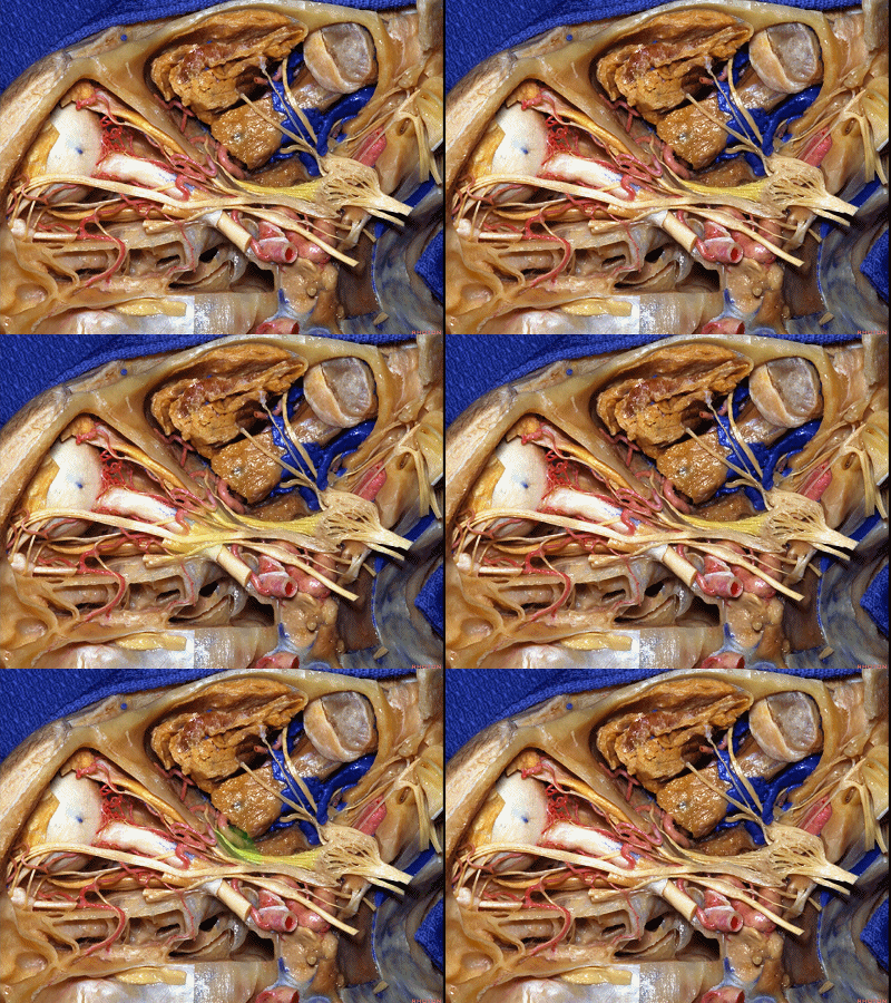
▼这是眶部的外侧观。眶外侧壁为病变侵犯眼眶的途径之一。病变也可经由眶上裂入眶,或通过视神经管,或经由内侧的鼻窦,也可经由眶下裂内侧部或者翼腭窝,向上通过眶下裂进入眶尖。
神经外科常需处理的情况包括眼眶及眶周病变。另外,越邻近眶尖的病变,需要神经外科处理的几率越高。
But we'll talk in greater detail about the anatomy within the orbit.But here we're looking at orbit from laterally and lesions can extend through the lateral orbital wall. They can access the orbit through the superior orbital fissure or the optic canal, or from the sinus medially,or they can pass upward here in the medial part of the inferior orbital fissure and pterygopalatine fossa up through the fissure into the orbital apex. But, commonly neurosurgery is involved when not only the orbit is involved but the adjacent structure. Also the closer the lesion is to the orbital apex,the more often the neurosurgery becomes involved in dealing with the pathology
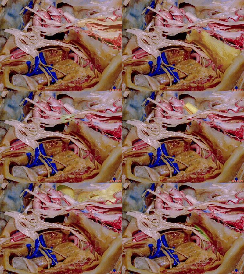
▼学习眼眶解剖,首先需了解眶壁骨性结构
In looking at orbit, we want to first of all understand the osseous wall of the orbit
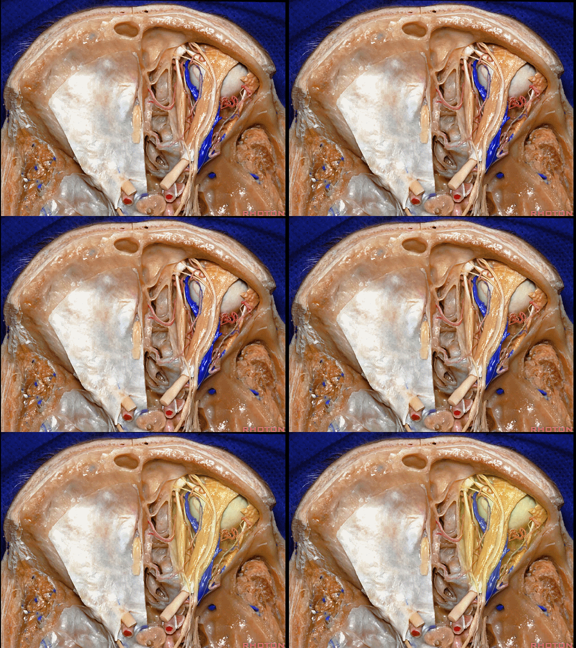
▼因为打开骨质,是我们从上方、从内侧的鼻腔及鼻窦、从眶外侧壁、或从下方的上颌窦顶壁,来进入眼眶的前提,其中上颌窦顶壁正是构成了眼眶底壁。
because we're doing osteotomy that will deliver us to the orbit from above,or from medially through the nasal cavity and the sinuses, or laterally through the lateral wall through a lateral orbitotomy, or from inferiorly through the roof of the maxillary sinus which forms the floor of the orbit.
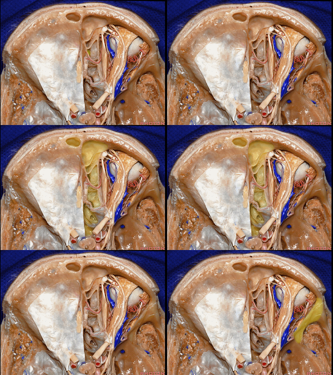
▼我们从骨性眶壁说起。蝶骨小翼构成了眶顶的后部。蝶骨大翼构成眶外侧壁的大部。
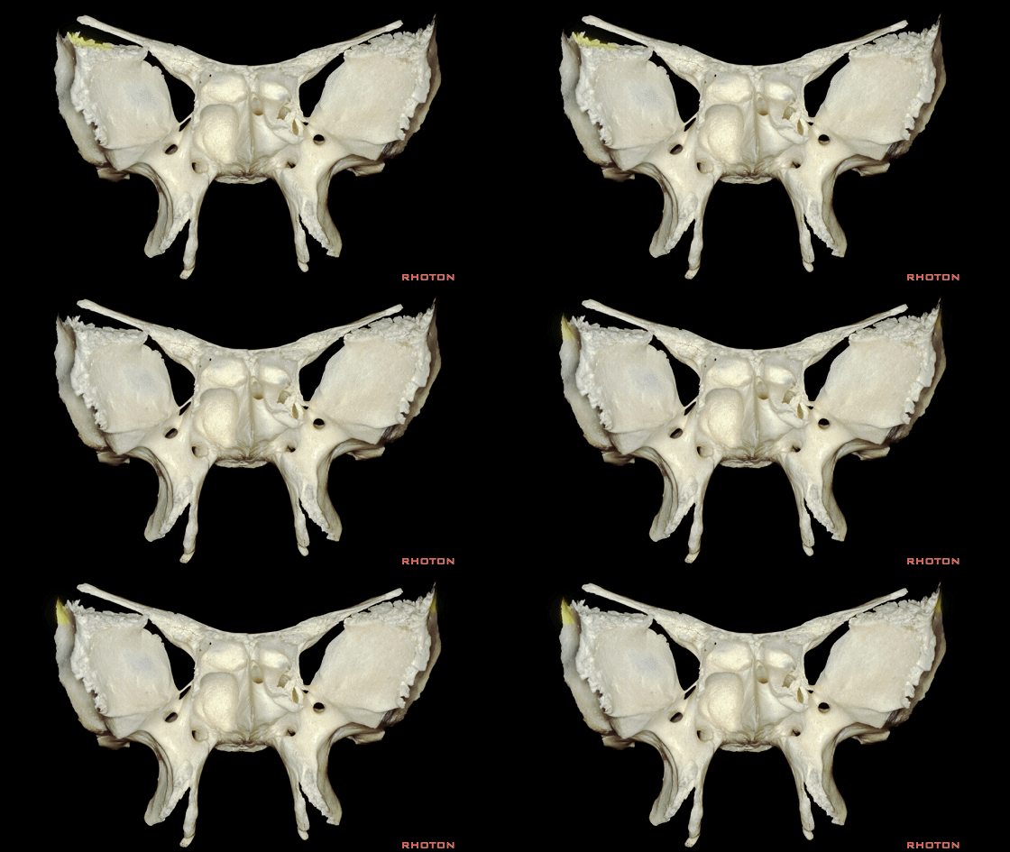
So we'll start out and talk initially about the walls of the orbit. And, the lesser sphenoid wing forms the posterior part of the roof of the orbit.The greater wing forms a large part of the lateral wall of the orbit.

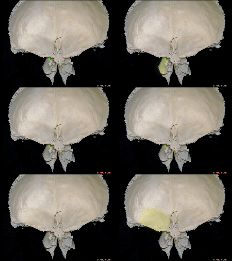
▼蝶窦于此处(下图黄色区域)与筛窦相连,构成了眶上裂的部分内侧壁。
And here, sphenoid sinus here that adjoins the ethmoid forms some of the medial wall of the superior orbital fissure. So that we wanna have a good understanding of the osseous anatomy because it's through the bony walls that we access the orbit.
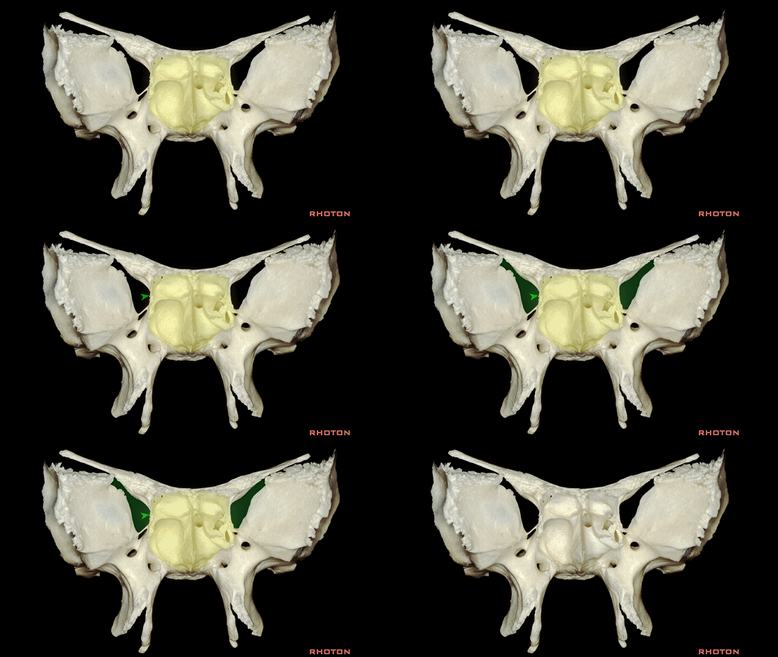
▼这是圆孔,上颌神经从中穿过而行于眶底。
Here we see foramen rotundum, through which V2 passes to course in the floor of the orbit.
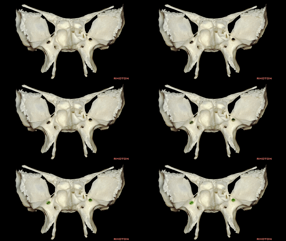
▼这是翼管,内有翼管神经走行,该神经包含副交感传出纤维,支配位于眶上外侧部的泪腺分泌。
Here is the vidian canal that transmits the vidian nerve that eventually provides the parasympathetic outflow for the lacrimal gland in the superior lateral orbit.
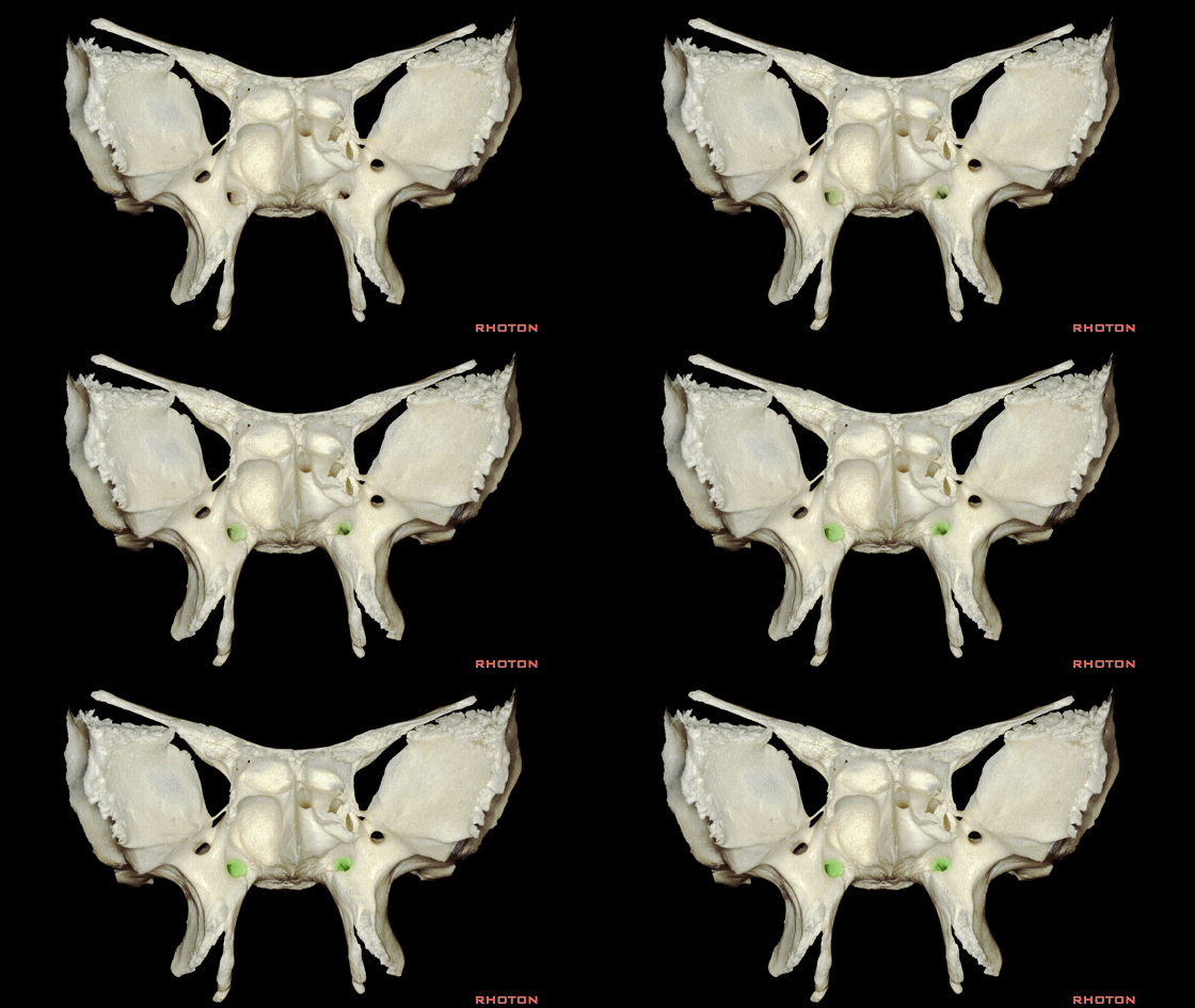
▼来看眶尖部,这是眶上裂、视神经管(箭头处)。
So when we look at the orbital apex, superior orbital fissure,optic canal area.

▼我们需要与周围的颅骨结合起来看,例如,蝶骨大翼的上部、与蝶骨小翼的前部分别参与组成眶外侧壁和眶顶,下图所示黄色区域与额骨结合一起构成完整的眶顶。
We wanna see the surrounding bony anatomy, such as the upper part of the greater and anterior part of the lesser wings that contribute to the lateral wall and roof of the orbit, and join the frontal bone here along this yellow area to complete the orbital roof.
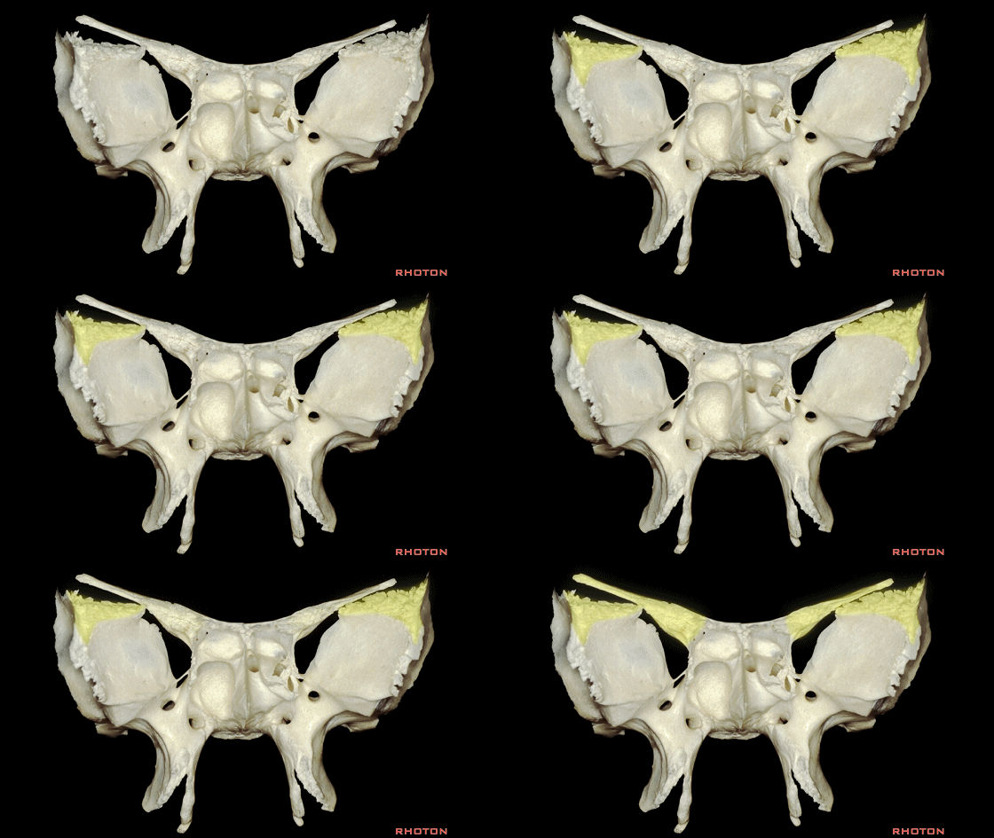
▼侧方的颧骨与此处的蝶骨(下图黄色区域)相结合,即构成了眶外侧壁的大部。
Laterally we want to see the zygomatic articulation with the sphenoid that completes the much of the lateral wall of the orbit.
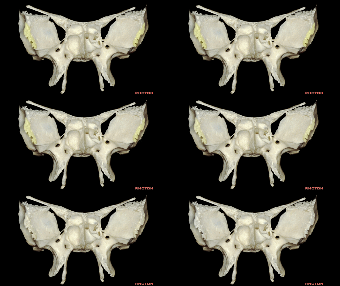
▼另外,此处(下图黄色区域)连于筛骨即构成眶内侧壁的大部,内含有气房,是到达眶内侧壁的一个手术通道。
And then, we have an articulation with the ethmoid bone that forms much of the medial wall of the orbit, as providing a group...as well as providing a group of air cells through which we can access medial wall of orbit.
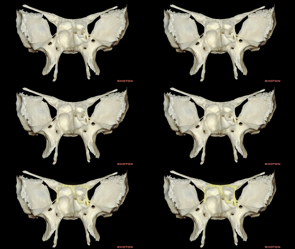
▼在下方与此处(下图黄色区域)相连的是腭骨,一起构成的是眶底。
And then, below we have an articulation with the palatine bone that contributes to the formation of the floor of the orbit.
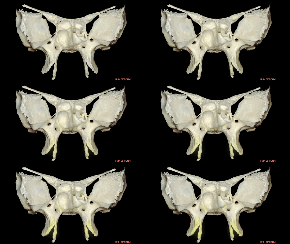
▼外侧的眶上外部为蝶骨嵴的外侧界,这一区域,为额、顶、颞、蝶四骨相交处,即为翼点,恰位于眶上外侧部。
以上就是我们需最先掌握的蝶骨解剖。
Laterally here at the superior lateral part of the orbit we have the lateral edge of the sphenoid ridge, and here in this area, the pterion where the frontal, parietal, temporal and sphenoid bone meet here right at the superolateral part of the orbit. So that we start with sphenoid bone.
▼接下来我们加入了筛骨。
So that we add in the ethmoid now.
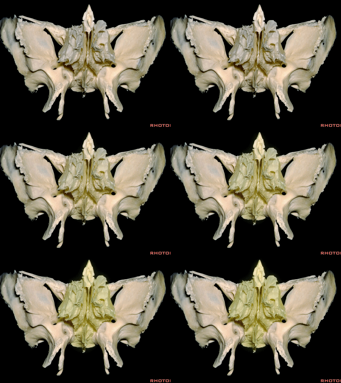
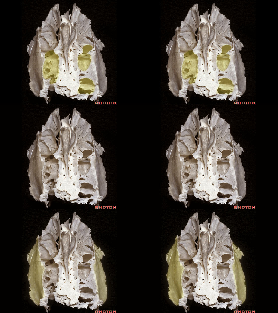
▼筛骨的外侧壁即为 眶板。
And the lateral wall of the ethmoid forms the orbit plate.

▼这是中鼻甲,其外侧为筛窦气房。
And here we see middle turbinate, and then laterally, these ethmoid air cells.
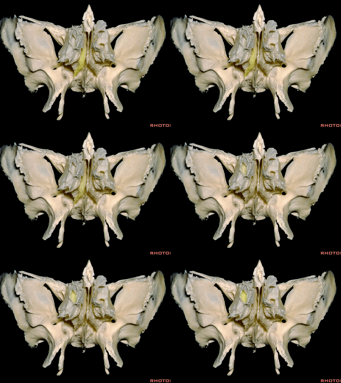
▼如果我们从中鼻甲下方,打开气房,打开筛骨纸板,即筛骨外侧壁、眶内侧壁,即可进入眼眶内侧部。
And if we get in under the middle turbinate here, and open the air cells, we have a route through the nasal cavity and through the lamina papyracea, the lateral ethmoid wall, the medial orbital wall,a route into the medial orbit.
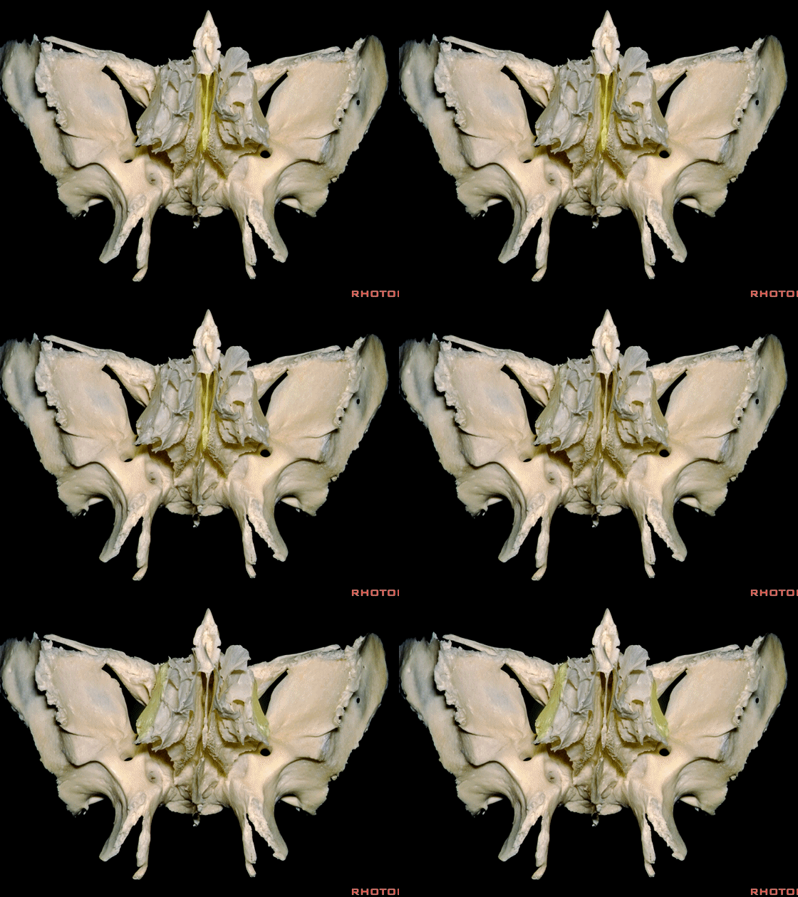
▼这是筛骨的上面观。筛骨与额骨连于此处,额骨构成了筛窦气房的顶壁。筛骨的外侧,即气房的外侧,即为筛骨的眶部,其构成了眶内侧壁,经此可进入眶内。
And here we see the ethmoid from above. But here it articulates, and the ethmoid air cells are roofed above by the frontal bone.And then laterally in the ethmoid, lateral to the air cells,we see the orbital plate of the ethmoid that forms the medial wall of the orbit through which we can approach it.
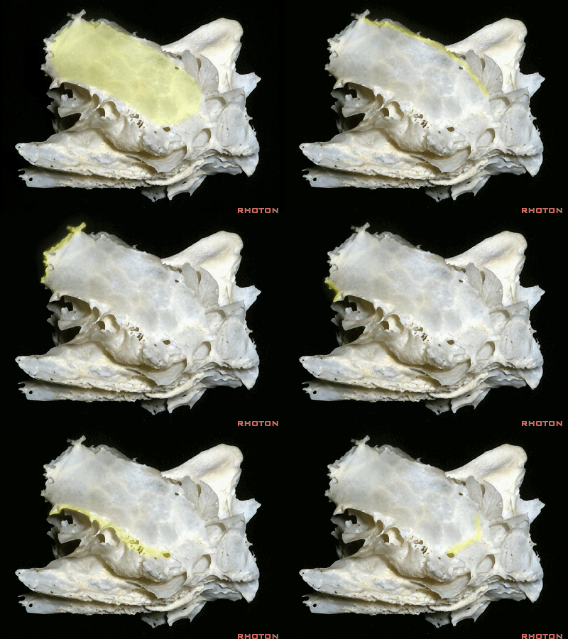
▼这是筛骨眶板的外侧观。上方与额骨相连。后方与蝶骨体外侧部相连,下方与一小部腭骨相连,这里与下方的上颌骨相连,前方与之相连的是泪骨 以及上颌骨的额突。
Just a lateral view of the orbital plate of the ethmoid. Above it articulates with the frontal bone.Posteriorly it articulates with the lateral part of the sphenoid body.Below it'll articulate with a little bit of the palatine bone.And then below with a maxillary,and anteriorly it'll articulate then with a lacrimal bone and the frontal process of the maxilla. But you wanna see all of these adjacent relationships.
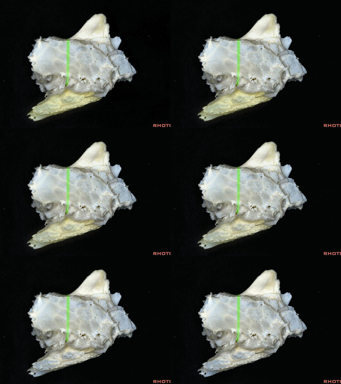
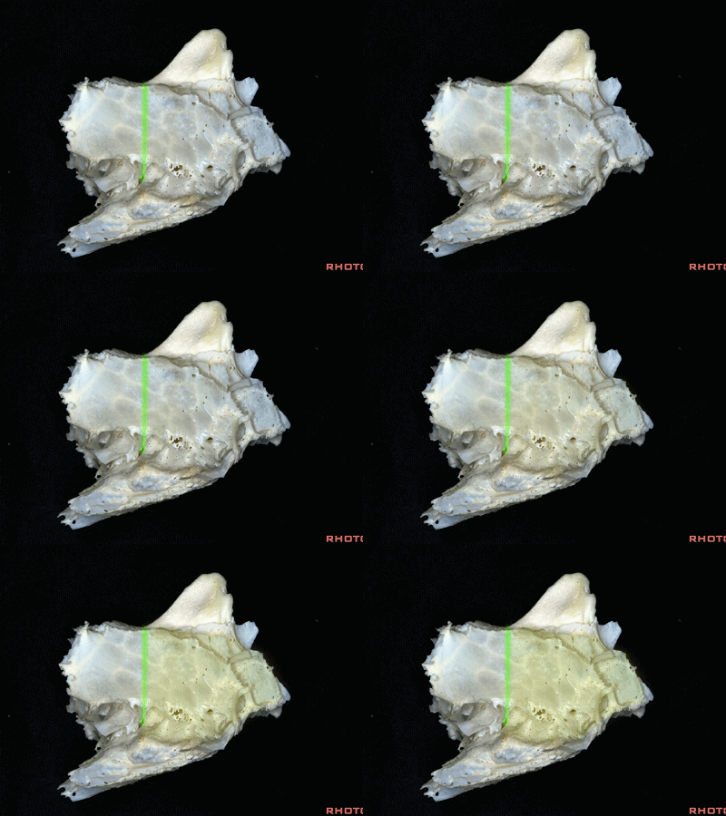
▼筛窦气房可分为前组和后组。分界线位于中鼻甲附着于筛骨的后部。
And the ethmoid air cells can be divided into a anterior group and a posterior group.And that division is right at the posterior part of the attachment of the middle turbinate to the ethmoid.
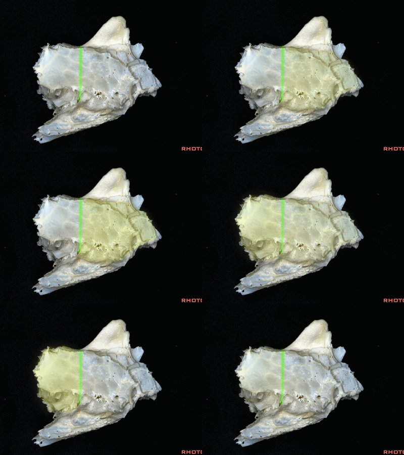
▼下图示中鼻甲
▼通过后组筛房,可至眶尖附近,或可进入蝶窦外侧部。
And if you go through posterior ethmoid, you end up near orbital apex and in the lateral part of sphenoid sinus.
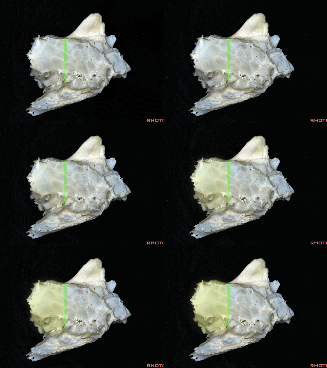
▼通过前组筛房向上,则可顺着筛板进入前颅窝。
If you work through the anterior ethmoid air cells above, you end up along the cribriform plate in the anterior fossa.
▼再换个角度来看筛骨。它实质上是两块骨在中线由筛板连接而成。
So here we see the ethmoid bone again. It's really two blocks of bone here connected across the midline by the cribriform plate.
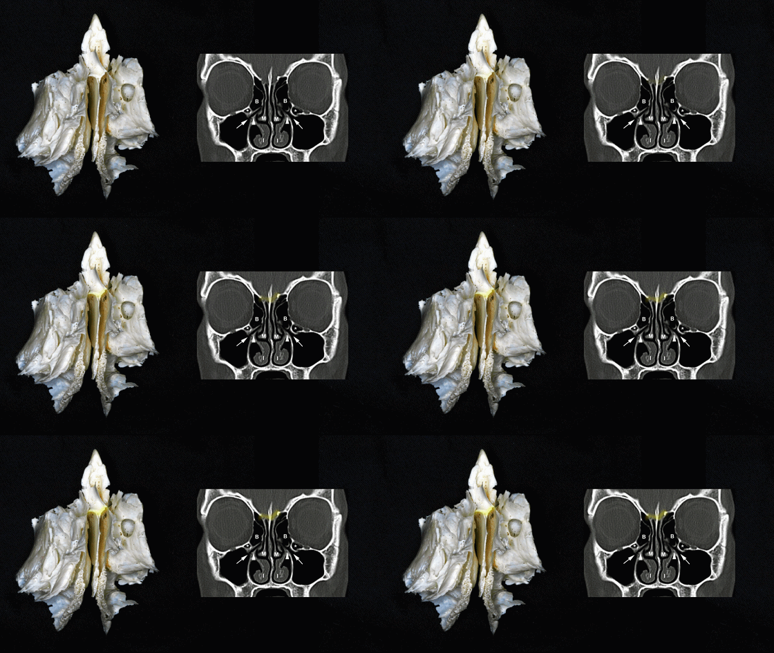
▼筛骨的这部分即为中鼻甲。
And as the part of it, we have the middle turbinate.
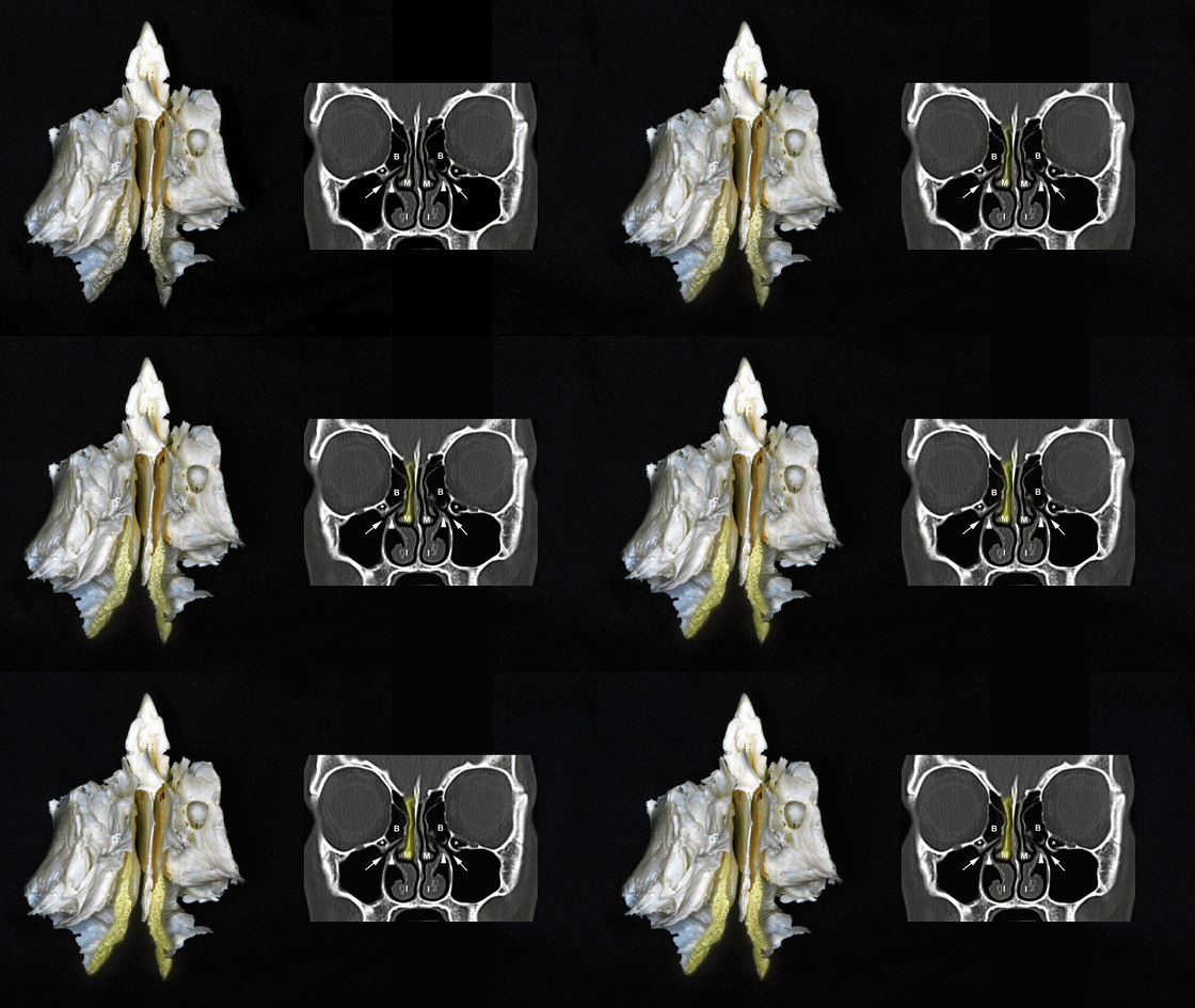
▼从中鼻甲的外侧即可进入筛房。进一步向外侧打开,即可经由眶内侧壁,实现眶内侧入路的其中一种。
And if you get in lateral to the middle turbinate, you're into the air cells. And then you can open laterally, through the medial wall of the orbit to complete one of the types of medial orbitotomy.
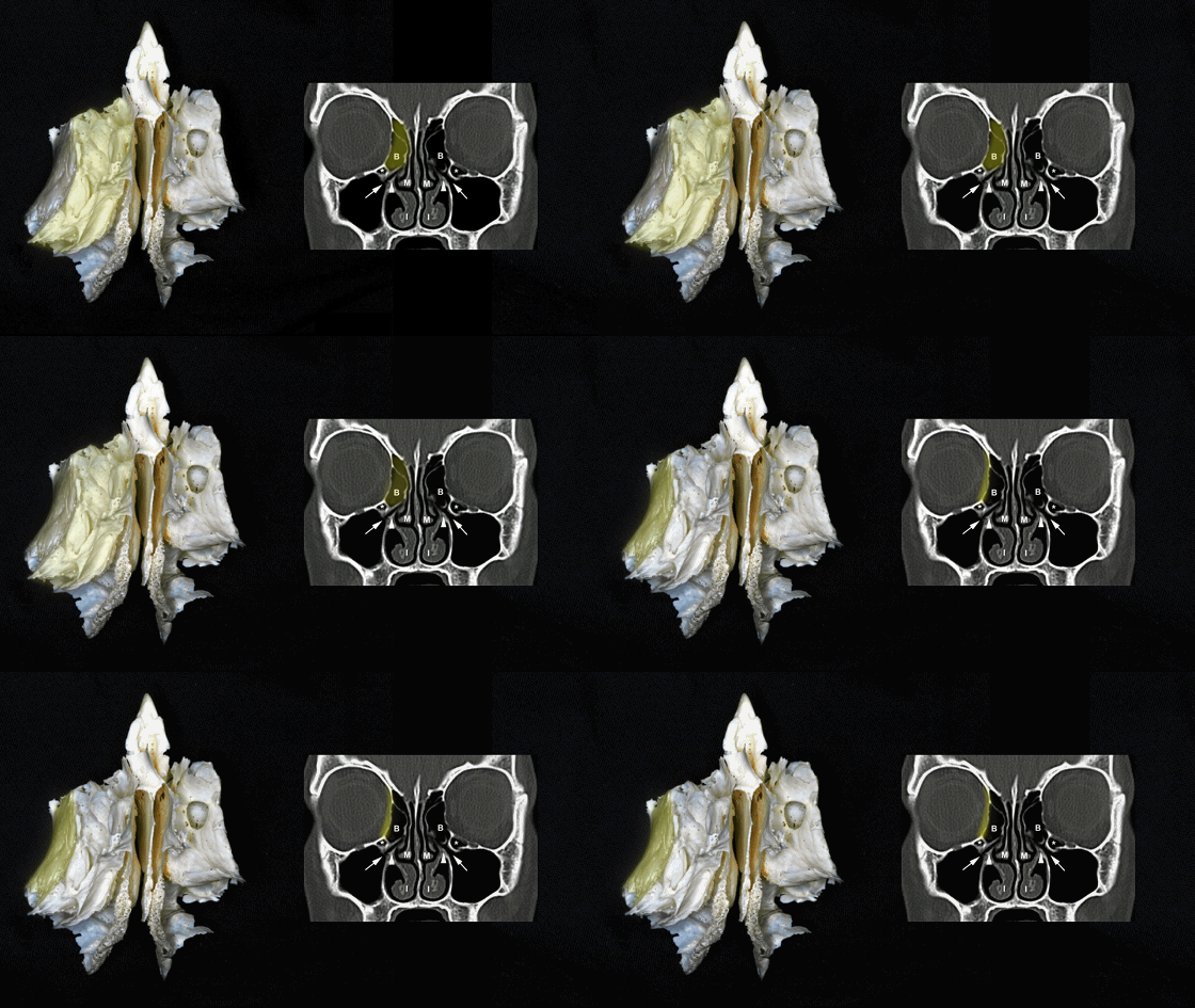
▼筛窦气房的顶壁由额骨这一裂隙处的骨质封闭
And roofing the ethmoid air cells above is the hiatus in the frontal bone
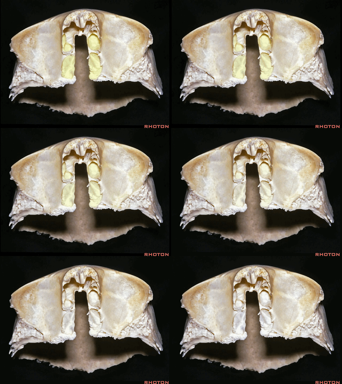
▼额骨构成了眶顶。
frontal bone that forms the orbital roof.
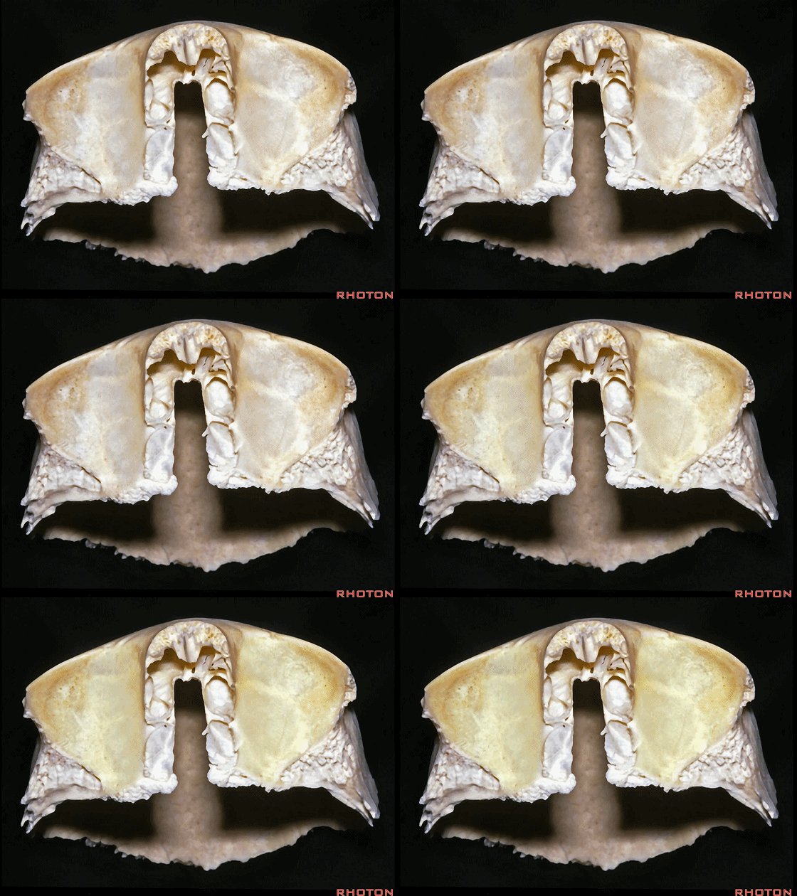
▼现在我们将筛骨嵌入额骨的筛切迹。
And here we fit the ethmoid into the ethmoid notch of the frontal bone.
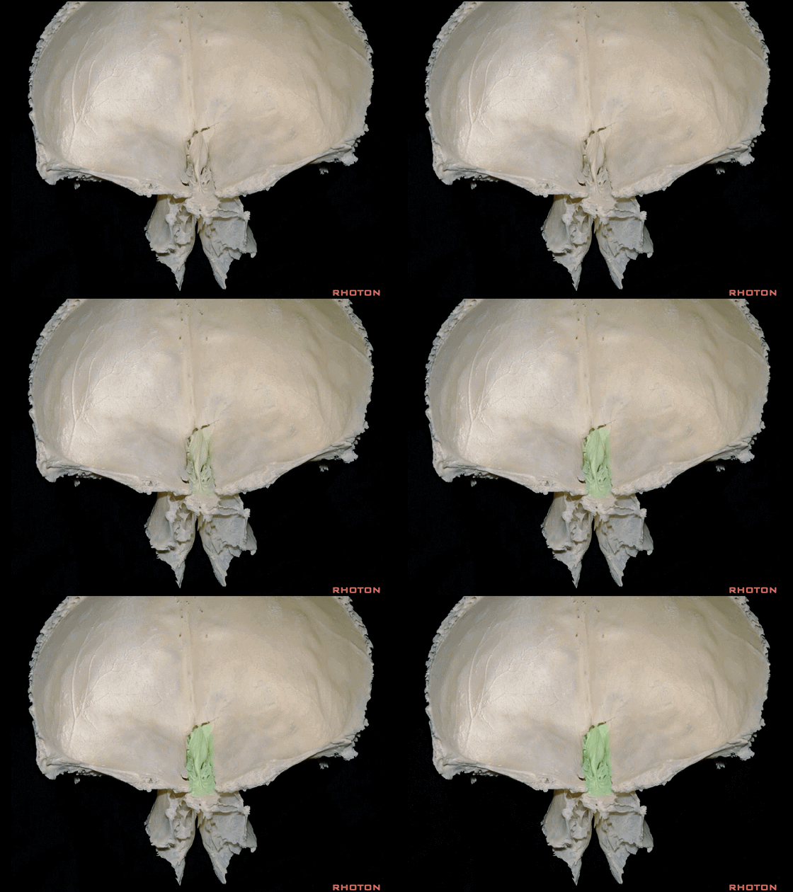
▼可见眶板的上界(箭头处)连于额骨,在其外侧即为眶顶。
And here the upper edge of the orbital plate articulates with the frontal bone, and then lateral to that is the orbital roof.
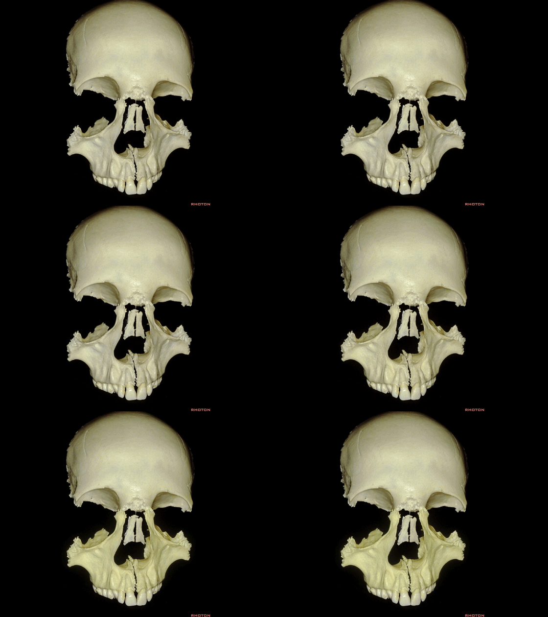
▼现在加入完整的上颌骨(下图)以重构完整的眶内侧壁、眶底及眶缘的下部。
And then we add in the full maxilla that complete the medial wall and floor or lower part of the orbital rim of the orbit.
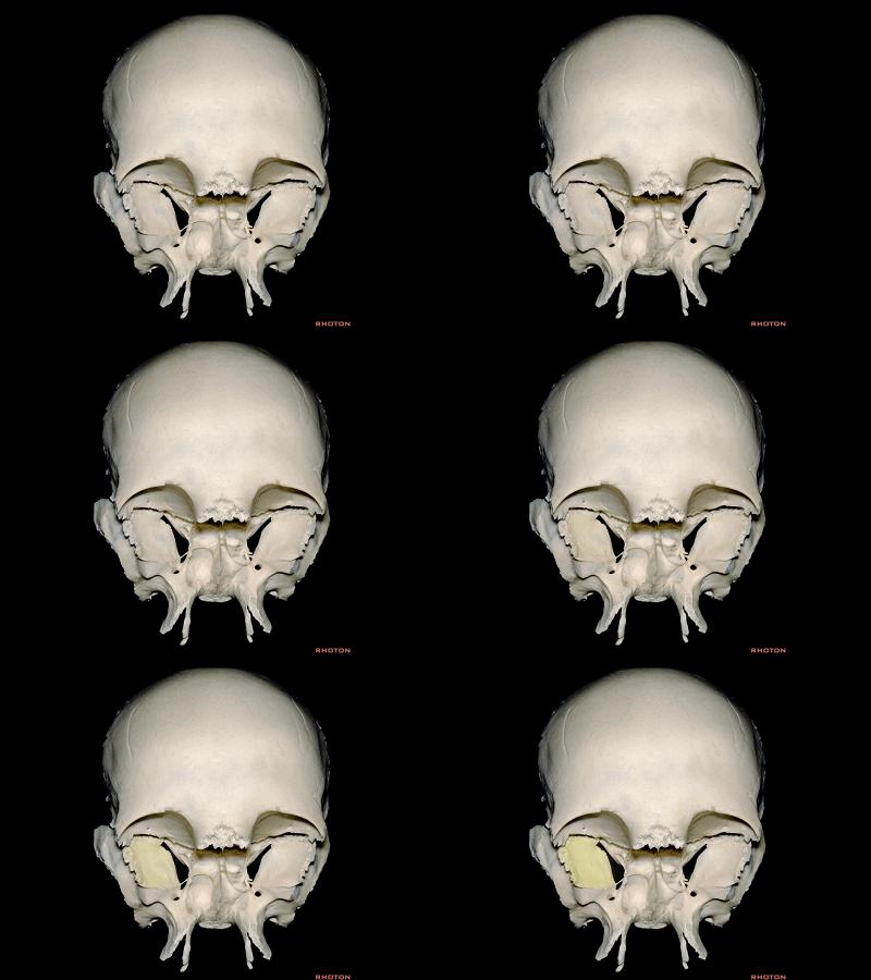
▼我们再加入蝶骨,蝶骨大翼构成眶外侧壁的后部
And then we can complete the lateral wall of the orbit,
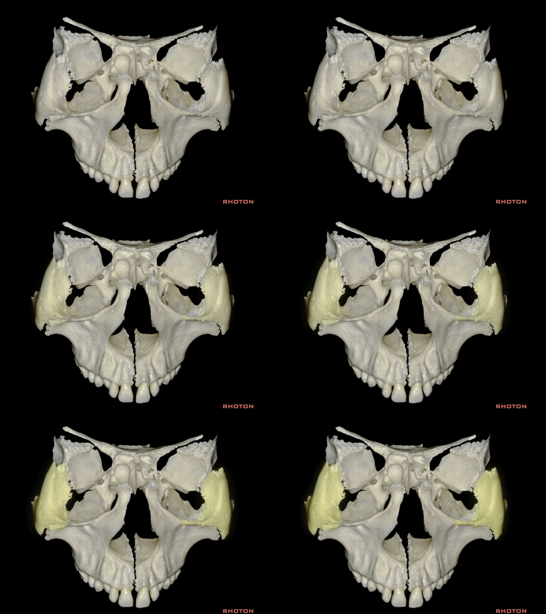
▼再加入构成眶外侧壁前部的颧骨
by adding a zygomatic bone which forms the anterior part of the lateral wall of the orbit.
▼下图示 上颌窦顶,其构成眶底。
And then below we have the roof of the maxillary sinus that forms the floor of the orbit.
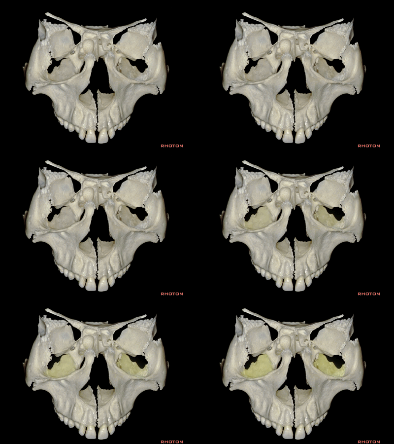
▼在上颌骨、蝶骨大翼和颧骨(下图)之间,即为眶下裂,其外侧部常在进行眶外侧入路或眶颧入路前打开。
Also, here between maxilla, greater wing of sphenoid and zygomatic bone we have inferior orbital fissure,which we often open into the lateral part of it before doing a lateral orbitotomy or an orbitozygomatic approach.
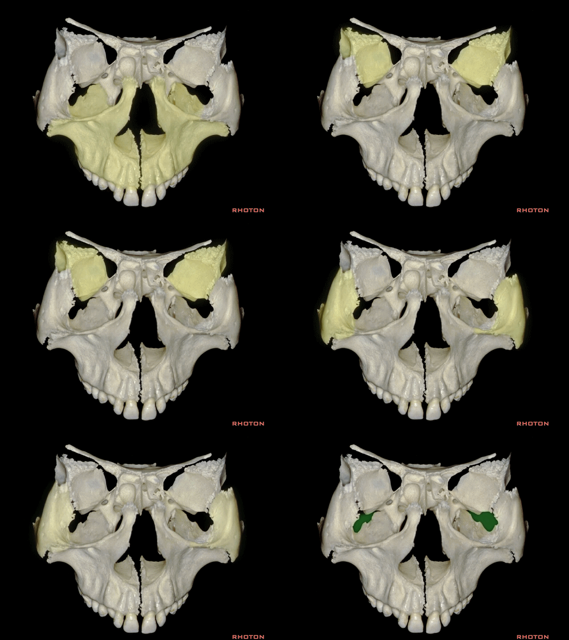
▼下图示腭骨的眶突,参与构成眶底的一小部分。毗邻于筛板及上颌骨构成的眶底后部。
we have the orbital process of the palatine bone that forms a small part of the floor of the orbit. And then forming part of the floor of the orbit here adjacent the ethmoidal plate and the posterior part of the floor formed by the maxilla,
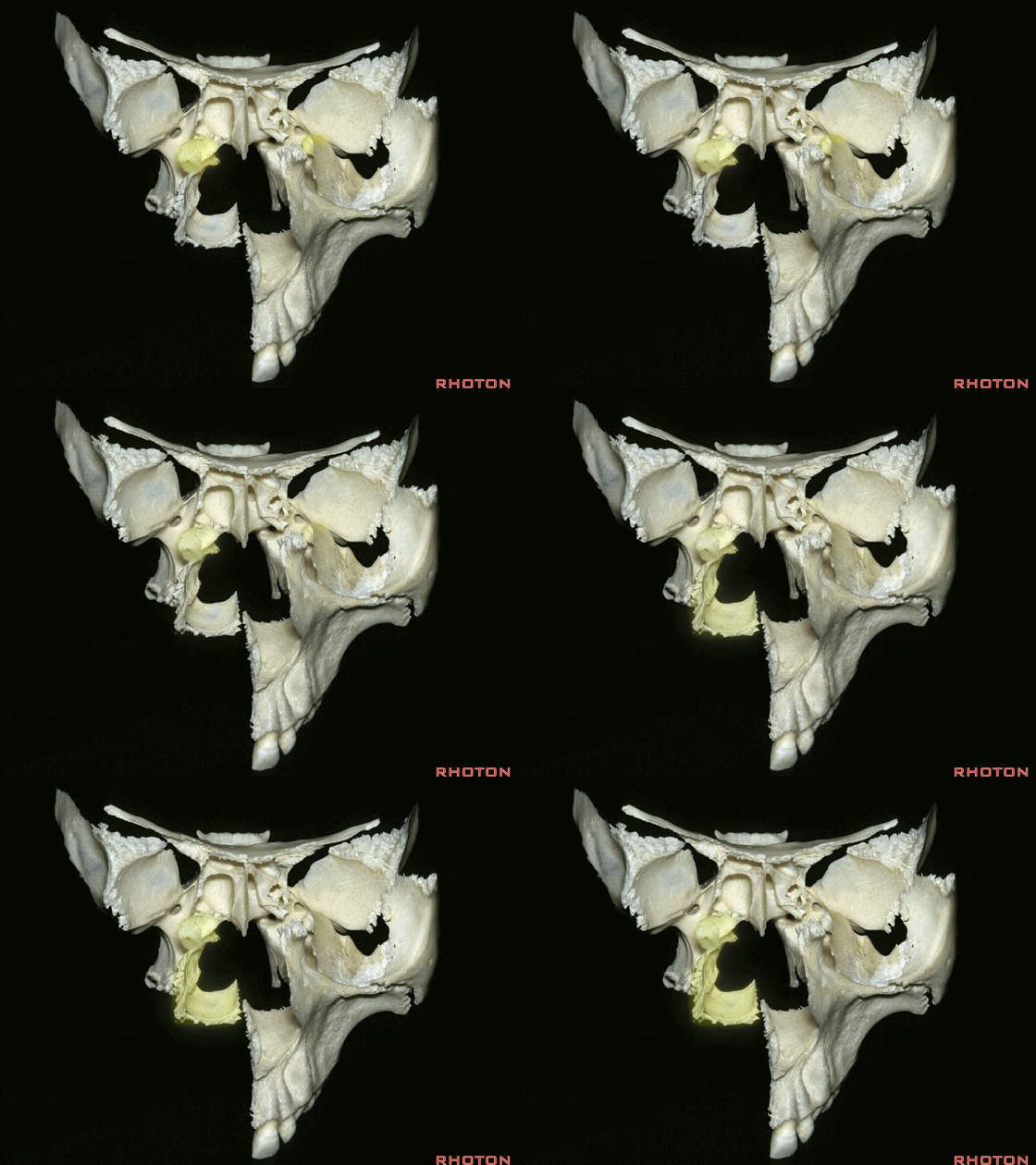
▼这部分腭骨,称为眶突
But, here's the part of the the palatine bone, the...
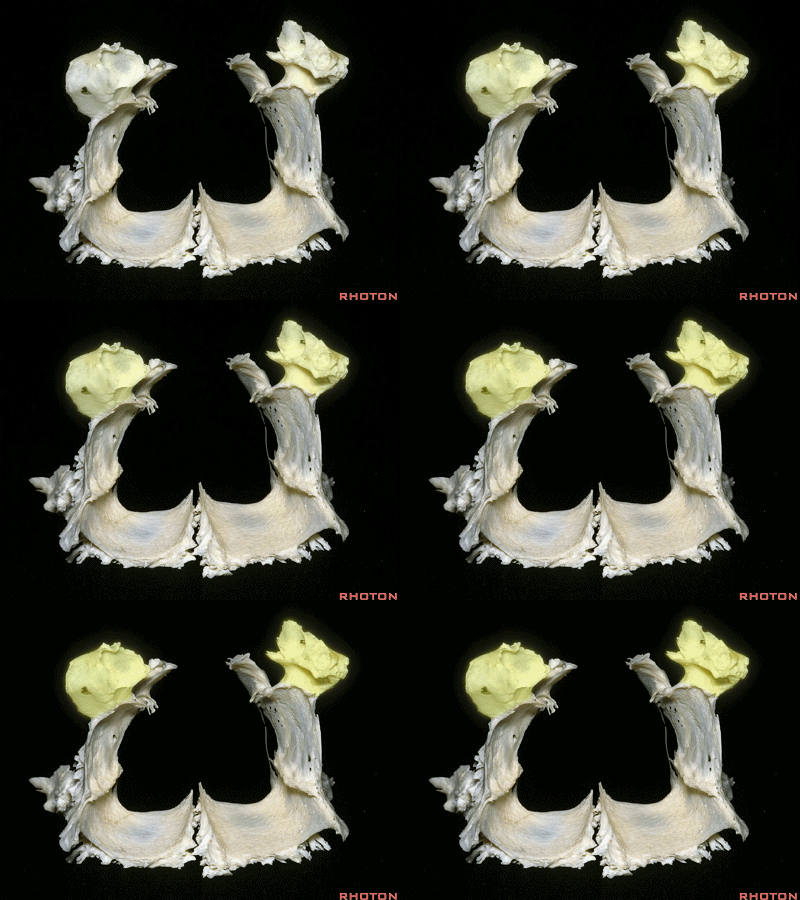
▼眶突,构成眶底的后内侧一小部分。
orbital process that forms the posteromedial part of the orbital floor
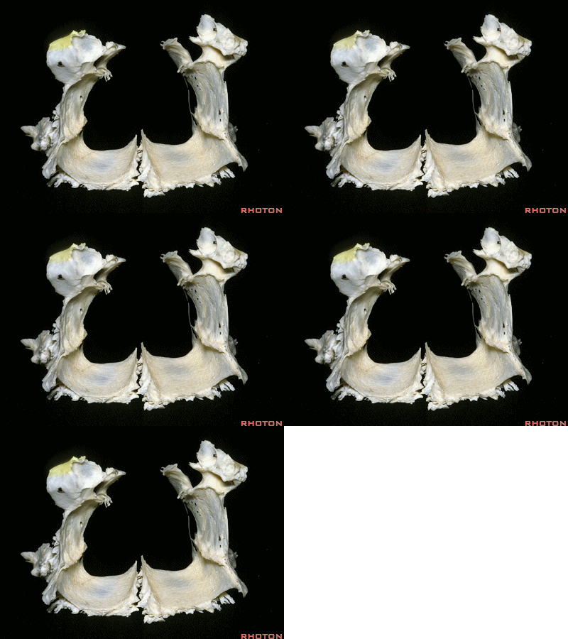
▼其后方为蝶腭孔,内有上颌动脉的分支穿行入鼻腔,供血于鼻中隔瓣,其常用于颅底的修补。
and behind that is the sphenopalatine foramen,through which the branches of the maxillary artery enter the nasal cavity that provide the vascularity for the nasoseptal flap that we use to close skull base.
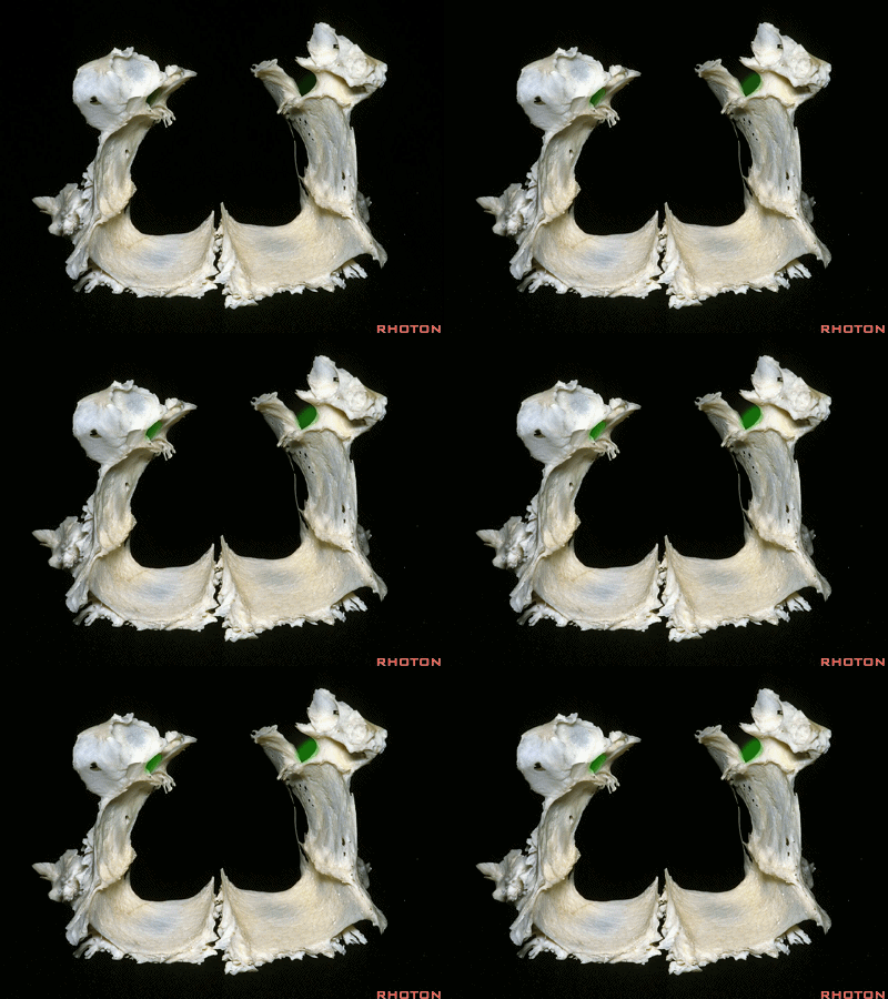
▼下图示上颌骨(正面观)。筛骨眶板下方的眶内侧壁由上颌骨构成。
But that medial wall below the orbital plate is formed by maxilla.
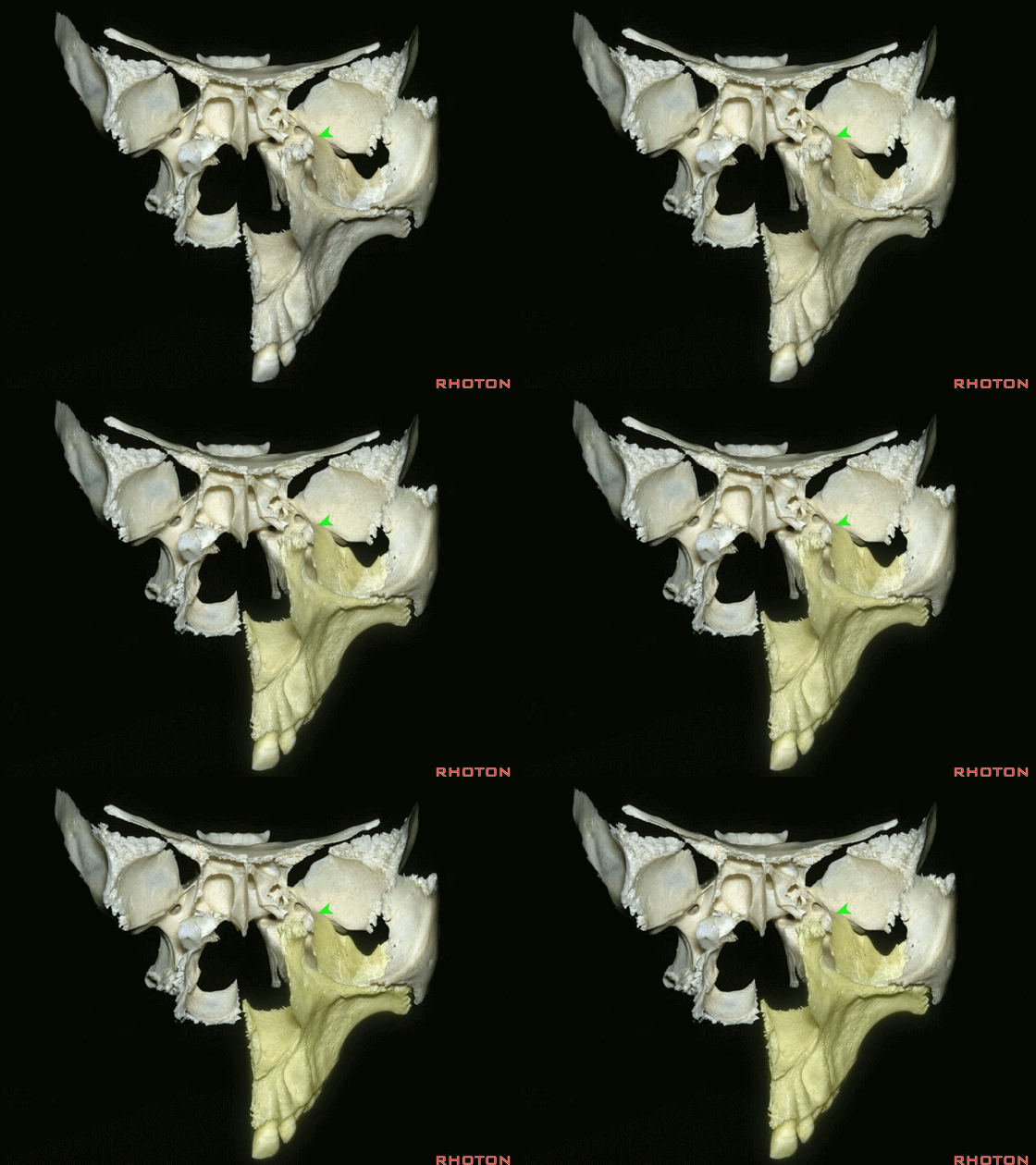
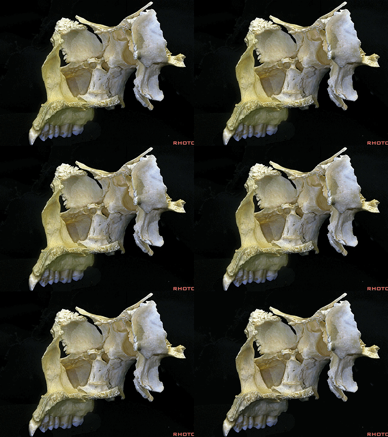
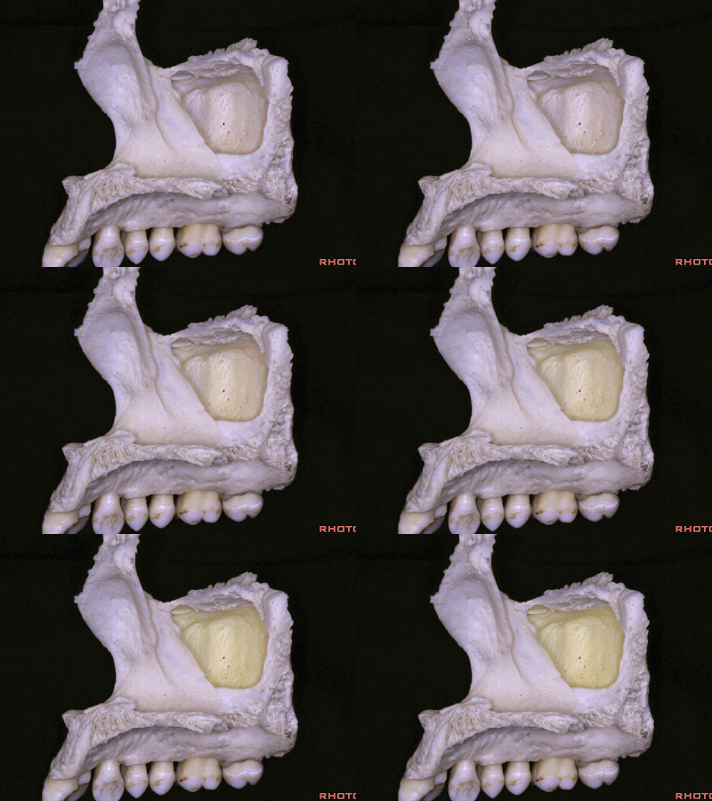
▼这是上颌骨(内侧观)
But, here's just maxilla
▼这是上颌窦内侧的自然开口
Here's the natural hiatus in the medial wall of the maxillary sinus
▼这是腭骨
palatine bone
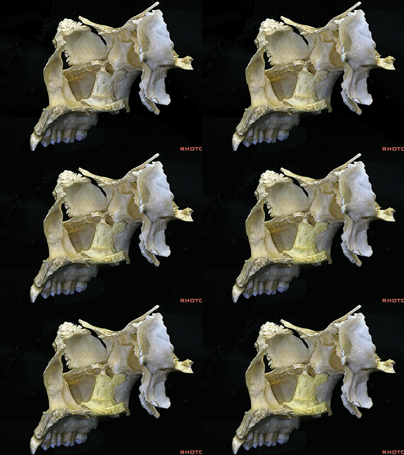
▼上颌窦内侧的自然开口被腭骨、下鼻甲遮蔽。
the hiatus that is closed in by palatine bone, by inferior turbinate.
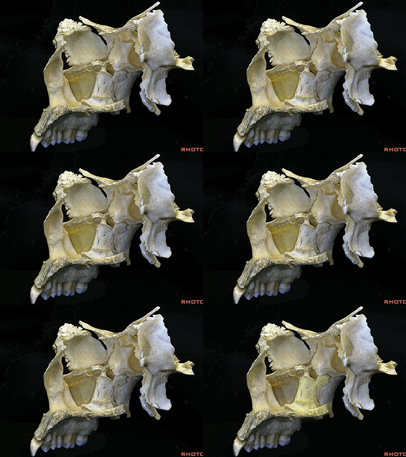
▼上颌窦内侧的自然开口被下鼻甲的这一部分(下图1)以及这部分腭骨的垂直板(下图2)所遮蔽。
by this part of the inferior turbinate and also by some of the perpendicular plate of the palatine bone to close the maxillary hiatus.
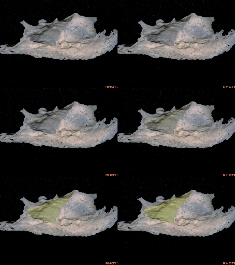
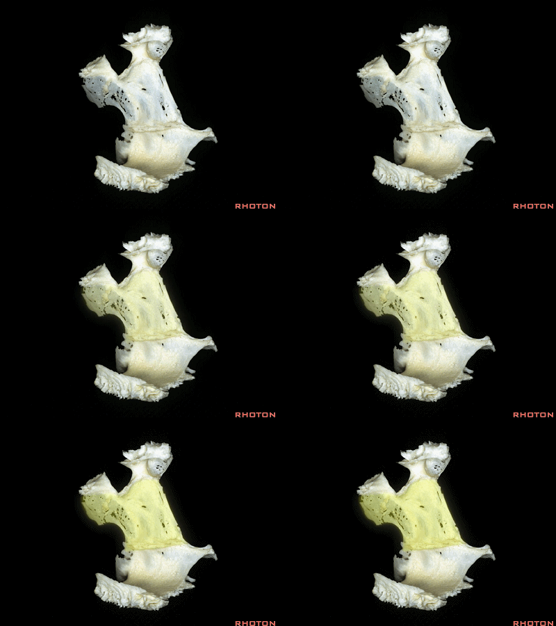
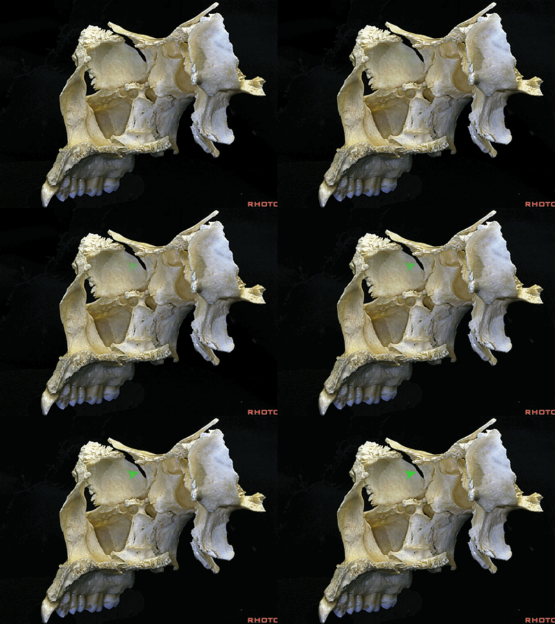
▼沿此处附着的是筛骨的眶板。
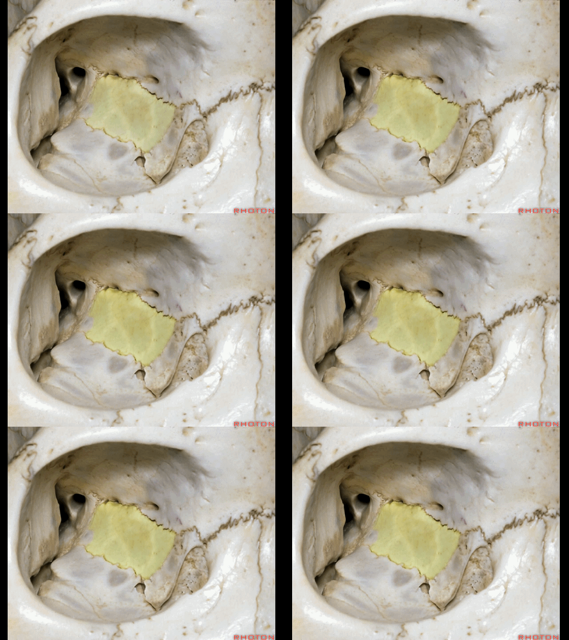
And then attached along all of this area, is the orbital plate of the ethmoid.
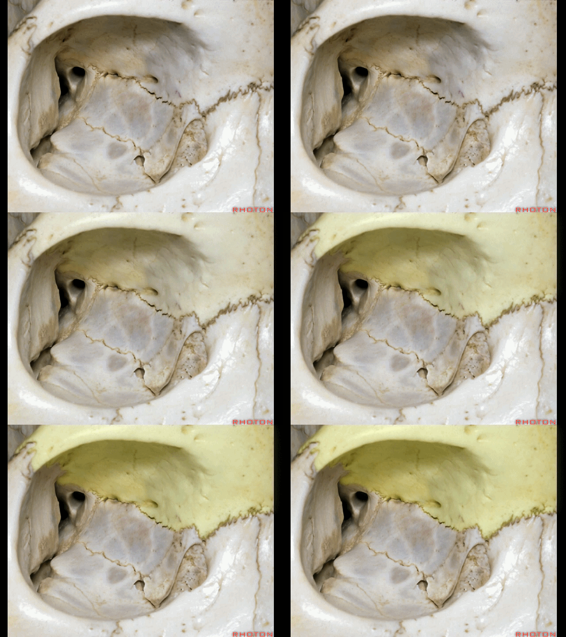
▼因此,眶由7块颅骨构成。包括 眶顶的额骨(下图),眶外侧壁的蝶骨大翼 和颧骨,眶底则由上颌窦顶构成,眶内侧壁由上颌骨额突、泪骨、筛骨眶板、腭骨眶突构成
So the orbit is made up of seven bones. We have frontal bone in the roof,the lateral wall formed by greater wing of sphenoid and the zygomatic bone,the floor of the orbit formed by the roof of the maxillary sinus,
▼下图示眶外侧壁的蝶骨大翼和颧骨
the lateral wall formed by greater wing of sphenoid and the zygomatic bone,
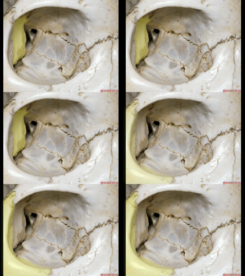
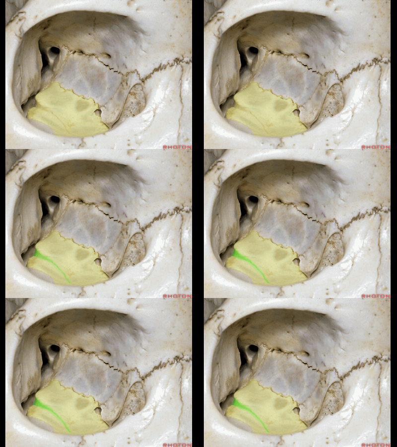
▼眶底则由上颌窦顶构成,沿此部位有眶下沟或眶下管,开口于眶下孔,内有上颌神经走行。
the floor of the orbit formed by the roof of the maxillary sinus, along which we have the infraorbital groove, canal here ending at the infraorbital foramen along which V2 passes.
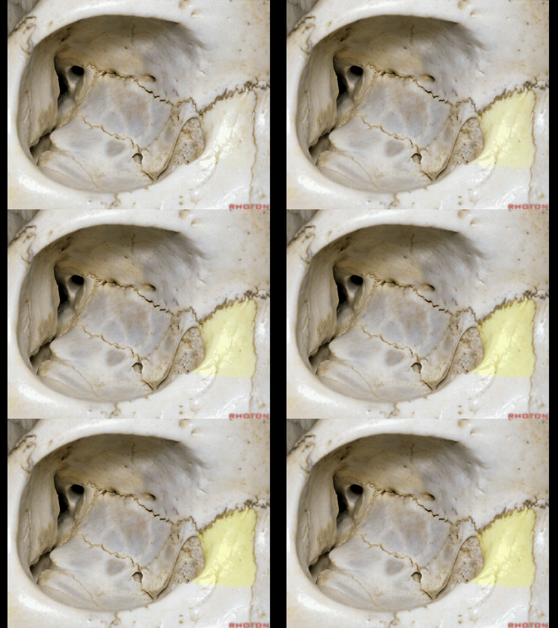
▼内侧,眶缘内侧部由上颌骨额突、以及泪骨构成。下图示上颌骨额突。
Medially, we have here medial part of orbital rim formed by frontal process of maxilla, and then the lacrimal bone
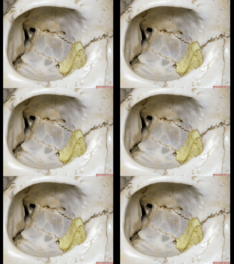
▼泪骨(下图)连于筛骨眶板的前界,并向上连于额骨,形成额筛缝。
lacrimal bone that articulates with the anterior edge of the orbital plate of the ethmoid, which articulates above with the frontal bone along the frontoethmoidal suture.
▼下图示筛骨眶板
▼下图示额骨,额筛缝、筛前、筛后管。
筛前管、筛后管内走行有眼动脉的筛前和筛后支,以及筛前和筛后神经。
We see the anterior and posterior ethmoidal canals that transmit the anterior and posterior ethmoidal branches of the ophthalmic artery, and the anterior and posterior branches of the ethmoidal nerves.
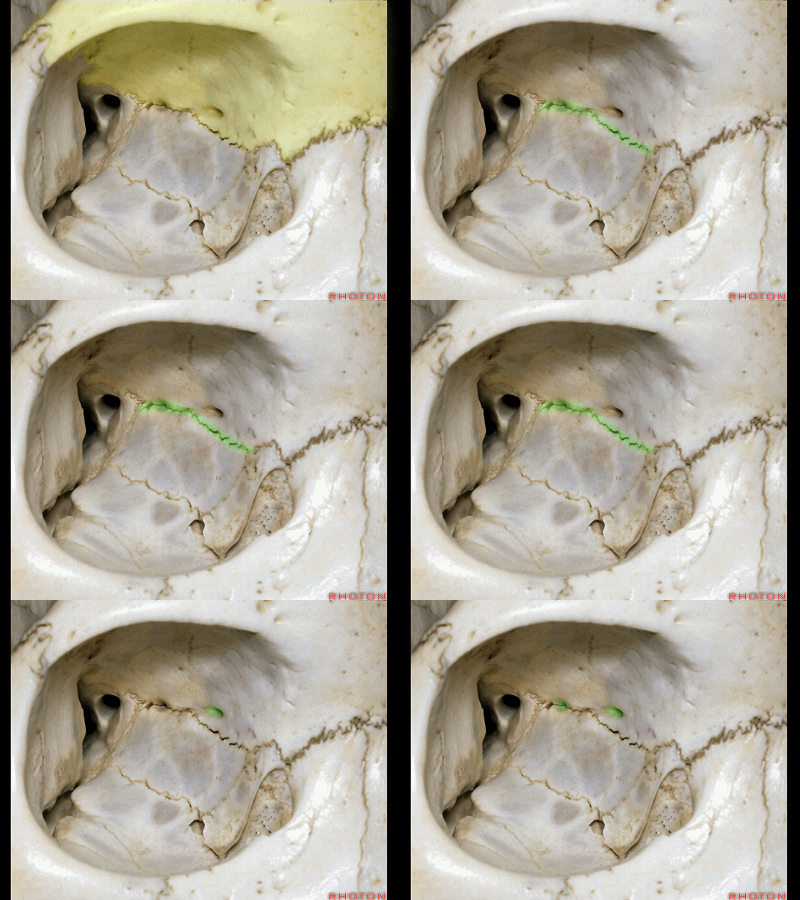
▼后方为视神经管(下图),其走行于蝶骨小翼内侧部。视神经管的外侧部由视柱构成,内侧部为蝶骨体。
Posteriorly, we have the optic canal that passes through the medial part of the lesser wing and the lateral part of the optic canal is formed by the optic strut which extends from the body of the sphenoid to the base of the anterior clinoid.
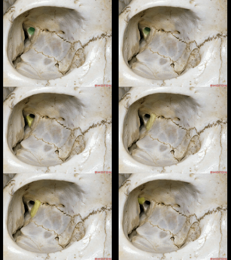
▼筛骨下方连于上颌骨。
Below the ethmoid we have the articulation here with the maxilla.
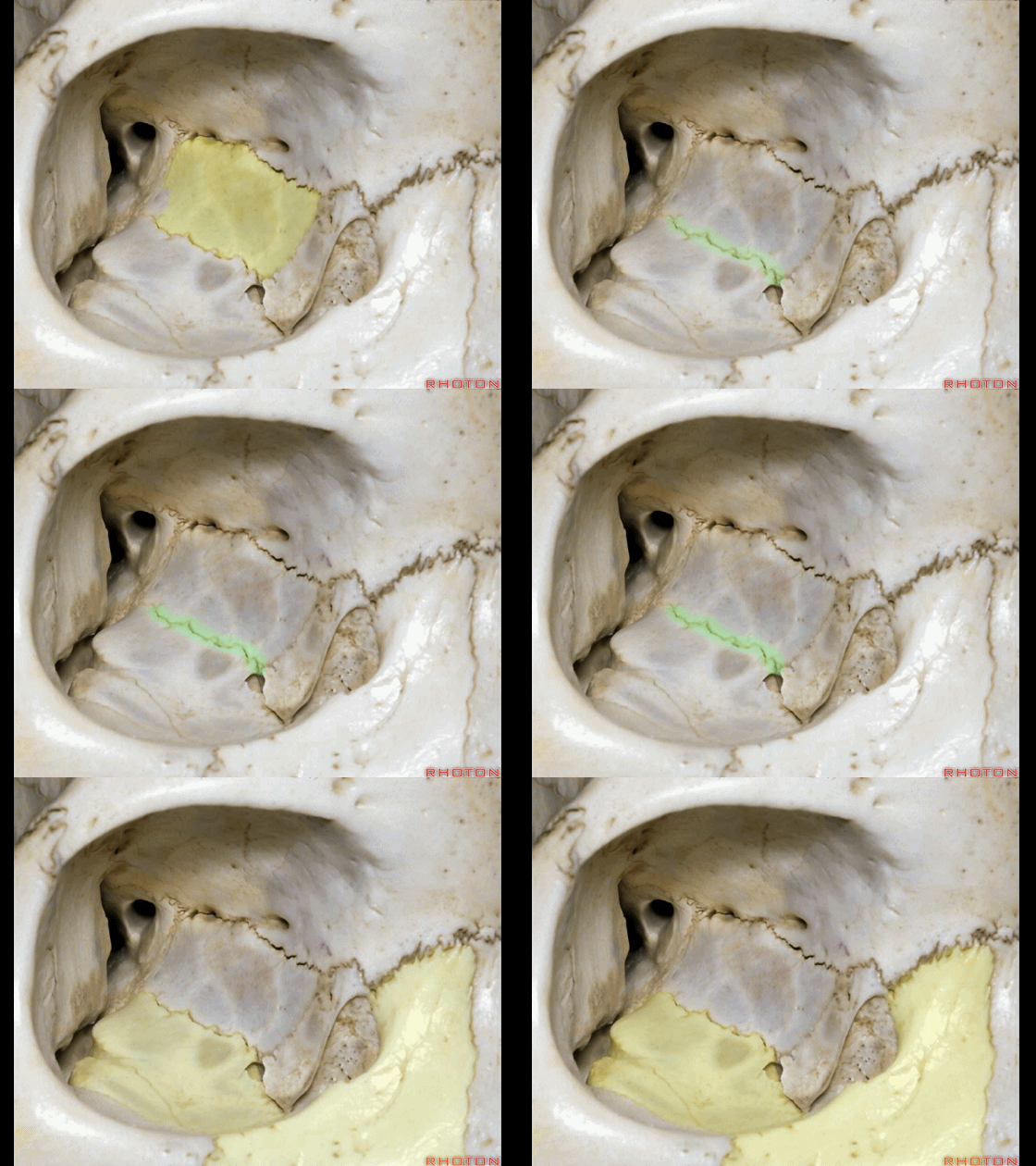
▼筛骨的后下界连于腭骨的眶突(下图),后者参与构成眶底。
And then at the posterior inferior edge,we have the part of the orbital floor formed by the orbital process of the palatine bone.
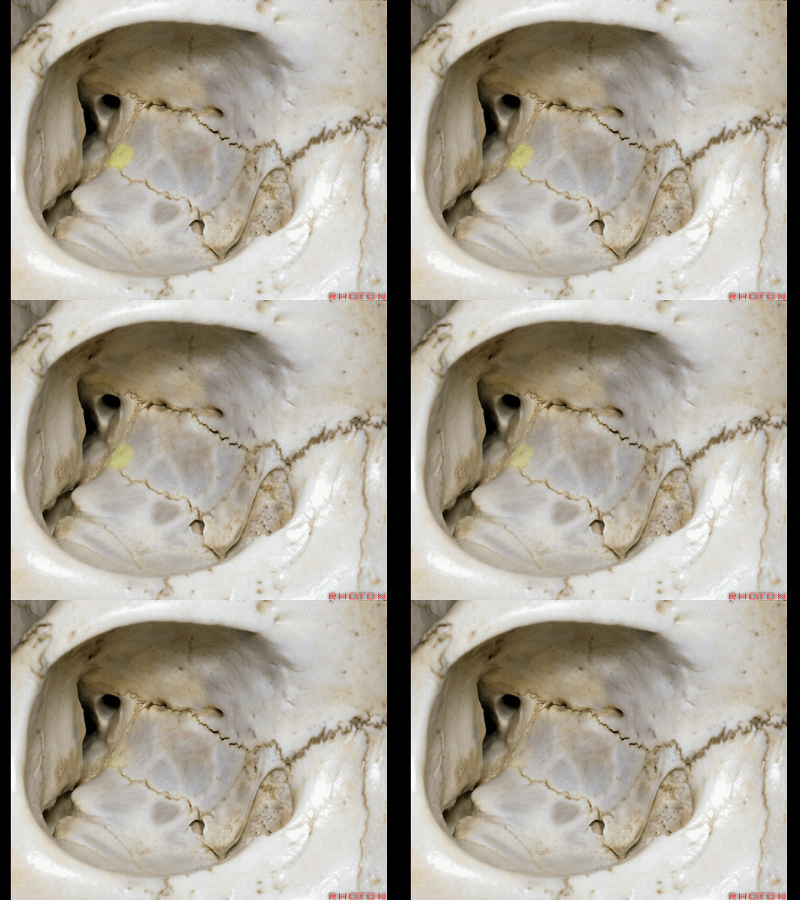
▼筛骨的后方连于蝶骨体(下图),此处为蝶窦的前界。
And then, along the posterior ethmoid, we have the articulation with the sphenoid body at the anterior edge of the sphenoid sinus in this area.

▼在充分掌握了这部分骨性解剖后,我们需进一步了解神经解剖。在眶尖,可见视神经管、视柱、前床突、眶上裂。
So that you wanna see and understand all of these articulations and begin to fit them together with the neuroanatomy. So that, at the orbital apex, we have optic canal, optic strut, anterior clinoid,and superior orbital fissure.
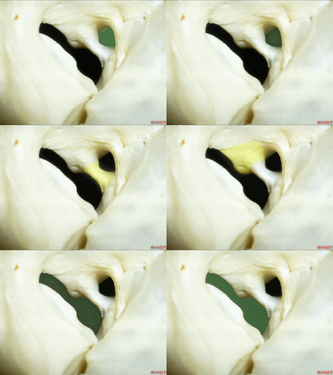
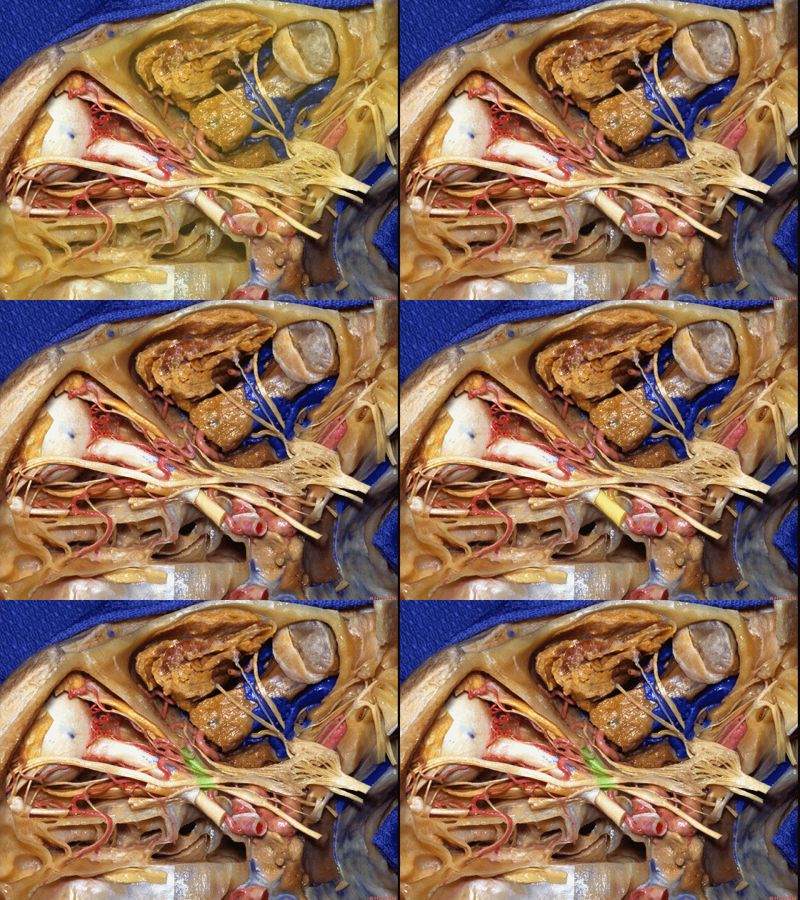
▼环绕于这一区域的是总腱环。总腱环是一环形腱环,为四块眼直肌的共同起点。其环绕于视神经管及眶上裂的中央部。
And surrounding this area we have the annular tendon. The annular tendon is the circular tendon from which the four rectus muscles arise. And the annular tendon surrounds the optic canal and the central part of the superior orbital fissure.
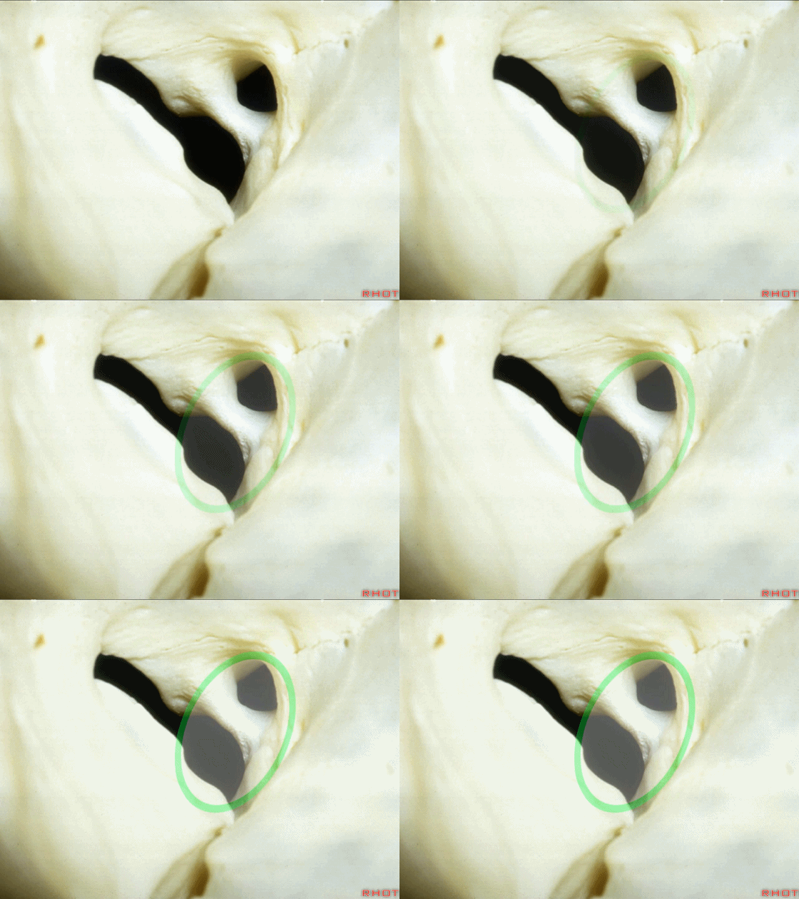
▼总腱环分为两部分,一部分内有视神经管,内有眼动脉穿行。另一个外侧部由这部分眶上裂围成,即为动眼神经孔,内有进出总腱环的神经穿行。
下图示视神经管及眶上裂的中央部。
And the tendon has two parts, a part through which the optic canal and the ophthalmic artery pass,and then the lateral part bordering the fissure is the oculomotor foramen, through which the nerves entering inside and outside the annular tendon pass.
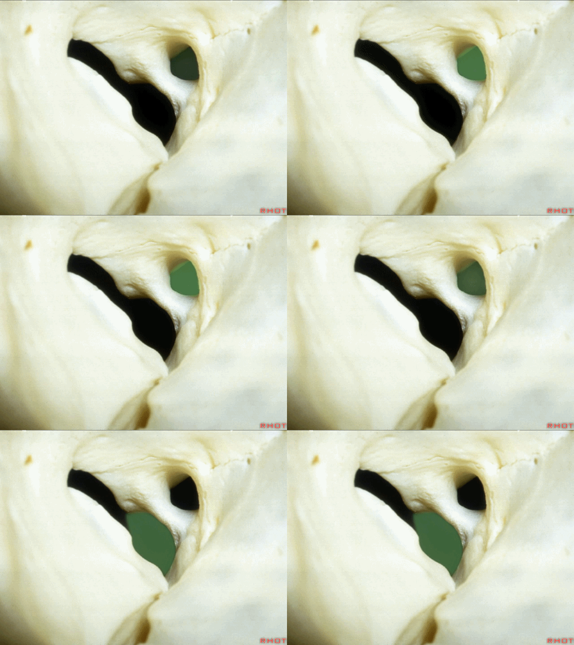
▼通常可见一小的突起位于眶上裂外侧壁,即为总腱环的附着点。
Usually there's a small prominence on the lateral wall of the fissure to which the annular tendon attaches.
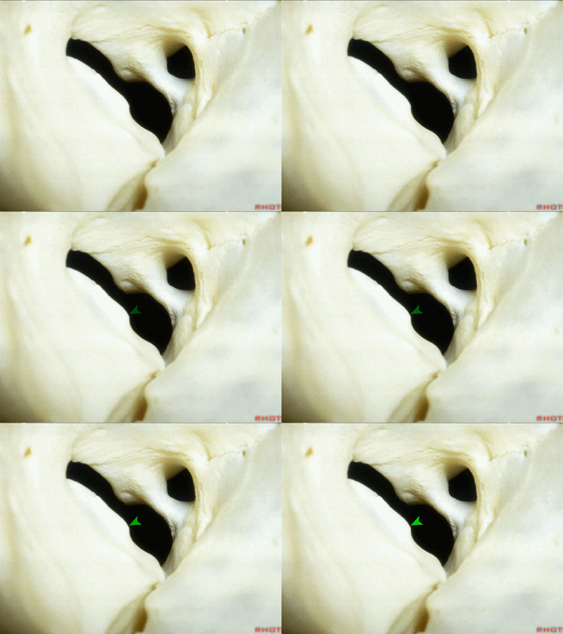
▼来看看四块眼直肌,均由总腱环发出。这是上直肌(下图)
Here we have the four rectus muscles,which arise from the annular tendon.superior rectus,
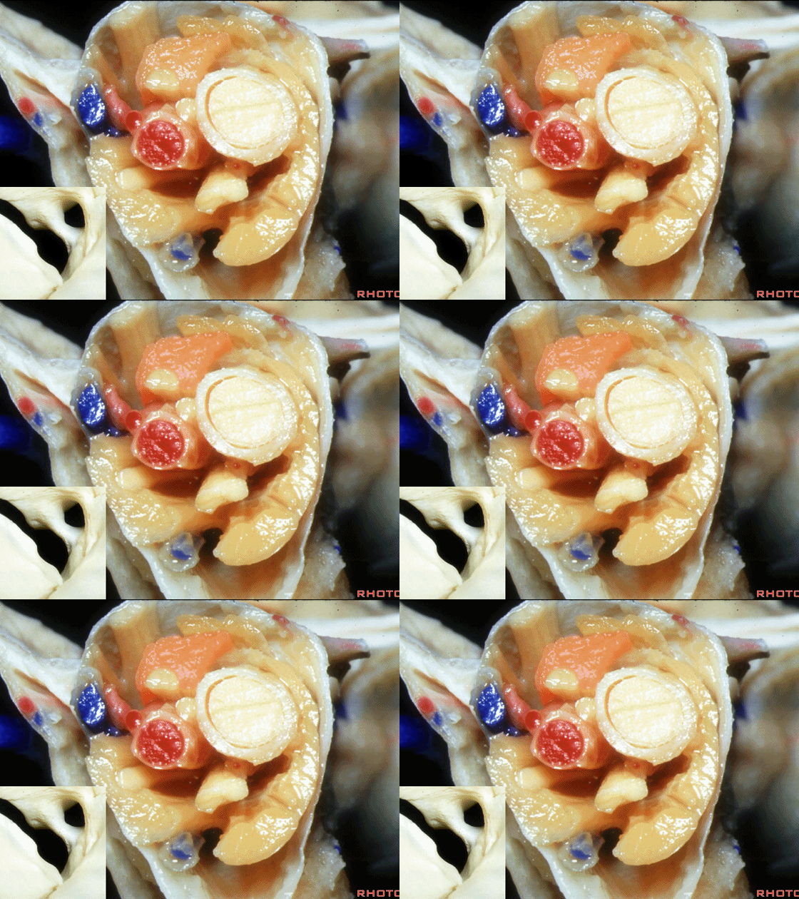
▼这是 内直肌
medial rectus
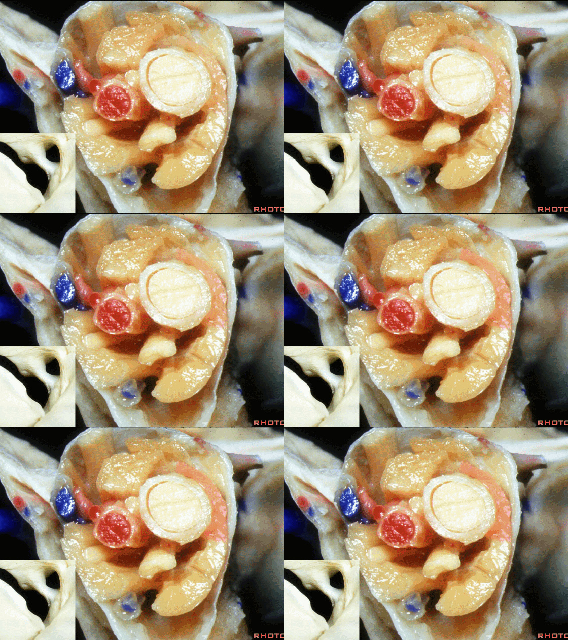
▼这是 下直肌
inferior rectus

▼这是 外直肌
lateral rectus
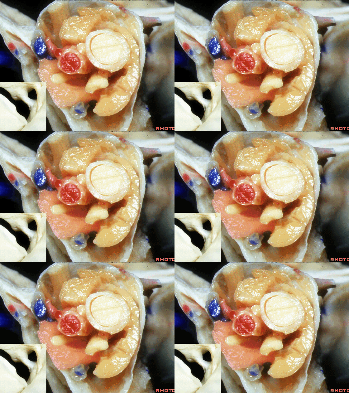
▼眼动脉和视神经均穿行于 视神经管。
And, the ophthalmic artery and optic nerve pass through the optic canal.
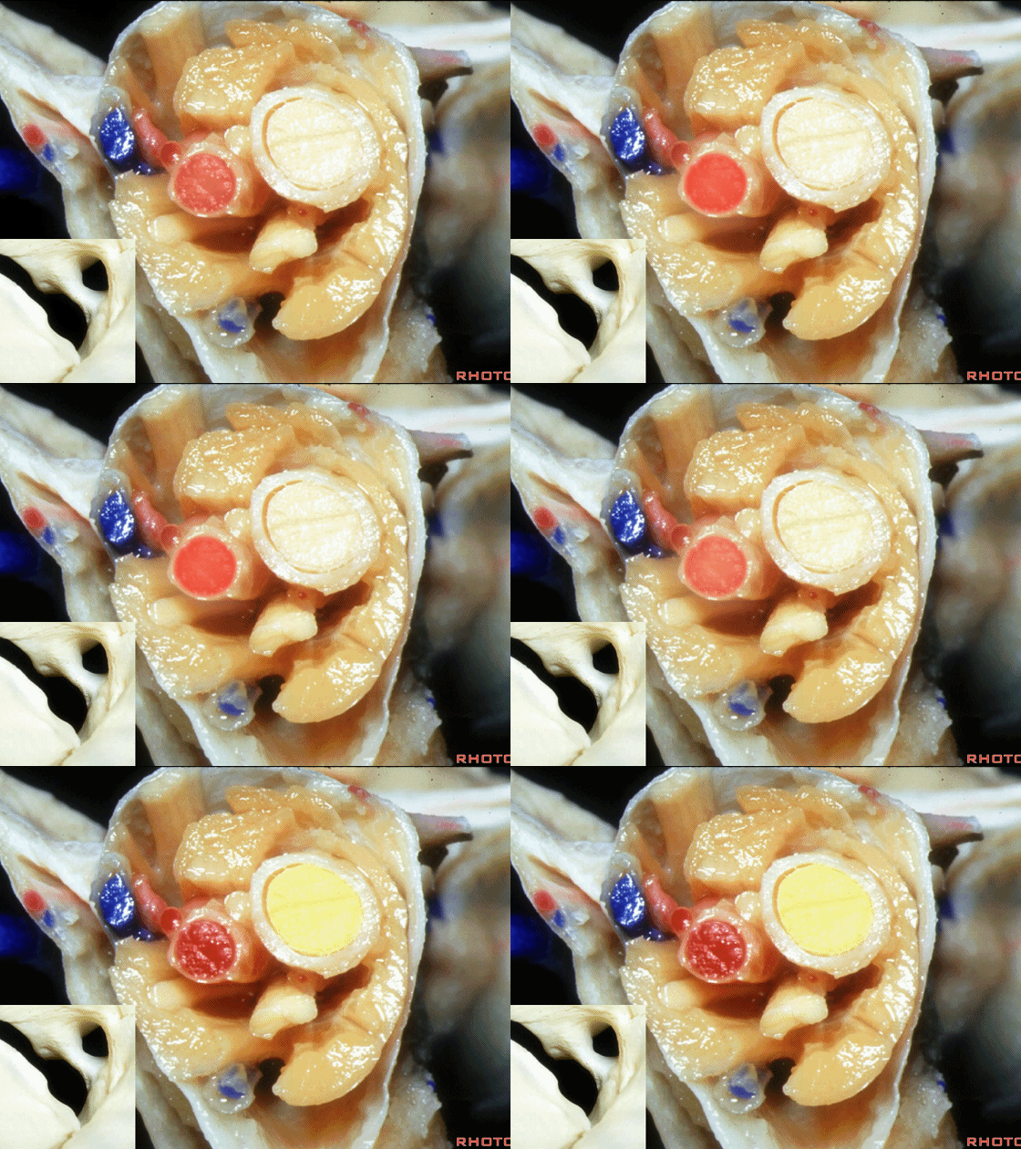
▼在眼直肌内,为肌锥区域。
But inside the rectus muscles, you have the intraconal area

▼下图示动眼神经的分支,即上支和下支
The oculomotor...branches or divisions of the oculomotor nerve, superior and inferior division pass
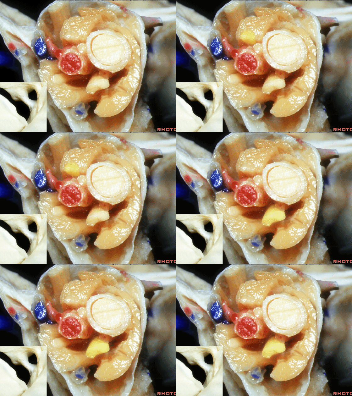
▼动眼神经与外展神经(下图)相伴行。上述结构穿行于总腱环内,另外穿行其中的还有一些三叉神经的分支。
along with the abducens nerve,They pass through the annular tendon as does some of the branches of the first trigeminal division.
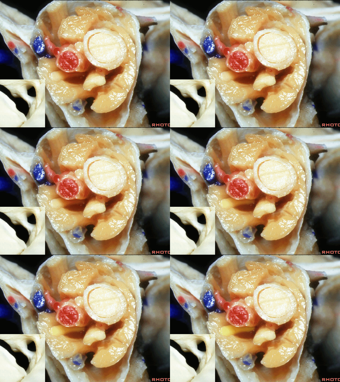
▼在眶尖部,走行于总腱环以外的是滑车神经(下图)、额神经的分支(包括眶上和滑车上神经),在外侧尚有发自眼神经的泪腺神经。
outside the annular tendon at the orbital apex, passes the 4th nerve, the branches of the frontal nerve, the supraorbital and supratrochlear, and then laterally from 1st division you have lacrimal nerve.
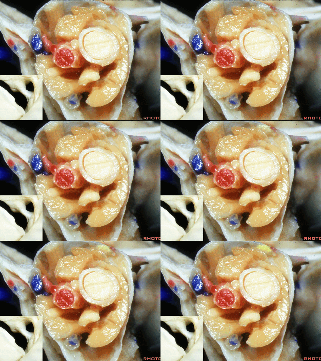
▼下图示额神经的分支,包括眶上神经、滑车上神经
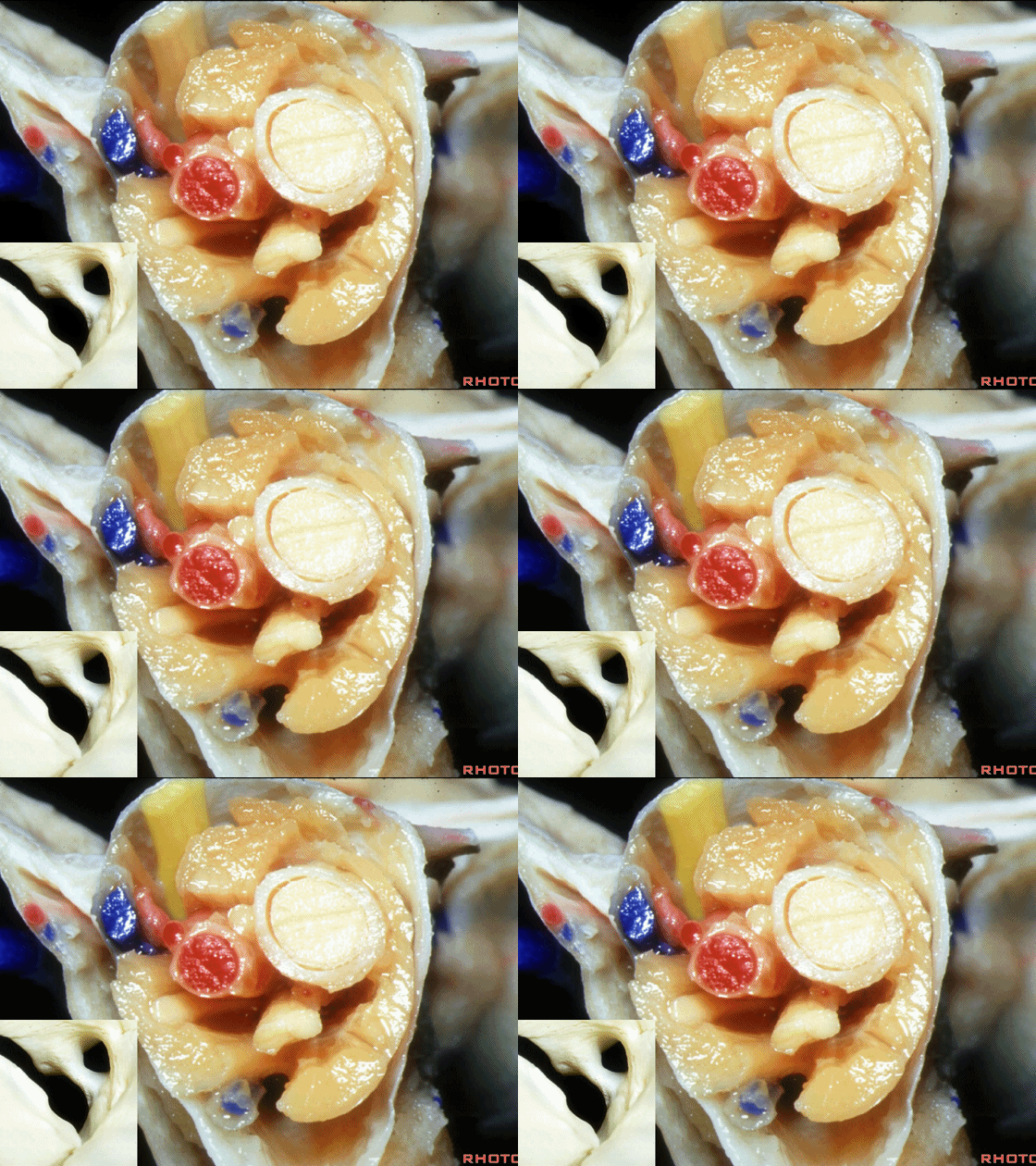
▼下图示发自眼神经的泪腺神经。
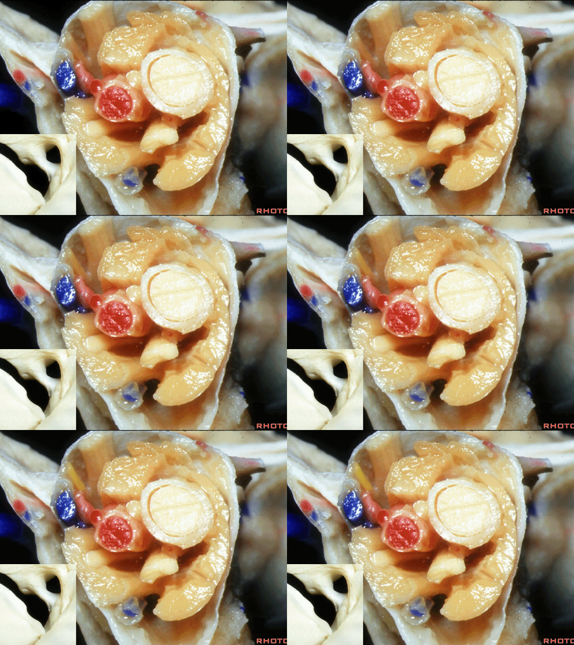
▼另外,眼上静脉和眼下静脉 从总腱环以外穿出眼眶,但在远侧的眼眶内,上述静脉位于肌锥内,即在四块眼直肌内走行。
And then the superior and inferior ophthalmic veins run outside the annular tendon as they exit the orbit, but further forward in the orbit these veins come into the intraconal area here between the four rectus muscles.
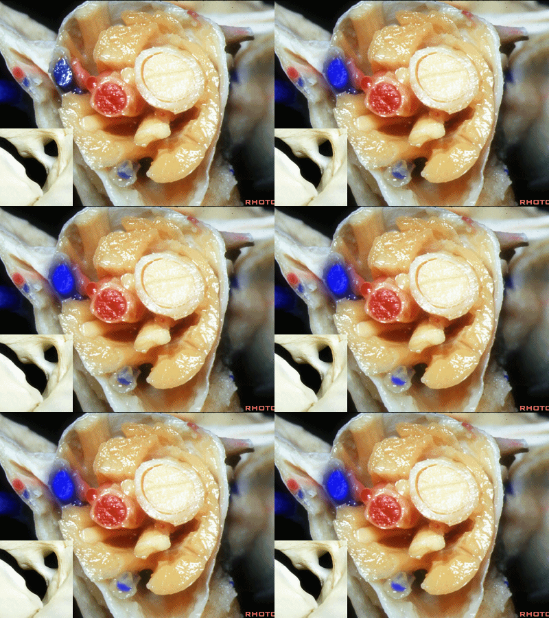
▼这是动眼神经上支,其支配上睑提肌和上直肌。
We have here superior division of 3rd nerve that supplies levator and superior rectus.
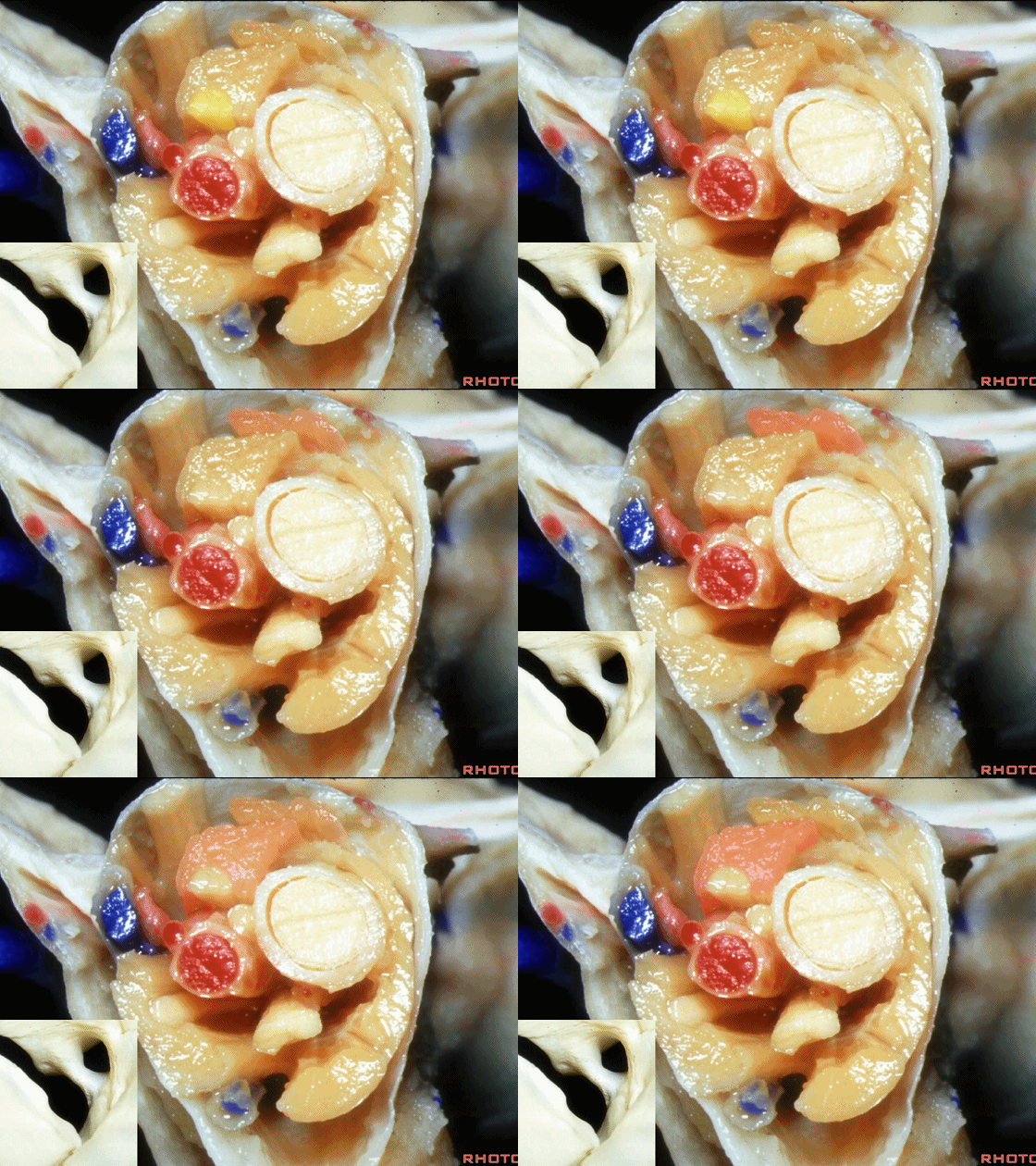
▼动眼神经下支 支配内直肌、下直肌、下斜肌,并内含副交感运动性纤维行至睫状神经节。
inferior division of 3rd nerve. The inferior division supplies the medial rectus, inferior rectus, and inferior oblique muscles, as well as giving rise to the motor, parasympathetic root of the ciliary ganglion.
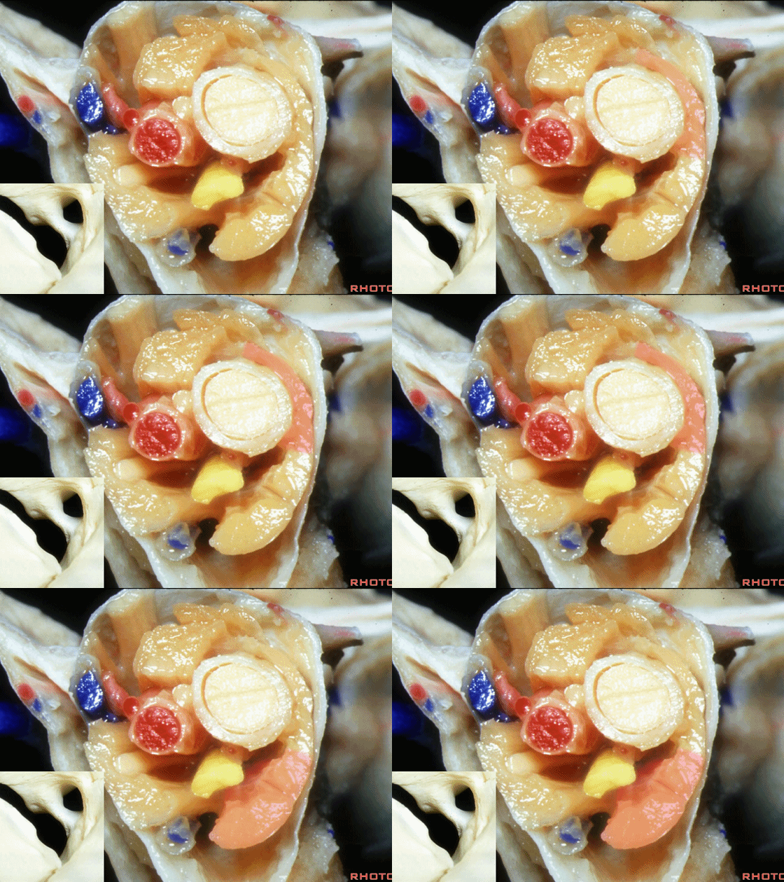
▼另外一支穿行于总腱环内的神经是三叉神经第一支(眼神经)发出的鼻睫神经。
And then passing through the annular tendon,we have the nasociliary branch of V1 that passes through the annular tendon.
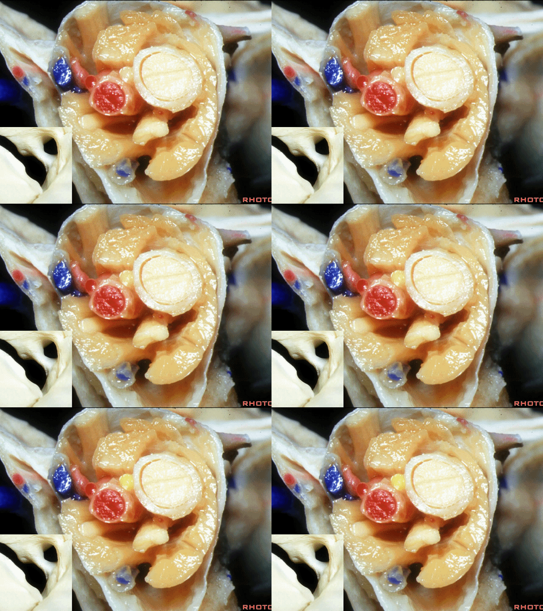
▼而眼神经发出的额神经和泪腺神经 则在总腱环外走行。
While, the branches of the frontal and the lacrimal nerve pass outside the annular tendon.
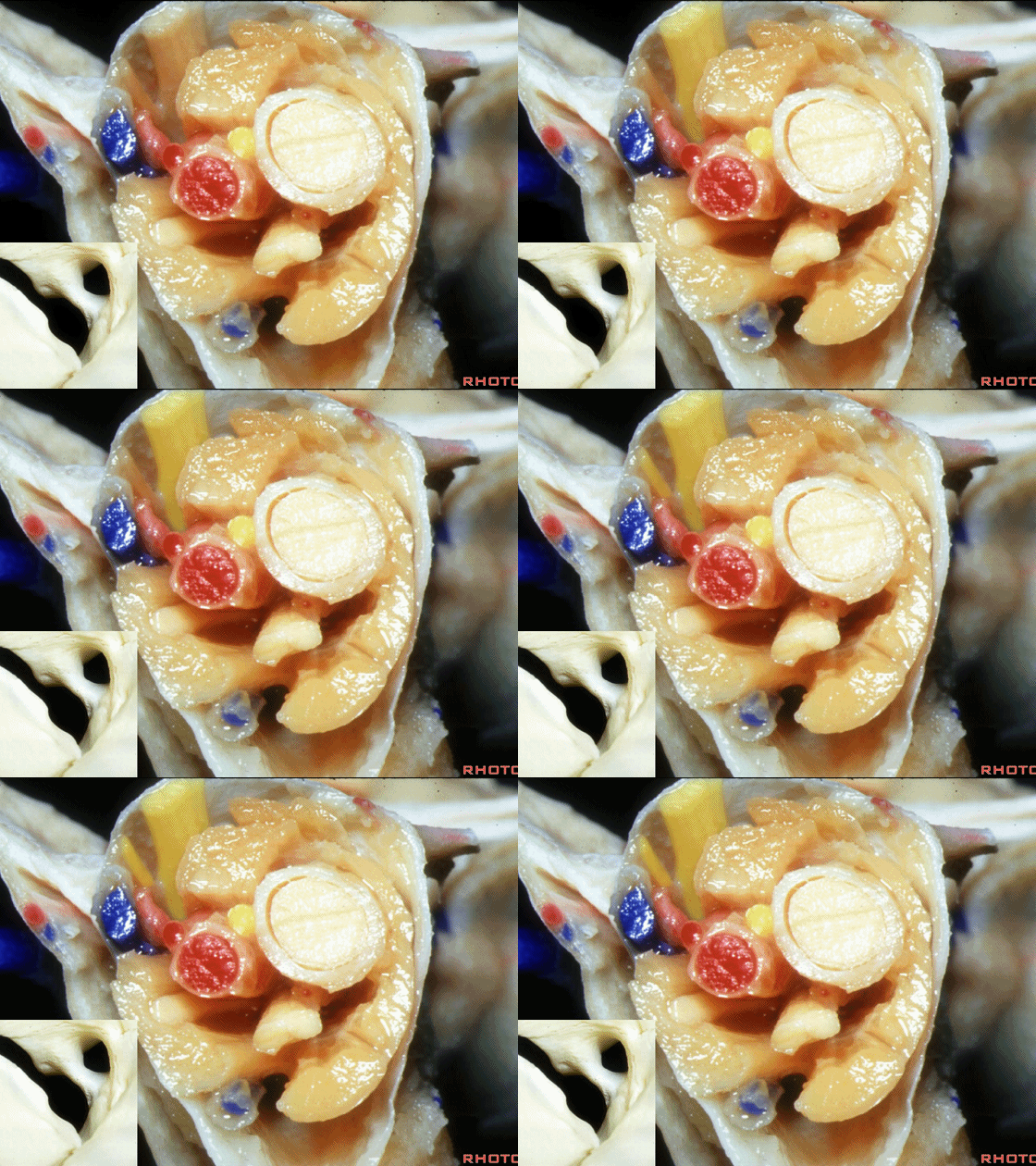
因此我们需掌握穿行于视神经管内、总腱环以外、以及动眼神经孔内的各个结构。
So you wanna think about the structures that pass through the optic canal, and then the group of structures that pass outside the annular tendon, plus have an understanding of the structures that pass through the oculomotor foramen, part of the annular tendon.
![]()


