


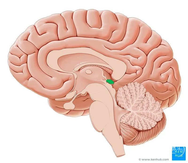
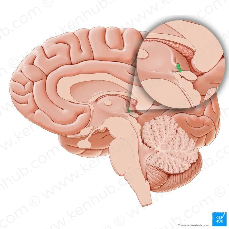
松果体(pineal gland or epiphysis cerebri ) 是一小的、灰红色器官,位于两侧上丘之间的凹陷内,胼胝体压部的下方, 被第三脑室脉络组织及大脑静脉分隔。
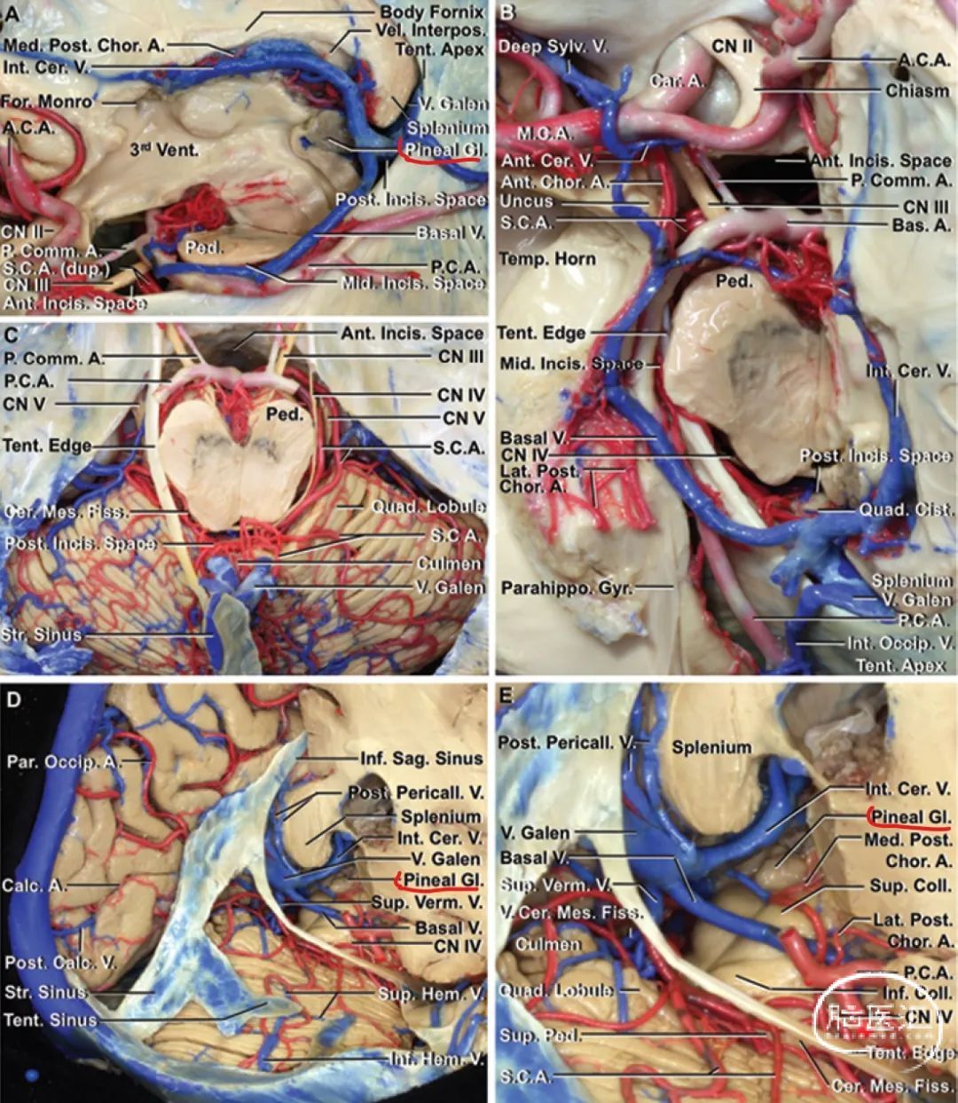
A.= artery; A.C.A.= anterior cerebral artery; Ant. = anterior; Bas. = basilar; Cale. = calcarine;Car. = carotid; Cent. = central; Cer. = cerebral, cerebellar; Cer. Mes. =cerebellomesencephalic; Chor. = choroidal; Cing. = cingulate; Cist. =cistern; CN = cranial nerve; Coll. = colliculus; dup. = duplicate; Fiss. =fissure; For.= foramen (of); Gyr. = gyrus; Hem.= hemispheric; Inds.=incisural; Inf. = inferior; Int. = internal; lnterpos. = interpositum; M.C.A. =middle cerebral artery; Med. = medial, medullary; Mid. = middle; Occip. =occipital; P.C.A. = posterior cerebral artery; P. Comm. = posterior communicating; Par. = parietal; Parahippo. = parahippocampal; Ped. =peduncle; Pericall. = pericallosal; Post. = posterior; Quad. =quadrigeminal, quadrangular; S.C.A. = superior cerebellar artery; Sag.=sagittal; Str. = straight; Sulc. = sulcus; Sup. = superior; Sylv. = sylvian;Temp. = temporal; Tent. = tentorium, tentorial; V. = vein (of); Vel. =velum; Vent.= ventricle; Verm. = vermian. (Images courtesy of ALRhoton, Jr.)
松果体由脉络组织下层包被, 该层再由腺体折返至中脑顶盖。
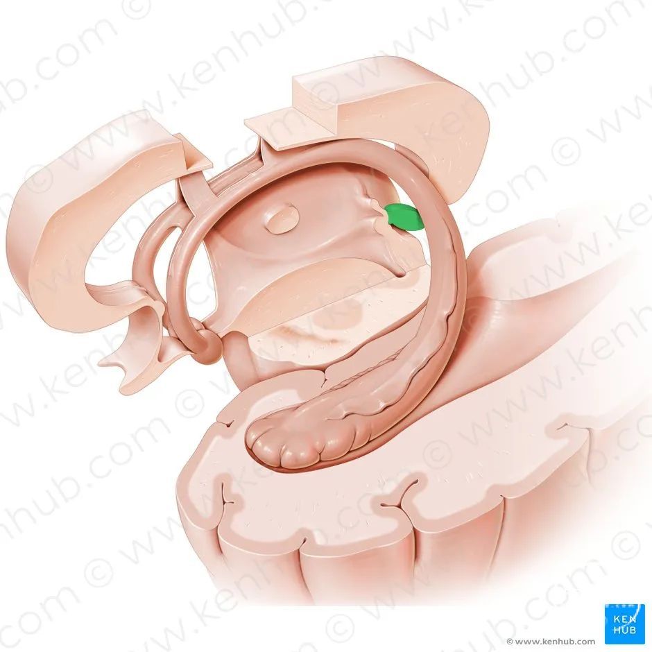
松果体长约8mm , 底部朝前,连着松果体脚, 脚分为上、下两层, 由第三脑室松果体隐窝隔开,其内分别含有后连合与缰连合。散在的连合纤维可能进入松果体,但不终止于附近的松果体细胞。
松果体
缰连合


隔从周边软脑膜伸入松果体,将腺体分隔为多个小叶,隔内携带有血管及细的无髓交感神经轴突。
松果体的血供


松果体有丰富的血供,松果体动脉是脉络膜后动脉的分支,而脉络膜后动脉是大脑后动脉的分支。

在腺体内, 动脉分支形成有孔毛细血管,毛细血管的内皮细胞的基板很薄,有的不连续。毛细血管回流入多条松果体静脉,这些静脉开口于大脑内静脉和(或)大脑大静脉。

Midline (A and B) and right paramedian (C and D) infratentorial supracerebellar microsurgical and endoscopic approaches to the pineal gland. A: Microsurgical midline approach to the pineal gland. Opening the quadrigeminal cistern exposes the superior vermian, internal occipital, basal, and internal cerebral veins and the vein of Galen. The superior vermian vein may need to be retracted or sacrificed to expose the pineal region in a midline approach. B: Endoscopic midline approach. C: Microsurgical right paramedian approach to the pineal gland. The pineal gland and the superior and inferior colliculi can be exposed between the superior vermian and the internal cerebral veins without venous sacrifice. The off-midline views of the gland open the viewing angle between the large veins around the gland. D: Endoscopic paramedian approach. The pineal gland can be exposed between the galenic tributaries. Retracting the cerebellum can provide access to the superior and inferior colliculi. Coll. = collicular, colliculus; Med. = medial; Tent. = tentorial, tentorium. (Images courtesy of AL Rhoton, Jr.)
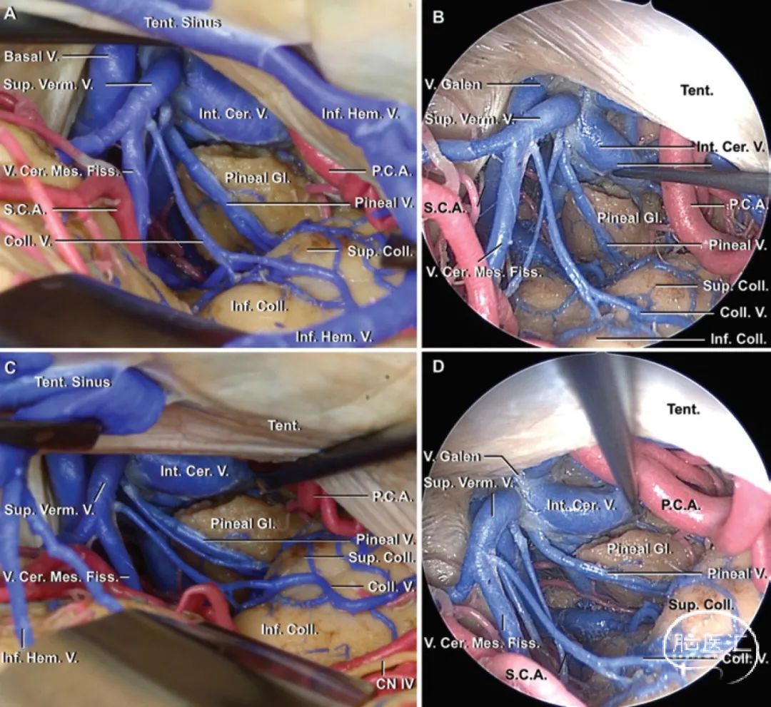
Right lateral (A and B), and far-lateral (C and D) infratentorial supracerebellar microsurgical and endoscopic approaches to the pineal gland. A: Microsurgical right lateral approach to the pineal gland. Note that the off-midline approaches permit exposure between the hemispheric veins emptying into the tentorial sinuses at a superficial level and anterior to the superior vermian vein at the level of the vein of Galen. B: Endoscopic lateral approach. C: Microsurgical right far-lateral approach to the pineal gland. The farther laterally the approach moves, the wider the approach between the superior vermian and internal cerebral veins becomes. D: Endoscopic far-lateral approach. Cer. = cerebral; Cer. Mes. = cerebellomesencephalic; Coll. = collicular, colliculus; Tent.= tentorial, tentorium. (Images courtesy of AL Rhoton, Jr.)

松果体细胞为高度修饰的神经元,含有多个突触带,随机地分布于相邻细胞之间,形成缝隙连接。每个细胞体伸出两个或更多的突起,以球形膨大形状终止于邻近的毛细血管,或少数情况下终止于松果体隐窝的室管膜细胞。这些终末球形膨大内含粗面内质网、线粒体、储存褪黑素的致密核心囊泡。松果体细胞将色氨酸合成为褪黑素及其前体5-羟色胺,并分泌到周围的有孔毛细血管网。
松果体是一个具有重要调节功能的内分泌腺, 调节腺垂体、神经垂体、胰腺内分泌部、甲状旁腺、肾上腺皮质、肾上腺髓质和性腺的活动。

松果体的作用主要是抑制性的。松果体细胞分泌的吲哚胺和多肽类激素,可通过直接作用于腺垂体分泌细胞或间接抑制下丘脑释放因子产物,从而减少腺垂体激素的合成和释放。松果体的分泌物可通过脑脊液或血流到达其靶细胞。部分松果体吲哚胺,包括褪黑素及其生物合成酶(如5-羟色胺、N-乙酰转移酶)的浓度呈现昼夜节律,浓度水平夜间升高,白天下降,分泌可被交感神经的活动所抑制。下丘脑视交叉上核内源性昼夜节律振荡器的内在节律被认为控制松果体的周期性行为。

从20 岁起,钙沉积物积聚在松果体细胞外基质内,集中沉积处为"脑砂(corpora arenacea or brain sand) "。

松果体及脉络丛钙化是颅内钙化最常见的部位,可在头颅断层扫描检查中被发现。当腺体由中线显著移位时,钙化能为空间占位性病变提供有用的标示。

所有分享仅供学习参考,如有错误欢迎指正。
