▼关于经胼胝体入路,我们常作这样的骨瓣,三分之一位于冠状缝后方,三分之二位于冠状缝前方。利用这一 桥静脉相对稀疏 的区域,
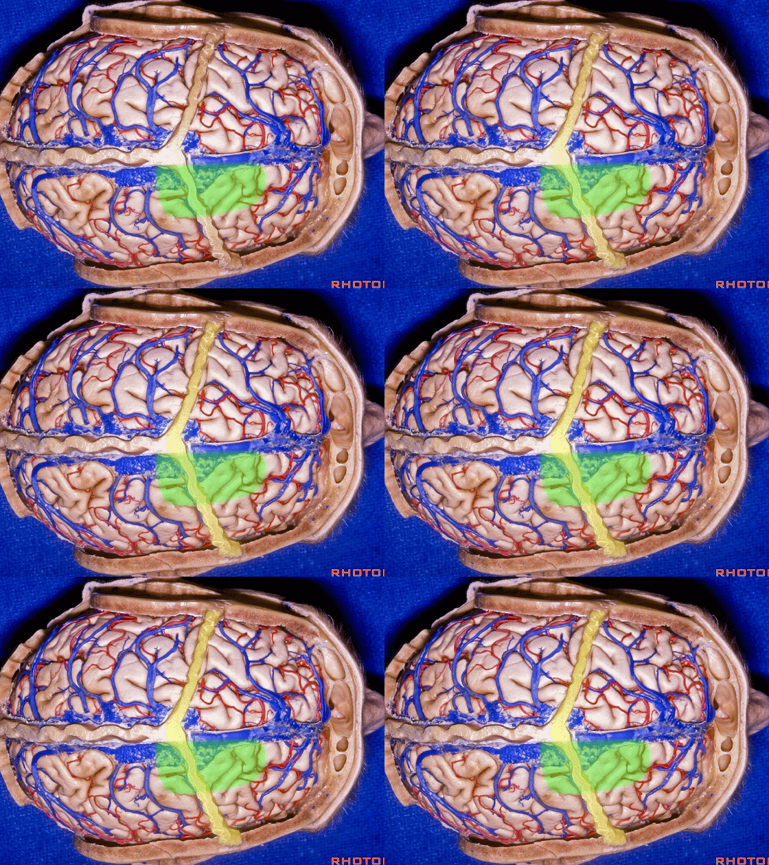
▼汇入上矢状窦前部的静脉走行向后。
The veins at the front of the sinus tend to be directed backwards.
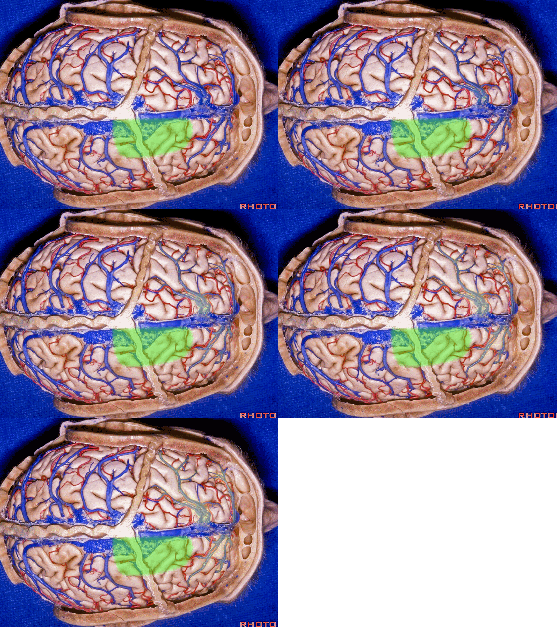
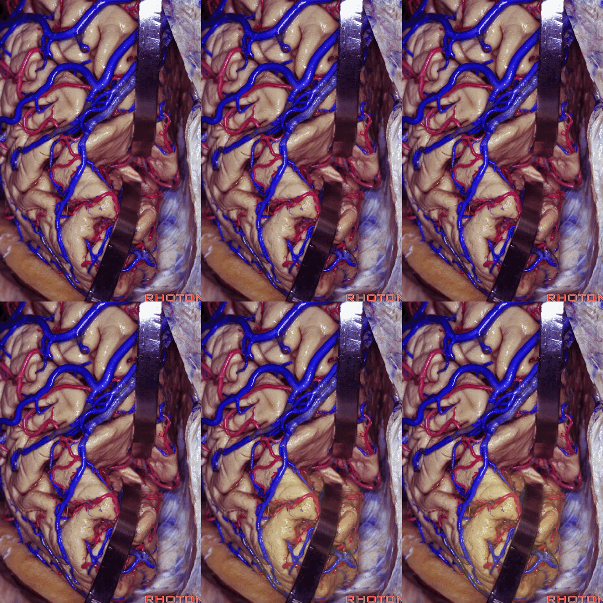
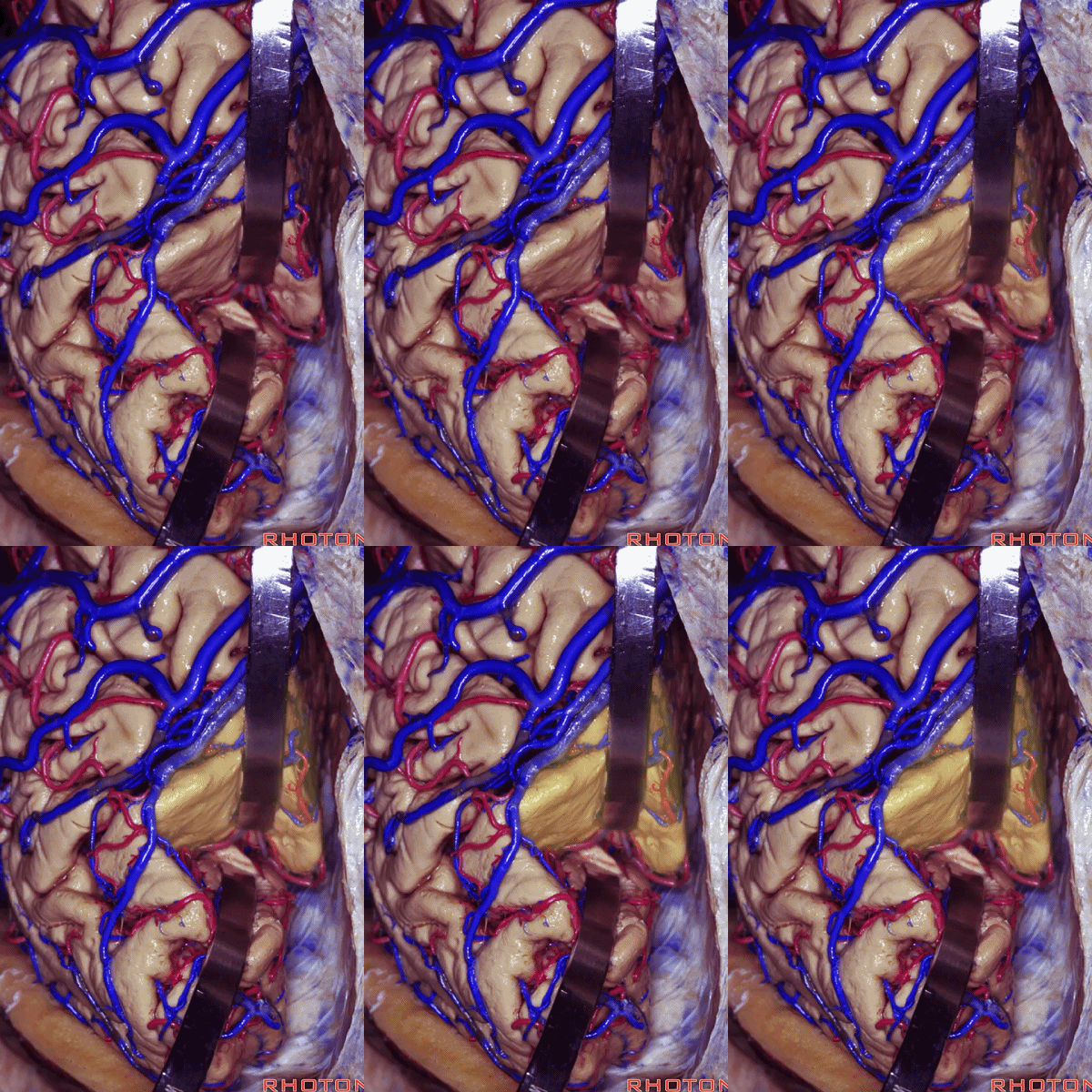
▼而越是到后方,桥静脉弯曲向前汇入上矢状窦的角度越是明显。很多教科书描述骨瓣为三分之一在前,三分之二在后。然而正如大家所见。这里会遭遇静脉间隙、大的桥静脉。
As you go progressively posterior, the veins entering the sinus take on a progressively forward curve before entering the sinus,A number of textbooks say go one third in front,two thirds in back for the bone flap.But here you see that there's...you often get into this area where there's venous lacuna,large bridging veins. So,one third back, two thirds forward is better.
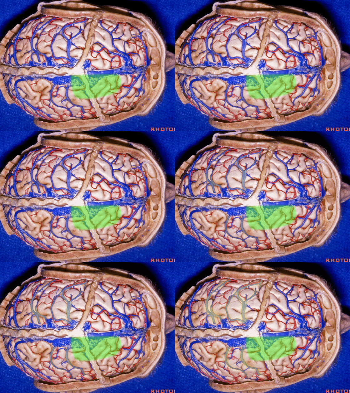
▼因此,三分之一位于冠状缝后方,三分之二位于冠状缝前方 更为妥当。
▼接下来,定位Monro孔的方法:向后作一个1英寸(2.5cm)的胼胝体切口。即可在Monro孔上方
▼切开胼胝体后,可见Monro孔,丘纹静脉位于脉络丛的右侧。因此我们在右侧的侧脑室。
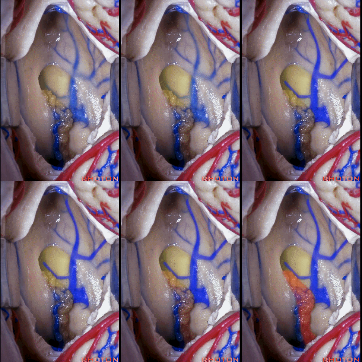
▼若要打开脉络裂,则需在穹窿(下图)边缘分离脉络丛。
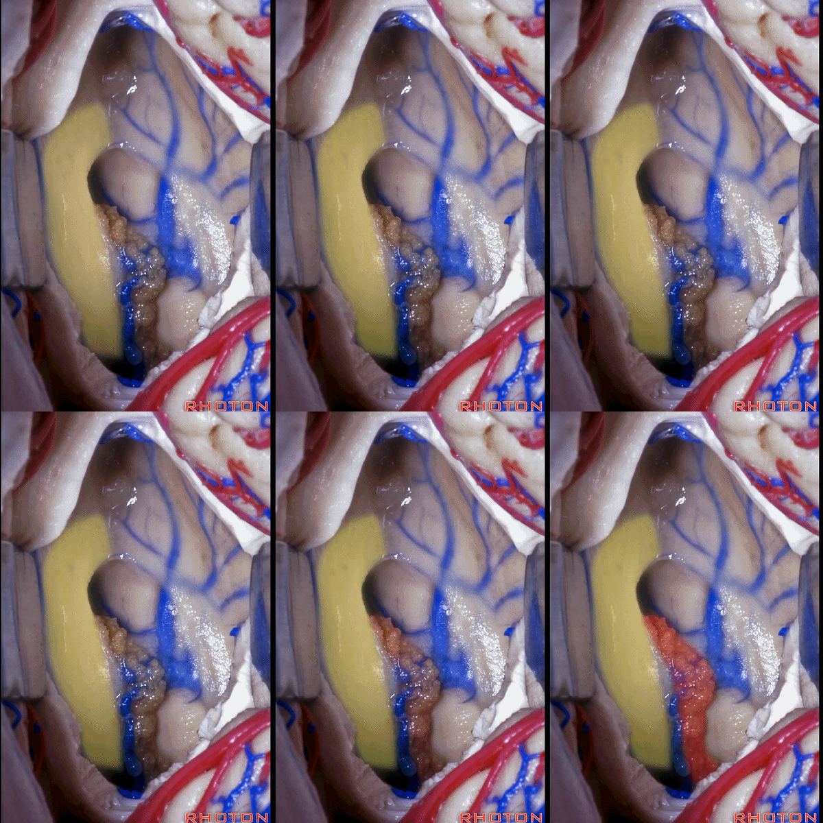
▼随后即可暴露中间帆。
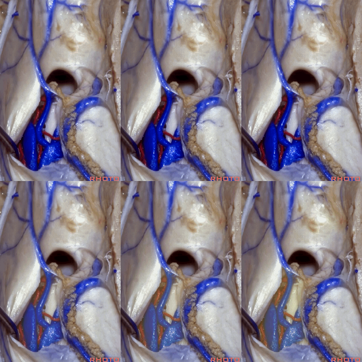
▼只需向后1cm,即可显露出中脑导水管(下图)。

▼不要在脉络丛和丘脑进行切开,因为容易损伤丘纹静脉。

▼通常,在大脑内静脉之间操作,比在静脉和丘脑之间操作来的简单。
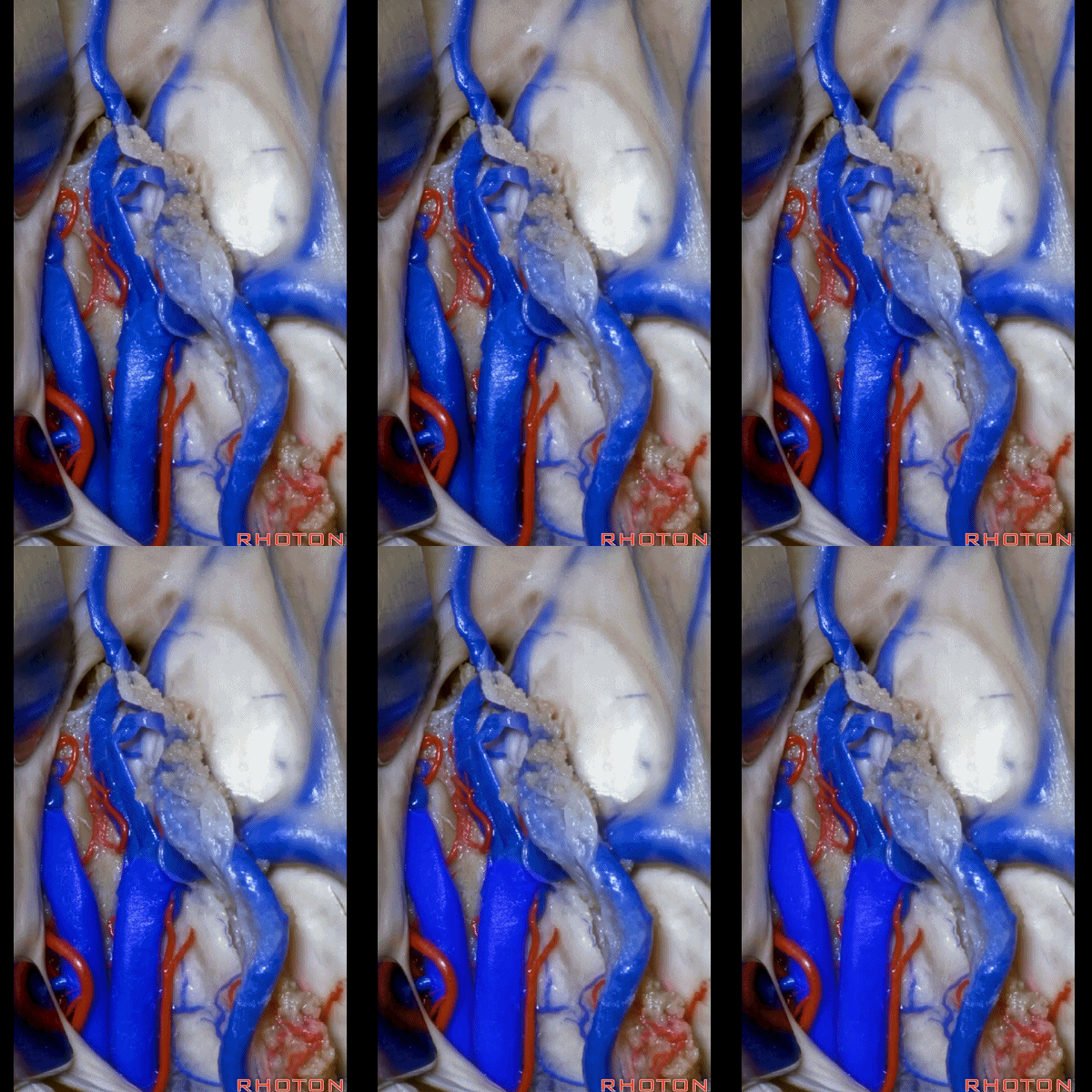
▼如果在静脉和丘脑之间操作,则有可能损伤引流丘脑和内囊的静脉。
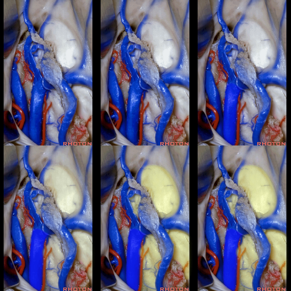
▼这是穹窿、丘脑。这就是经穹窿带切开的 经脉络裂入路。
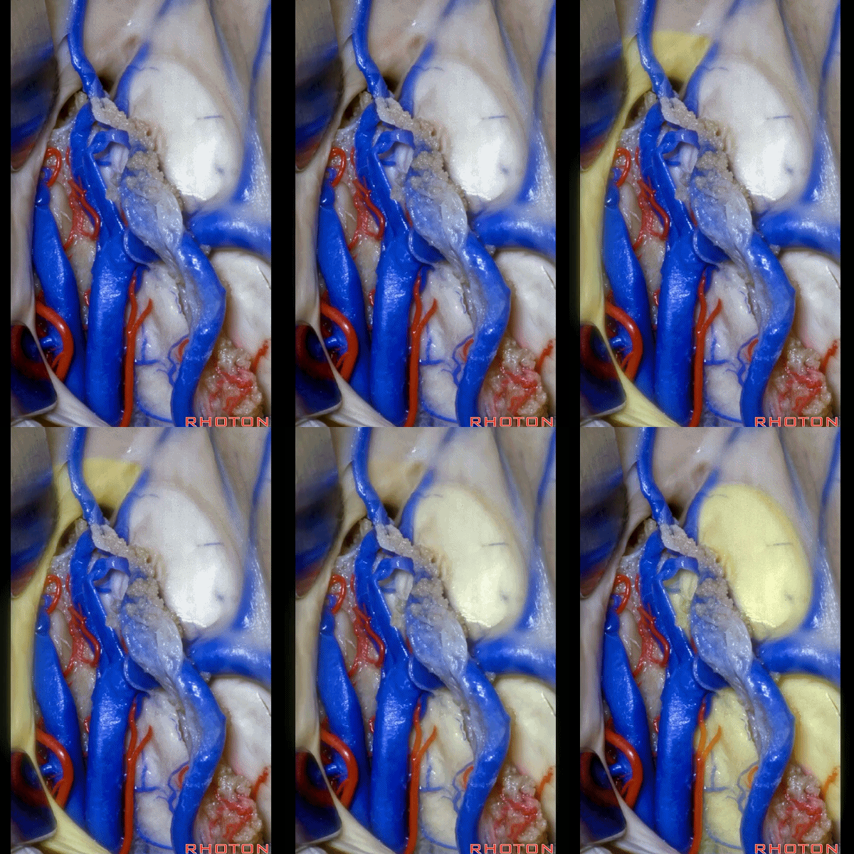
▼这是下层的脉络膜。
Here's the lower layer of tela.
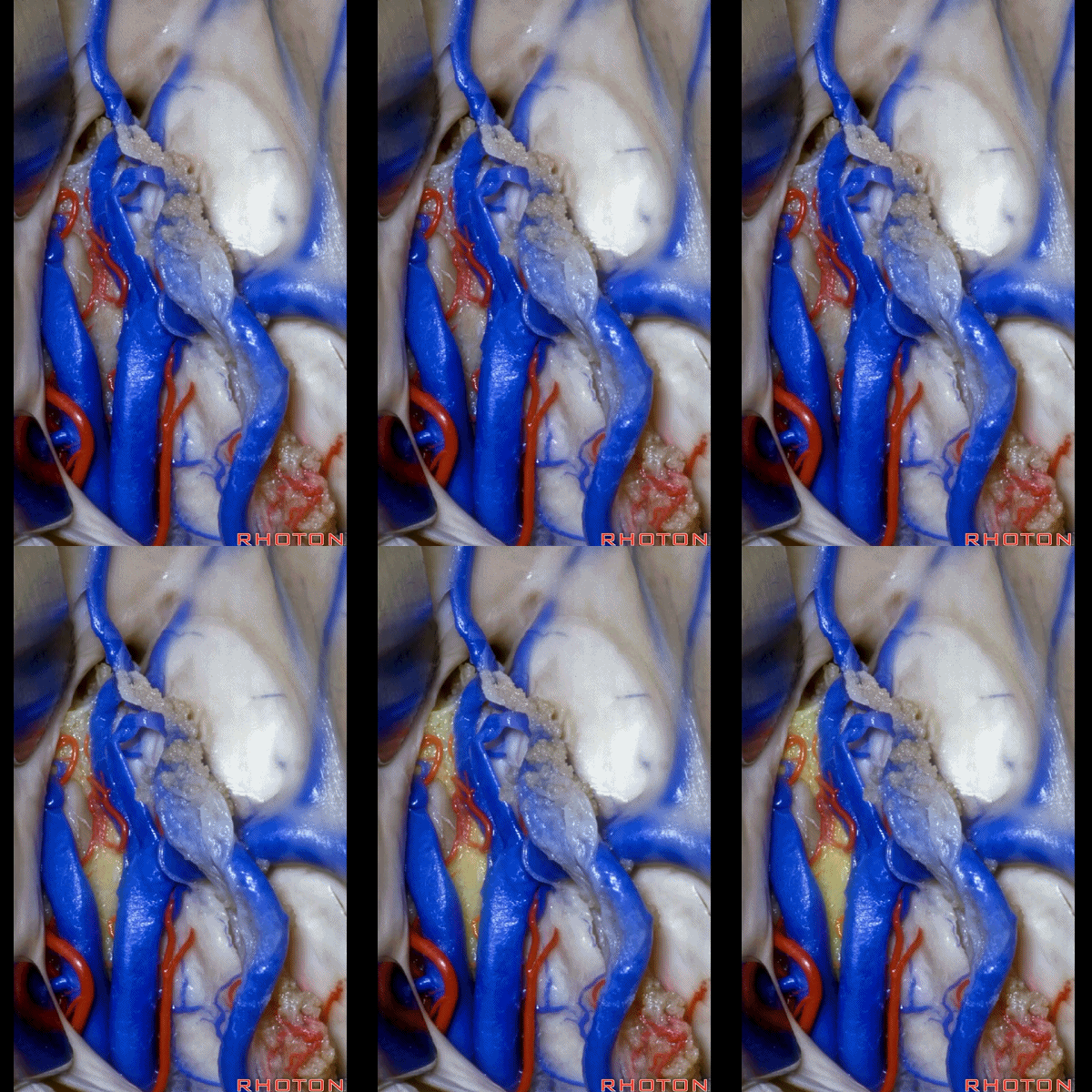
▼此处可见很大的中间块。
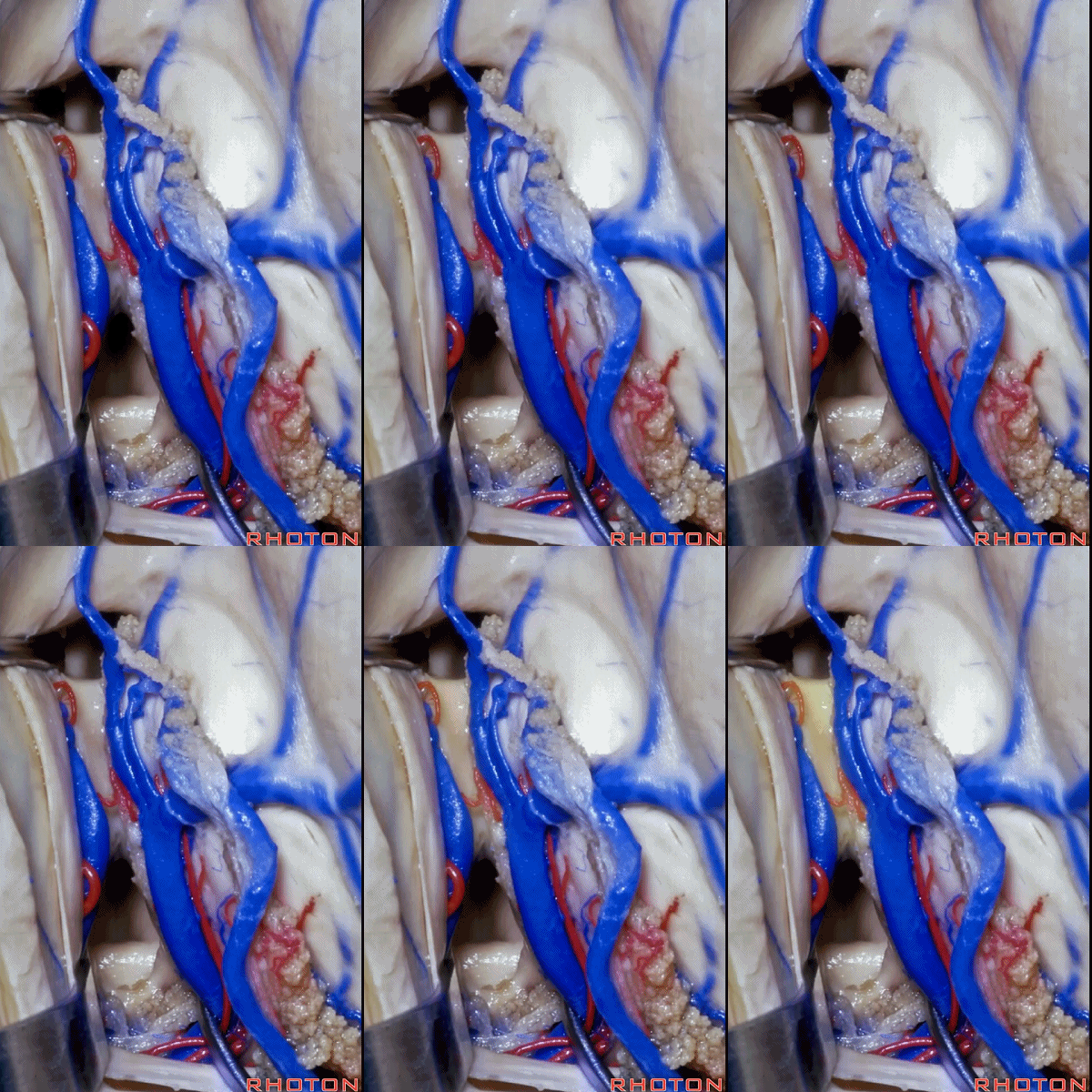
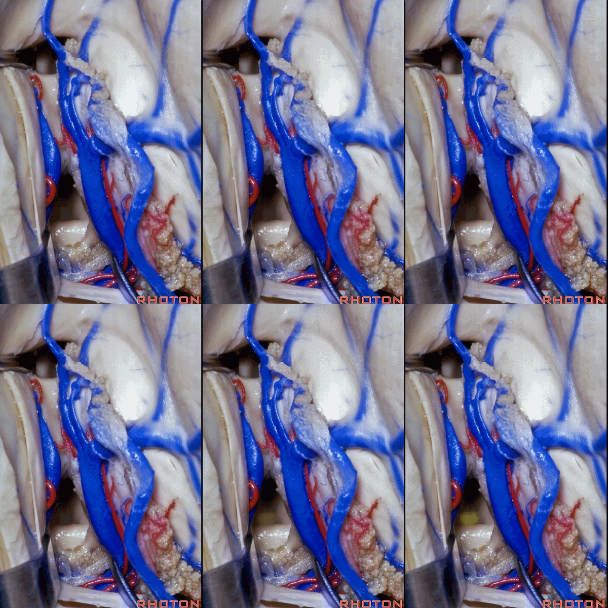
▼但通常只需向后打开1cm的脉络裂,即可获得向后至中脑导水管的视野。
![]()
▼这里我们经由扩大了的Monro孔,进入第三脑室。这是穹窿柱(下图)
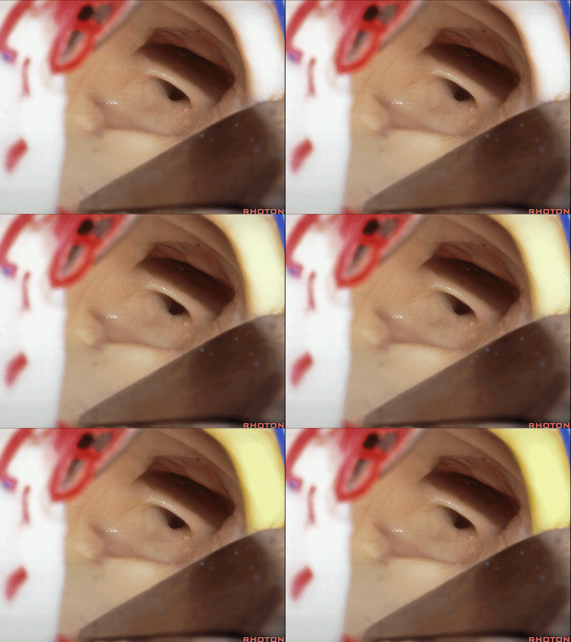
▼位于第三脑室前壁的是 前连合(下图)。
What is...here in anterior wall of the third? Anterior commissure.
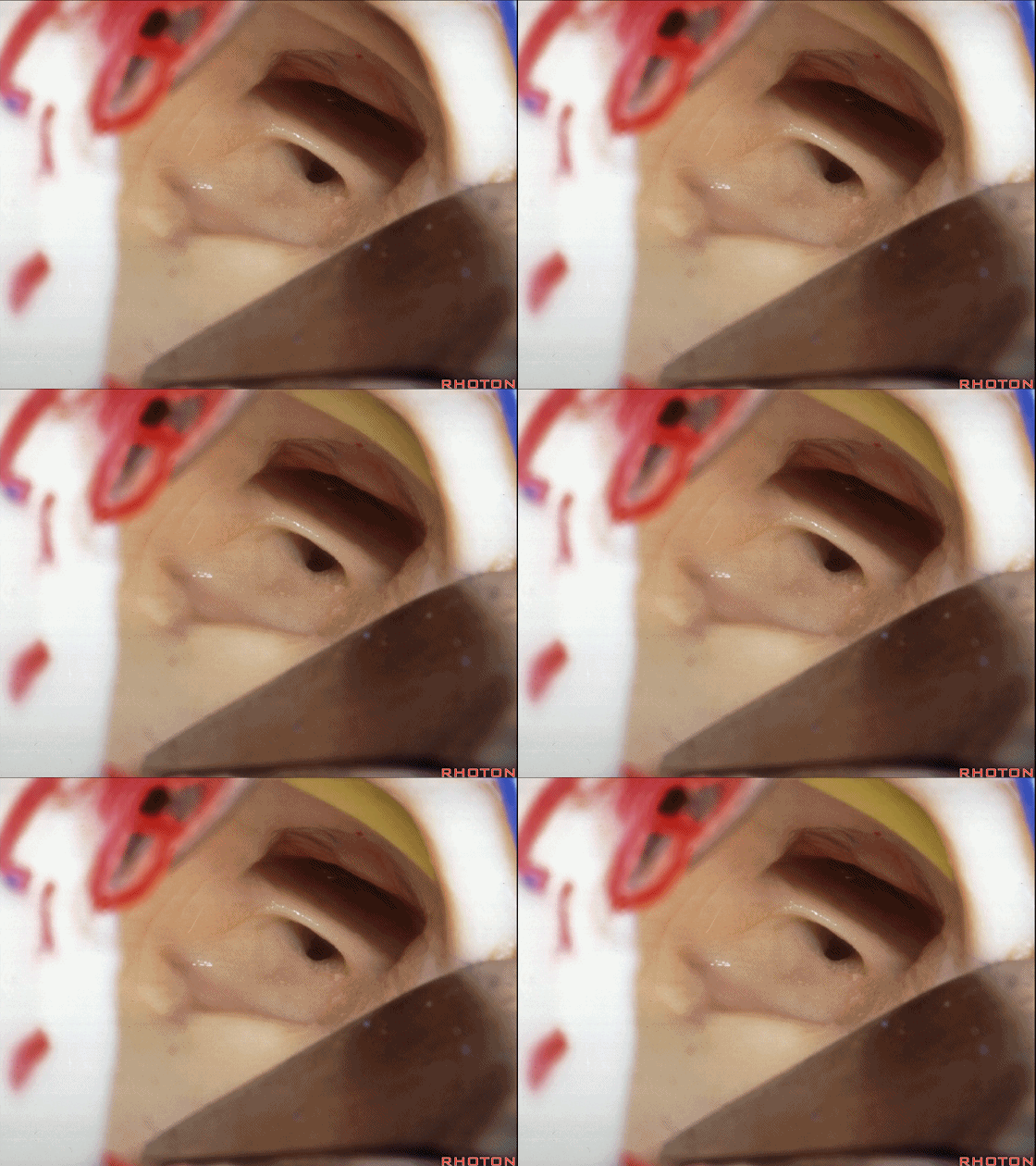
▼再往下是 终板(下图)。
And then below it, lamina terminalis.
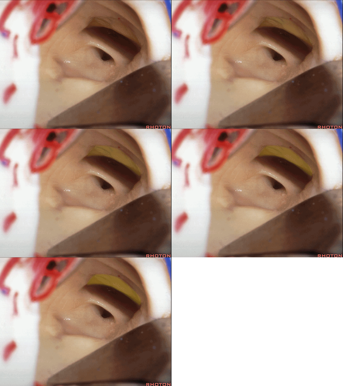
▼这是 视交叉隐窝
And this recess? Chiasmatic recess
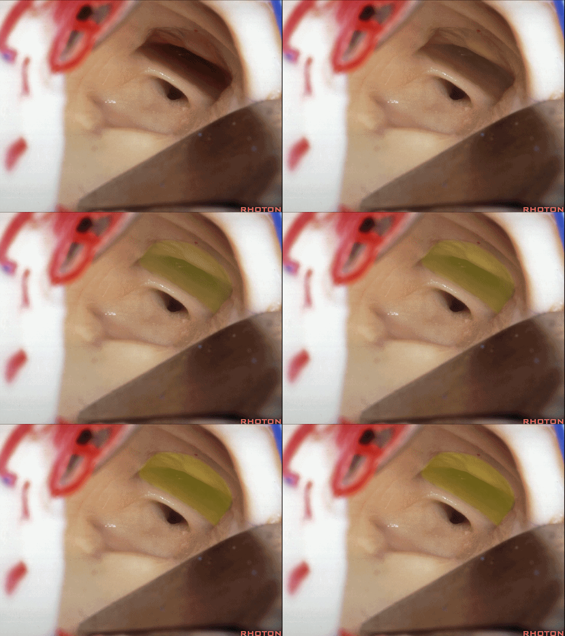
▼这是 视交叉后缘。
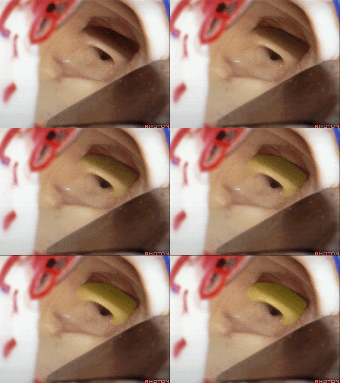
▼这是 漏斗隐窝。
What recess? Infundibular recess
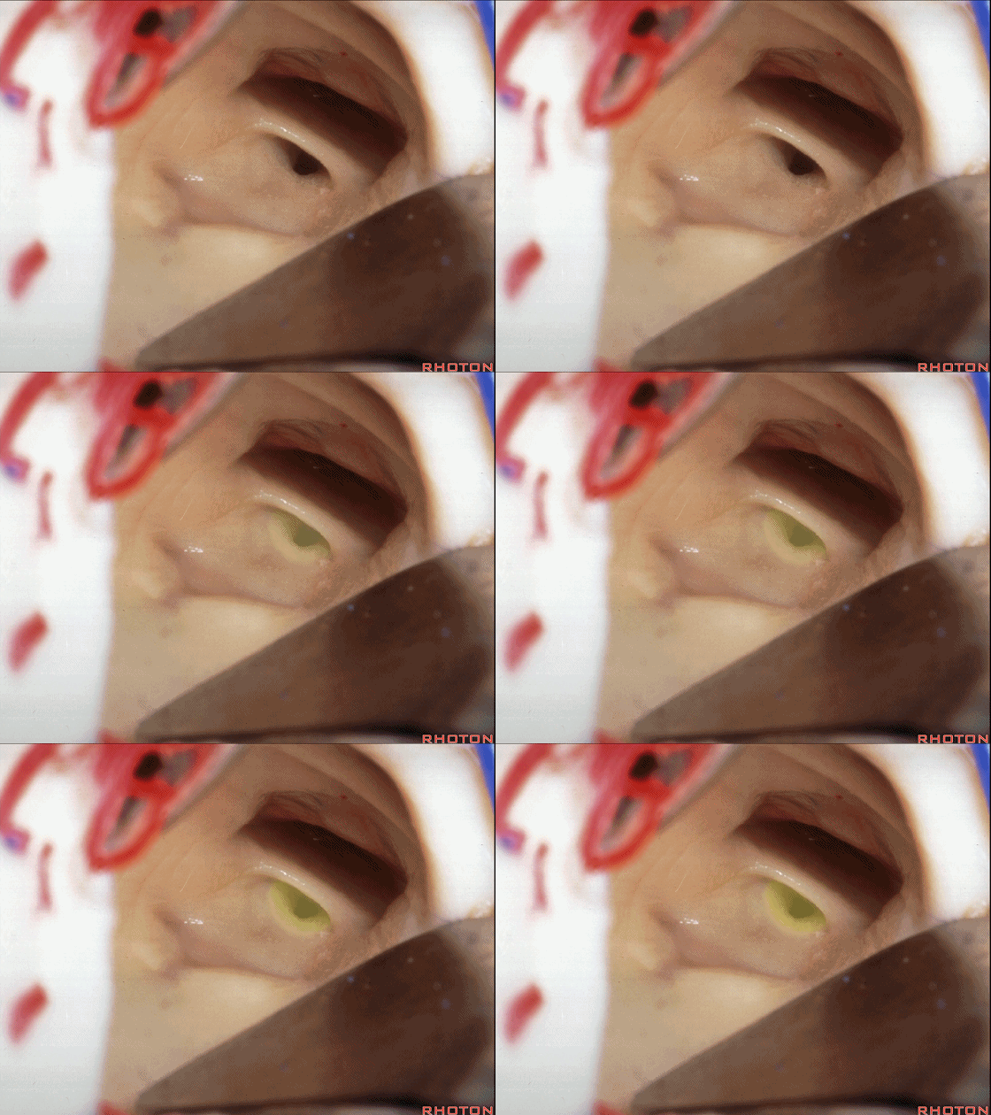
▼这是 乳头体。
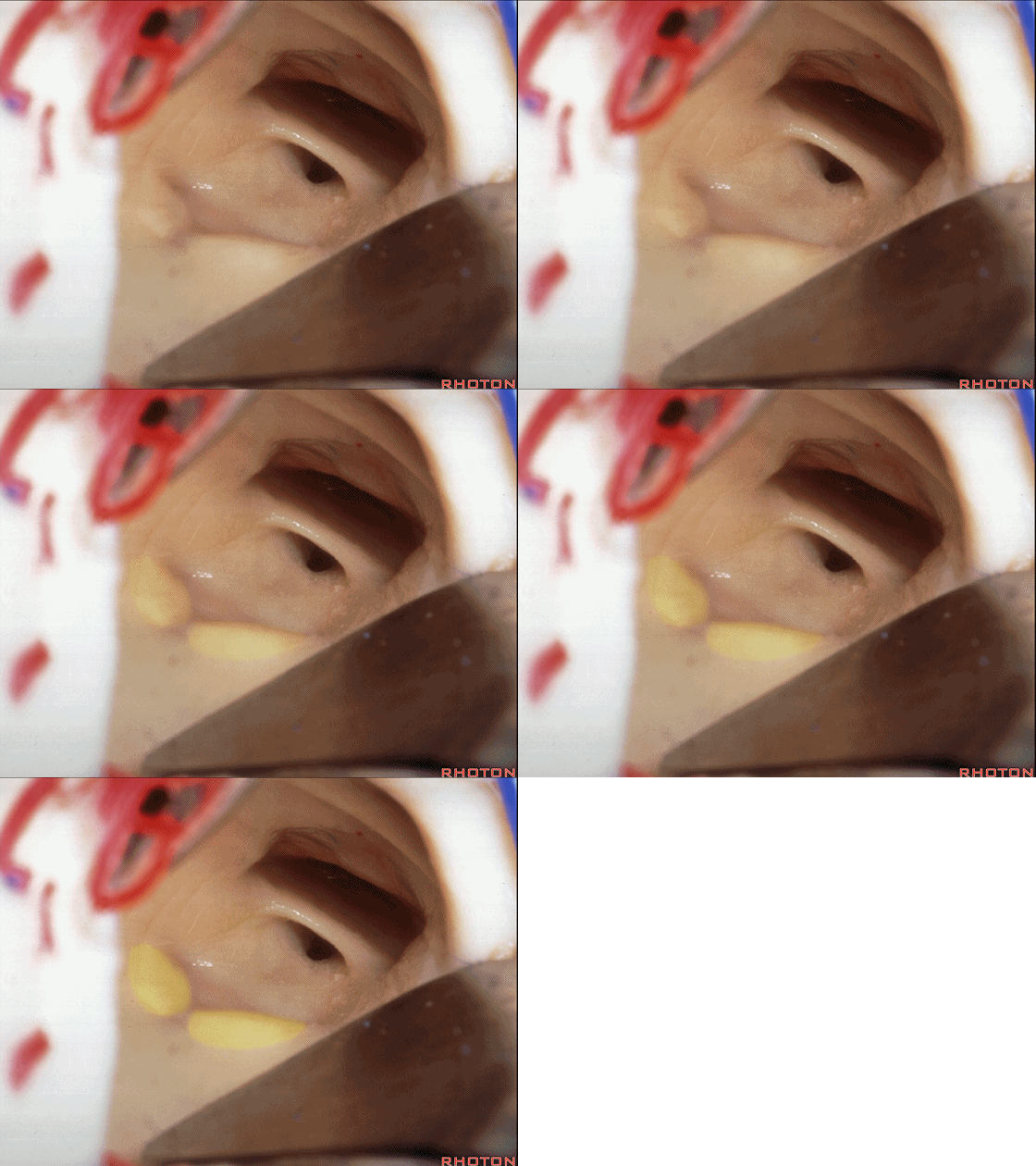
▼三脑室底造瘘术,是在乳头体前方操作。
And we do third ventriculostomy.We go behind the mamillary bodies in this area.Yes? No.We go in front.
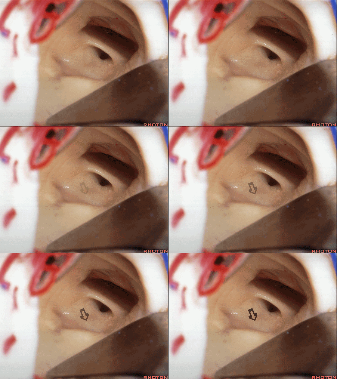
▼从矢状位来看 第三脑室底的这个区域。
Well, if you look at this area below the floor of the third in this area,
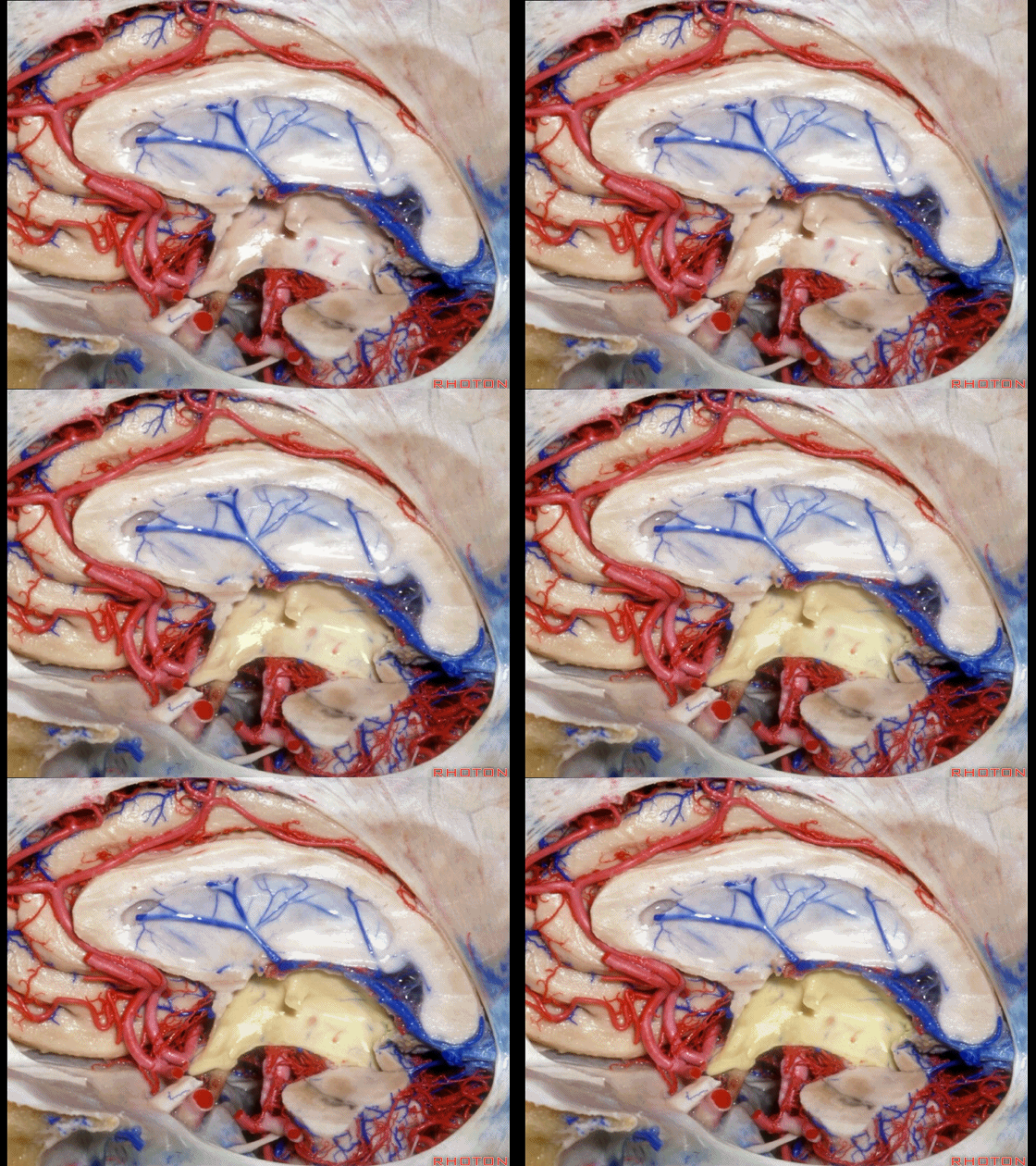
▼这是 乳头体。
here are mamillary bodies.
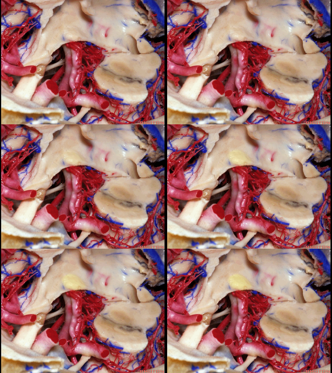
▼这是 基底动脉尖部
So if you go back here, you get into basilar apex,
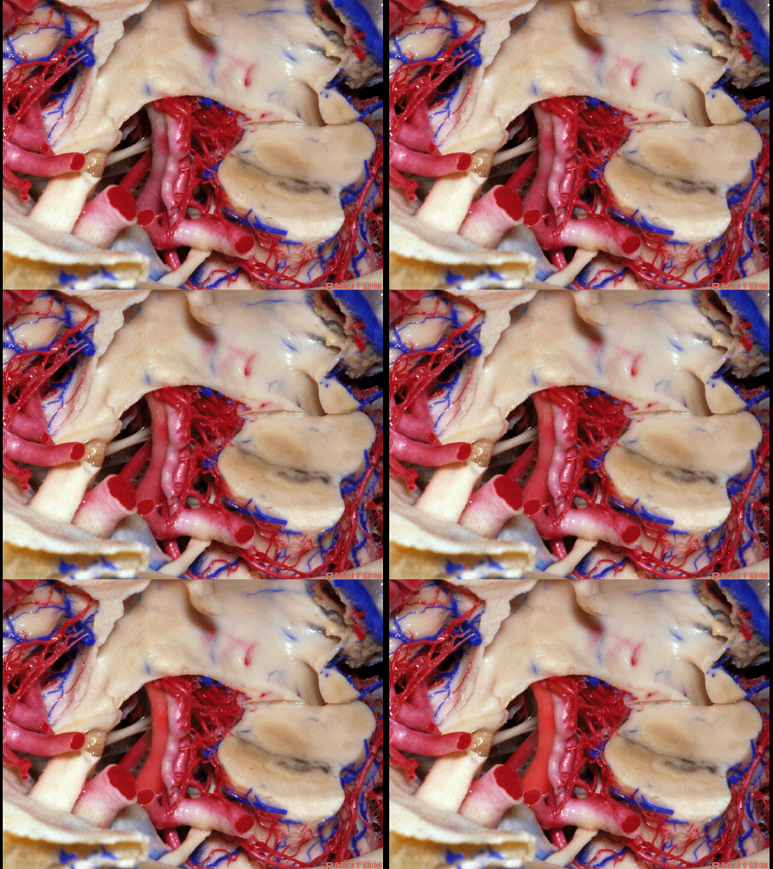
▼这是 脚间窝
interpeduncular fossa,
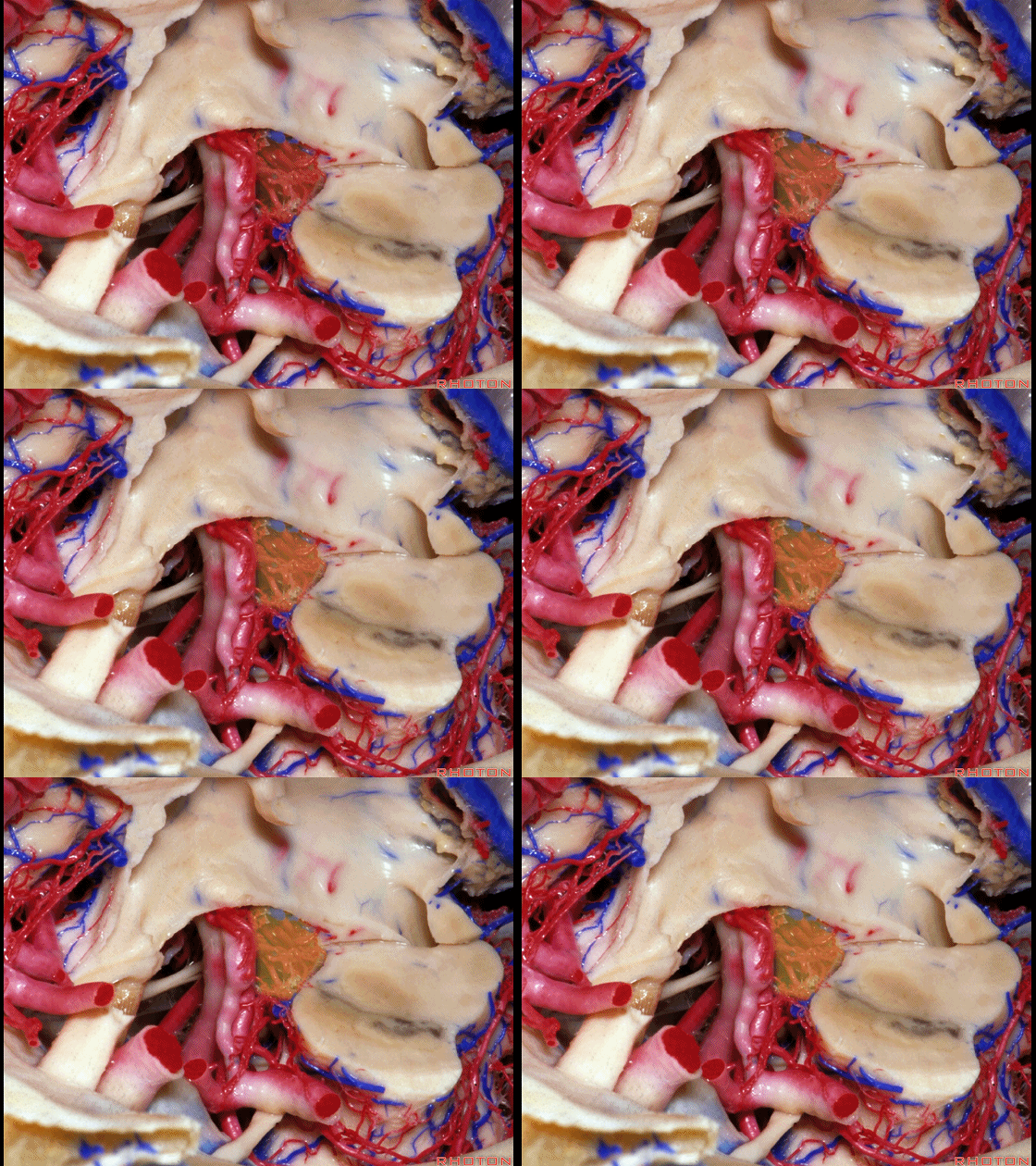
▼这是 丘脑穿支动脉
thalamoperforating arteries
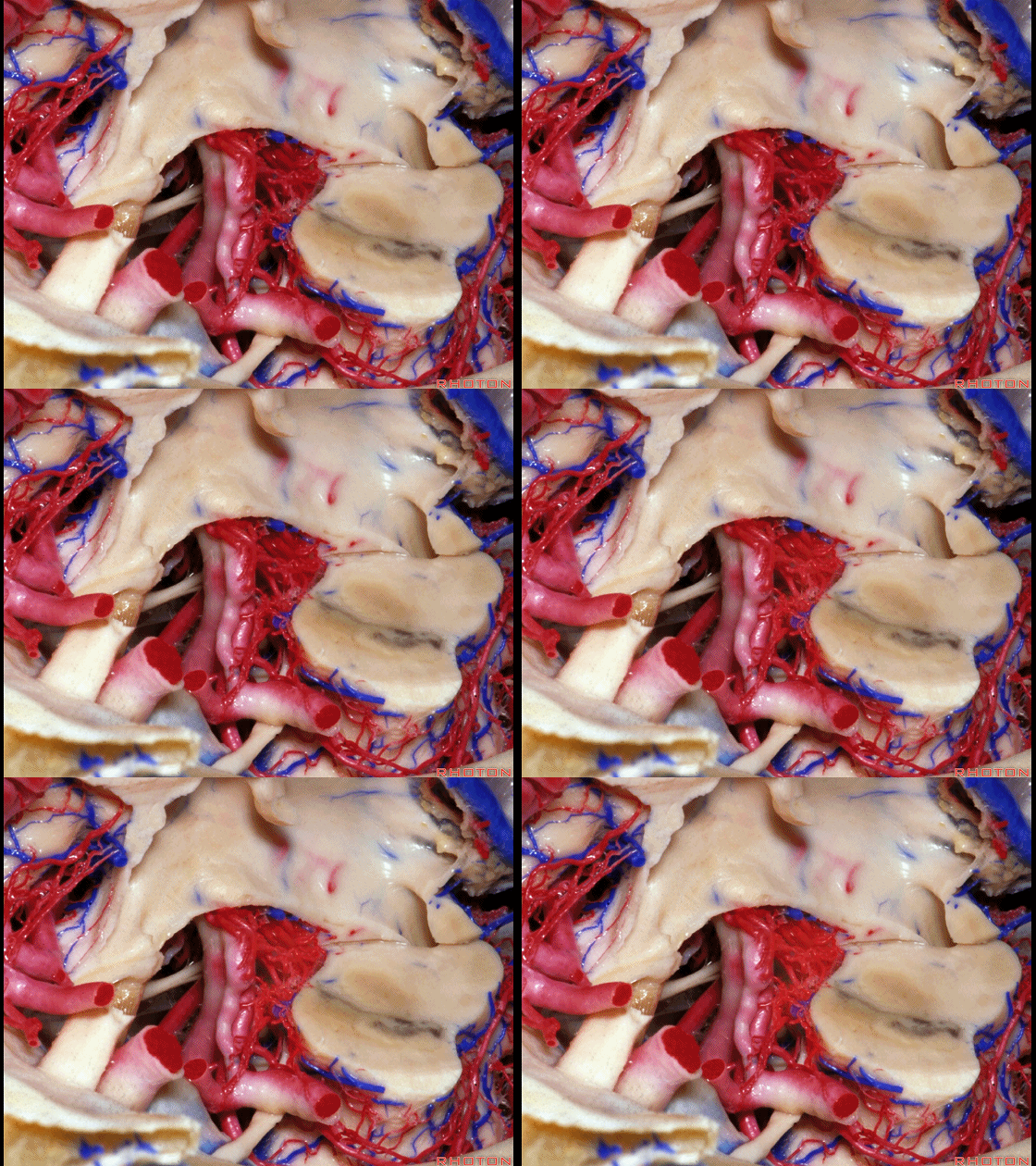
▼这是 中脑
midbrain
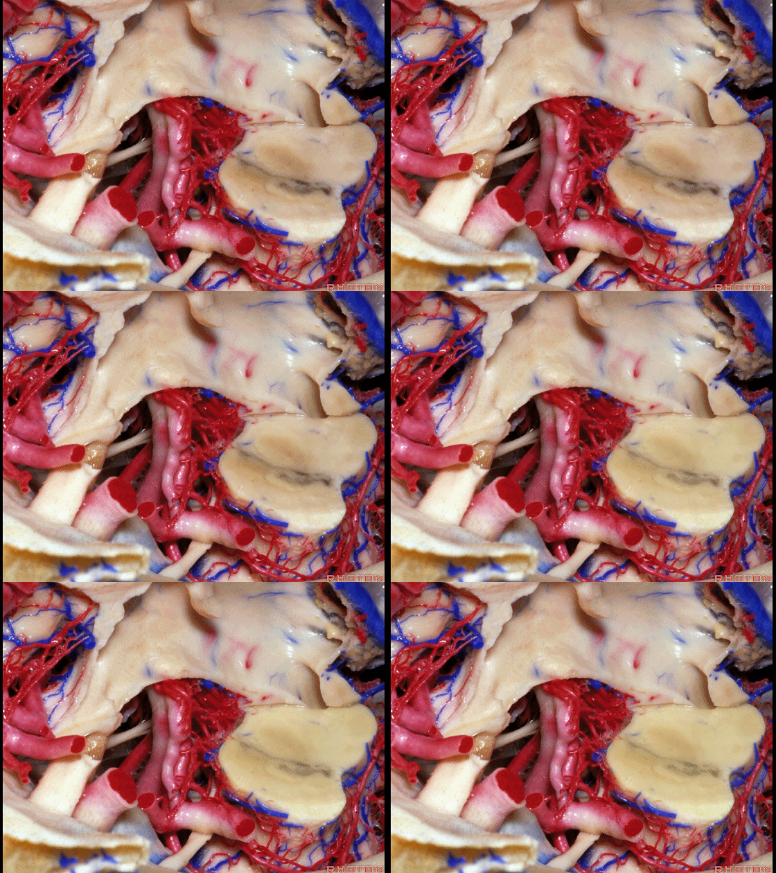
▼这是 动眼神经核
3rd nerve nuclei.
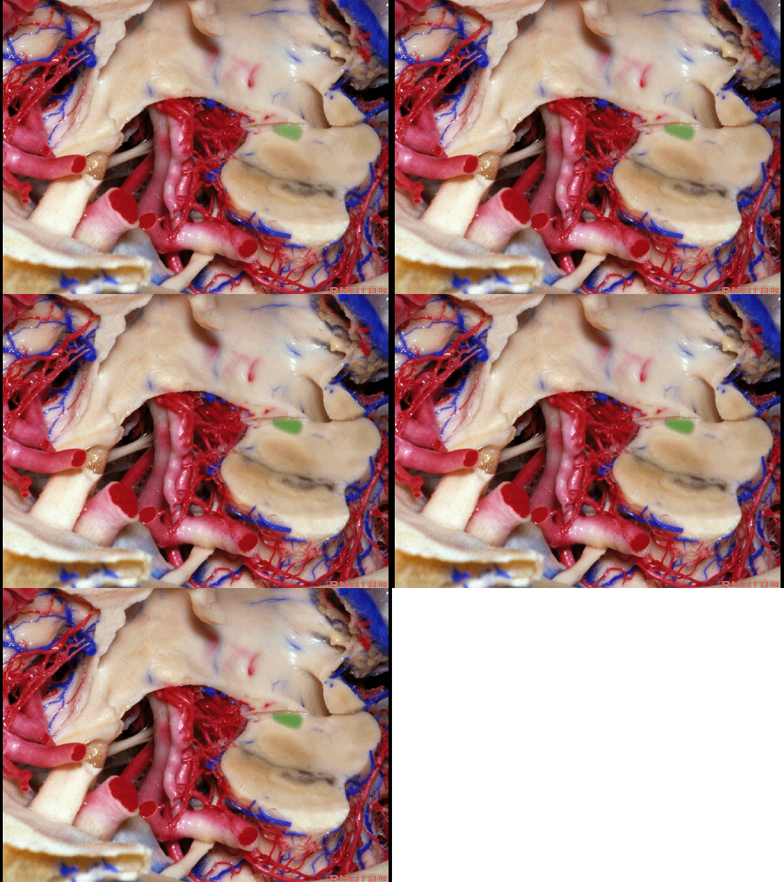
▼因此需在其前方,即在乳头体和漏斗隐窝之间 进行 三脑室底造瘘术。
So you always want to go forward here between mamillary bodies and infundibular recess, through this area for third ventriculostomy.
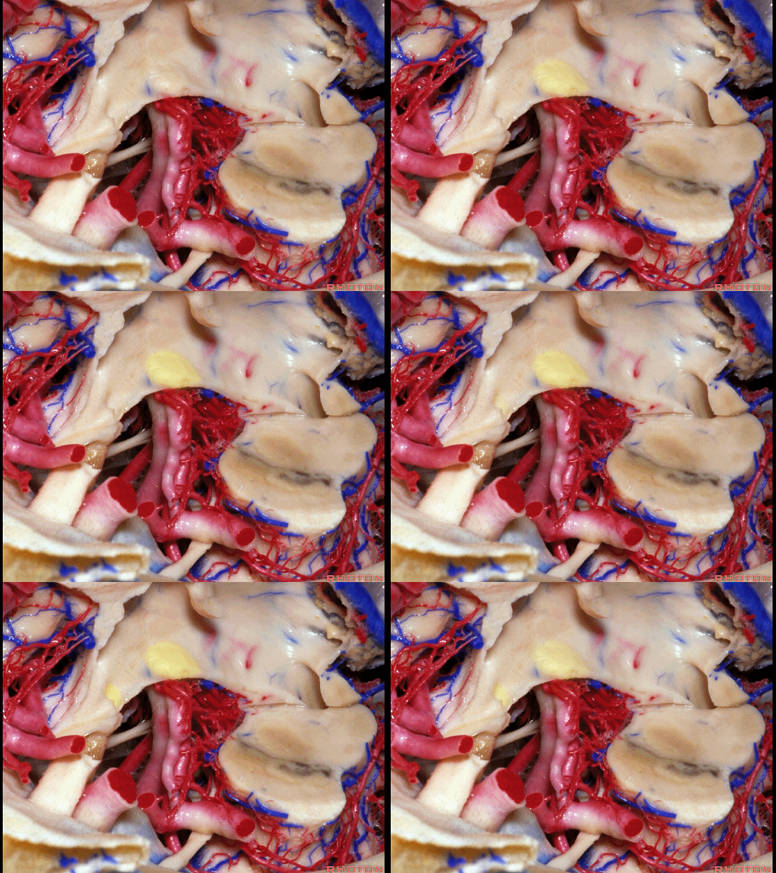
▼这是 动眼神经
What is this?3rd nerve
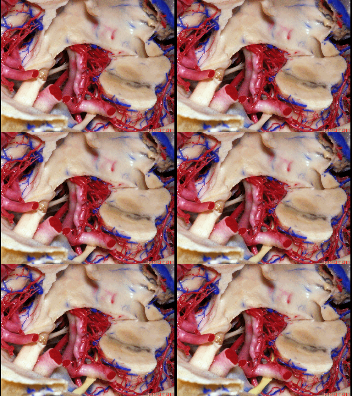
▼这是 大脑后动脉 P1段
This is..P1,
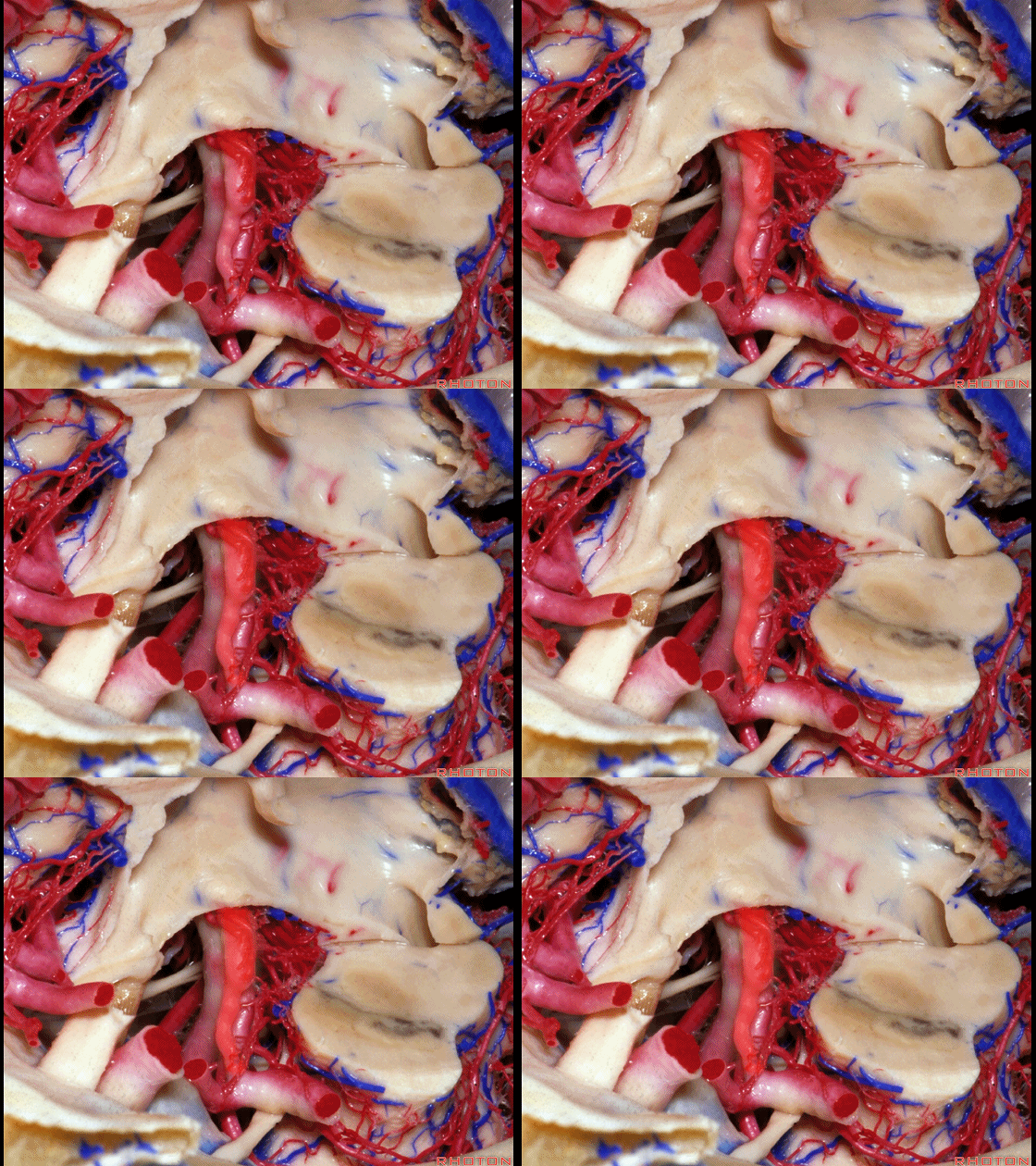
▼这是大脑后动脉 P2段

▼这是 脚池
crural cistern
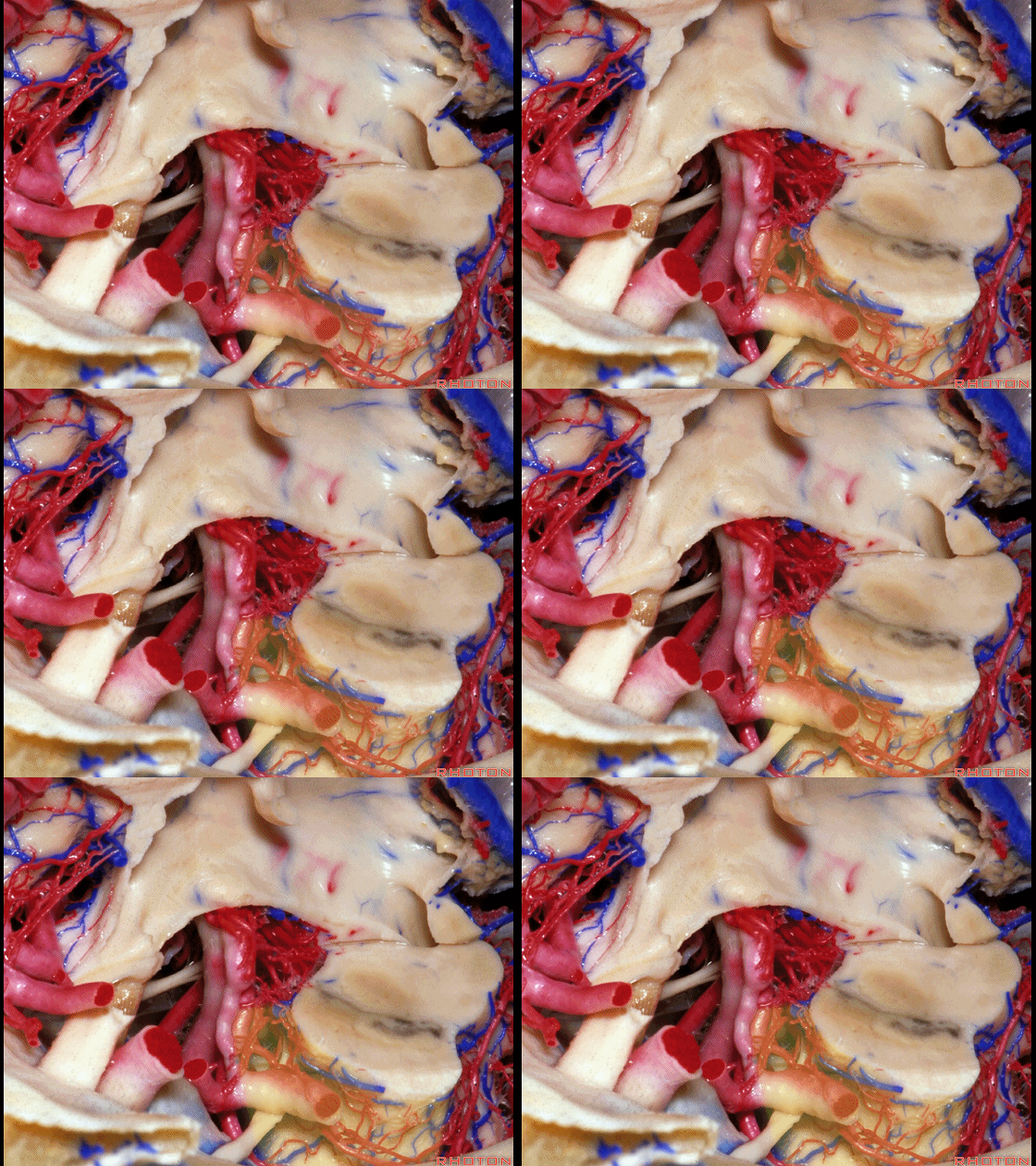
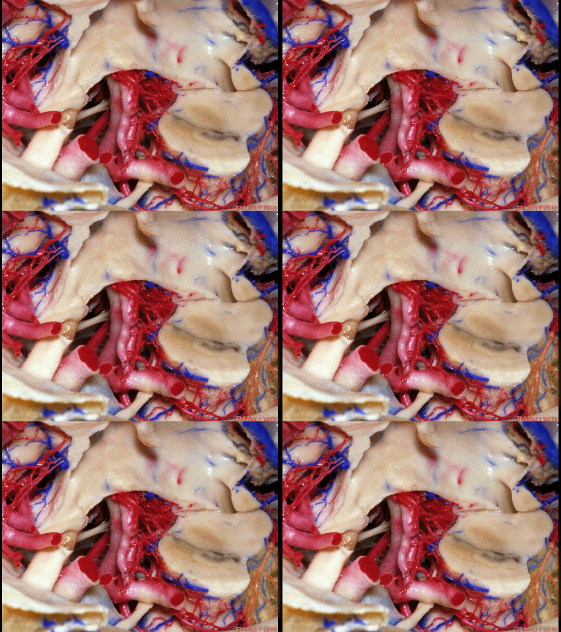
▼这是 环池
ambient cistern.
▼有一种情况下,我们可轻易地通过穹窿间入路在两侧穹窿之间(下图)进入第三脑室。
Now, the one situation where it's easy to get in to the...through the fornix in the midline to do an interforniceal approach,
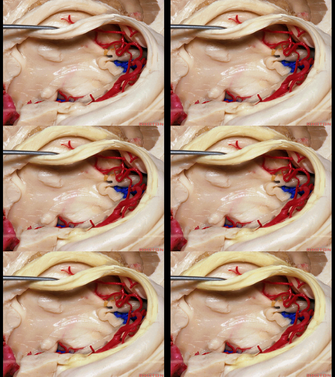
▼即当有一巨大腔隙(下图)分隔于两侧的穹窿体部之间时,我们可利用这一腔隙暴露第三脑室顶壁,在双侧的穹窿体部之间进行操作。
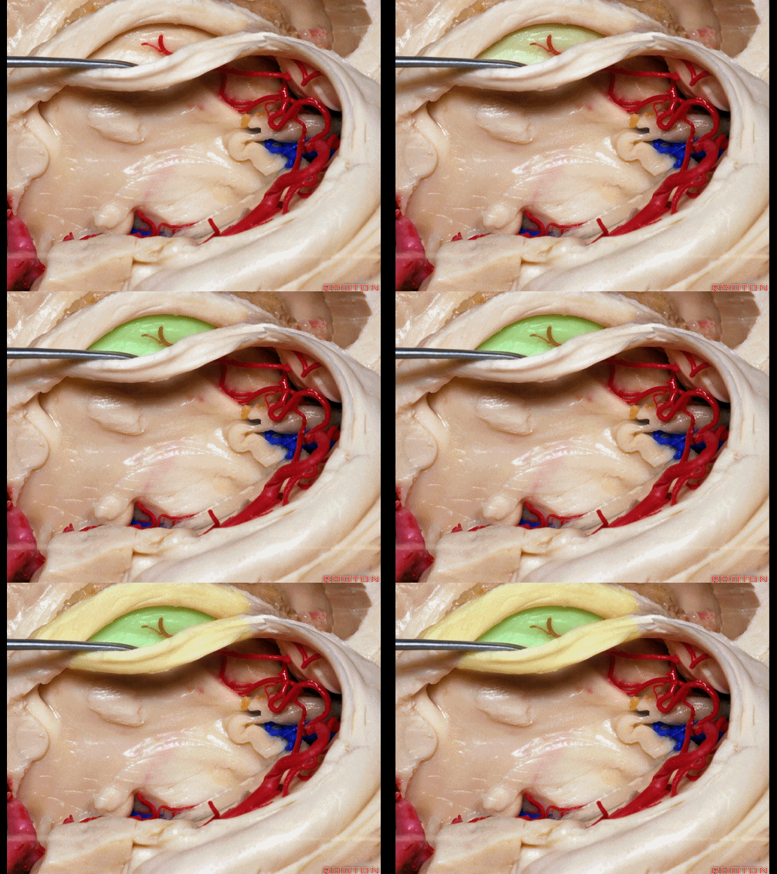
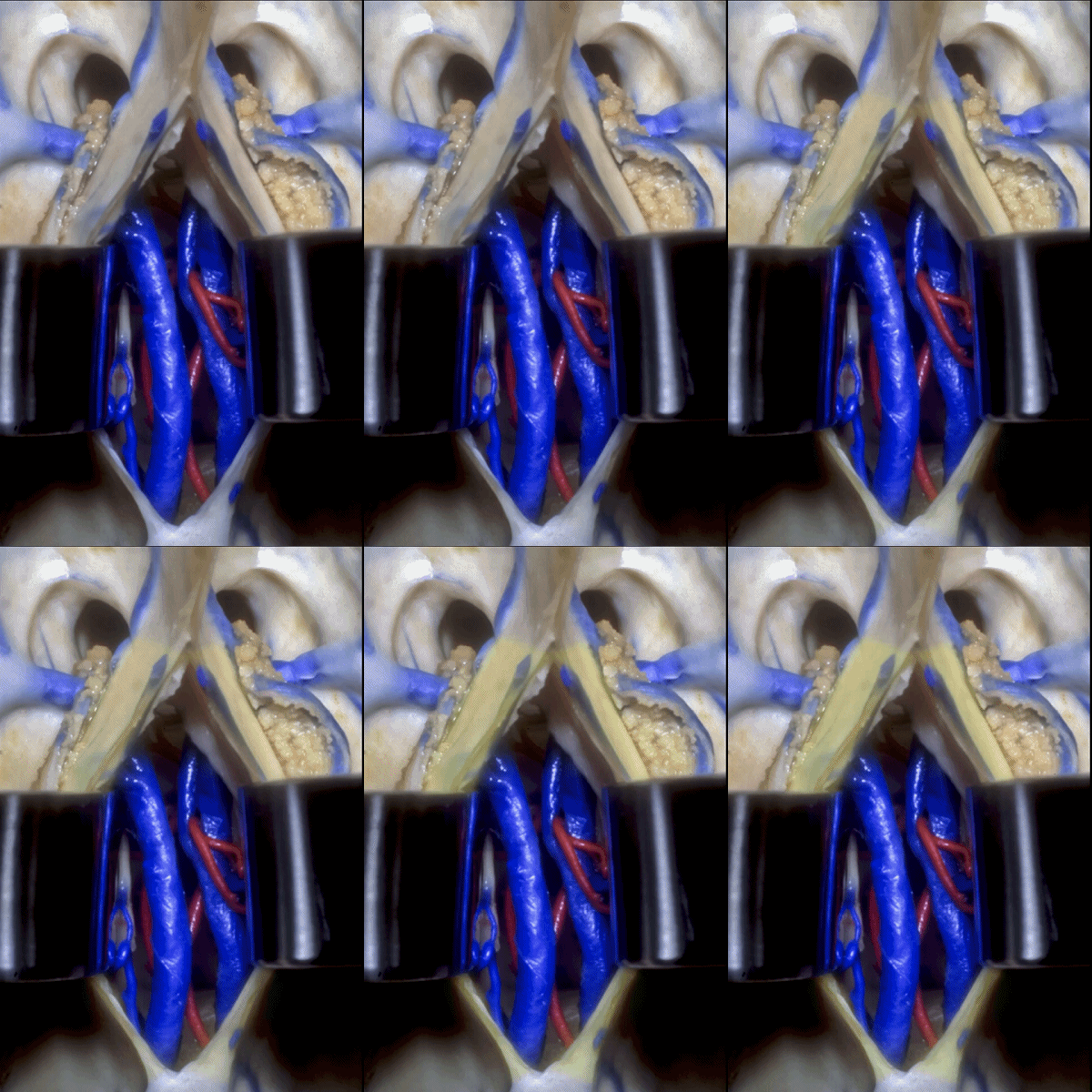
▼这时可通过穹窿间入路,经由穹窿体部,位于Monro孔的后方。这种情况下,我们会使用穹窿间入路,而非经脉络裂入路。
And that makes it easy to do an interforniceal approach that comes through then, through the body of the fornix here, behind the foramen of Monro. So that's one situation where we are now using interforniceal approach instead of coming through the choroidal fissure.
![]()
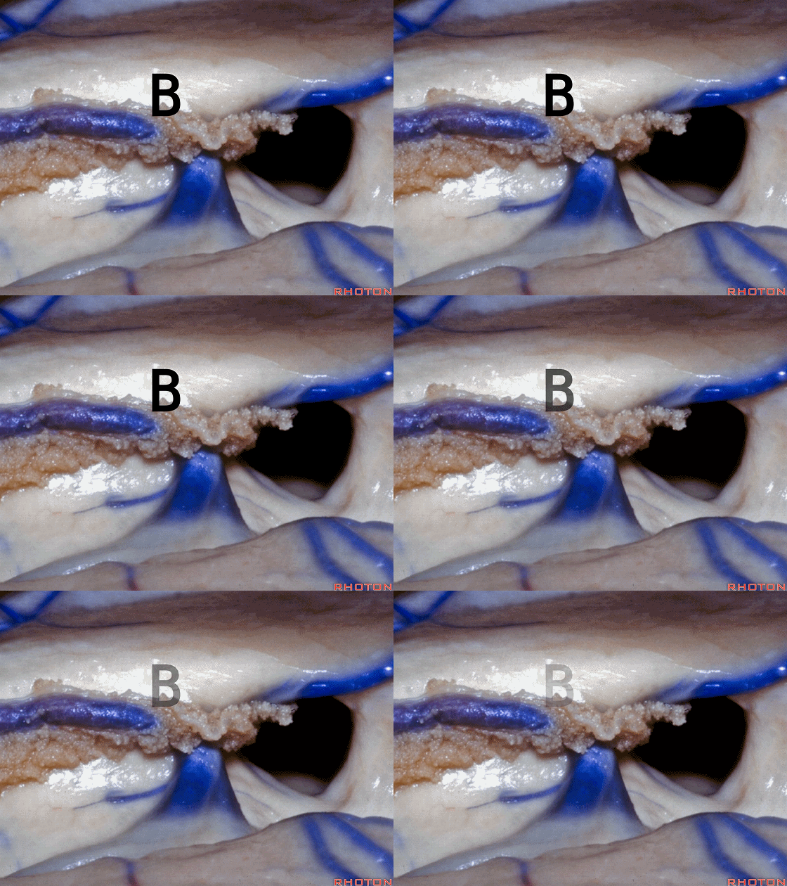
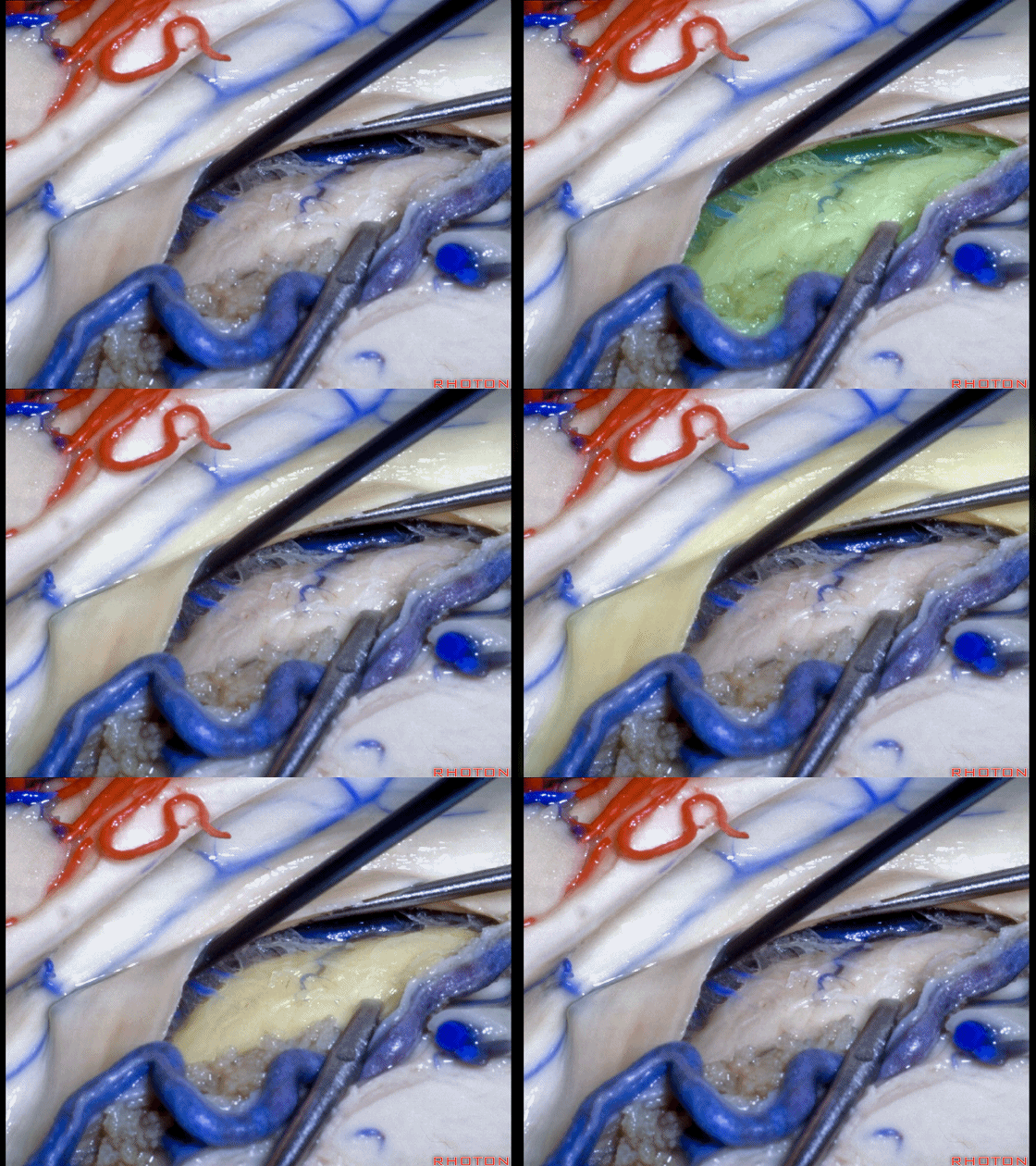
▼接下来我们还是要重点讨论脉络裂的解剖。 我们应该在Monro孔后方、穹窿侧的B点 打开脉络裂。
Now we're gonna work our way around the fissure. And...here were foramen of Monro. And we're gonna open the fissure, are we going to open the fissure behind the foramen at A or B?At B on the forniceal side.
▼继续沿着脉络裂向后暴露,穹窿、丘脑。
So we work our way back along the fissure,fornix,thalamus.
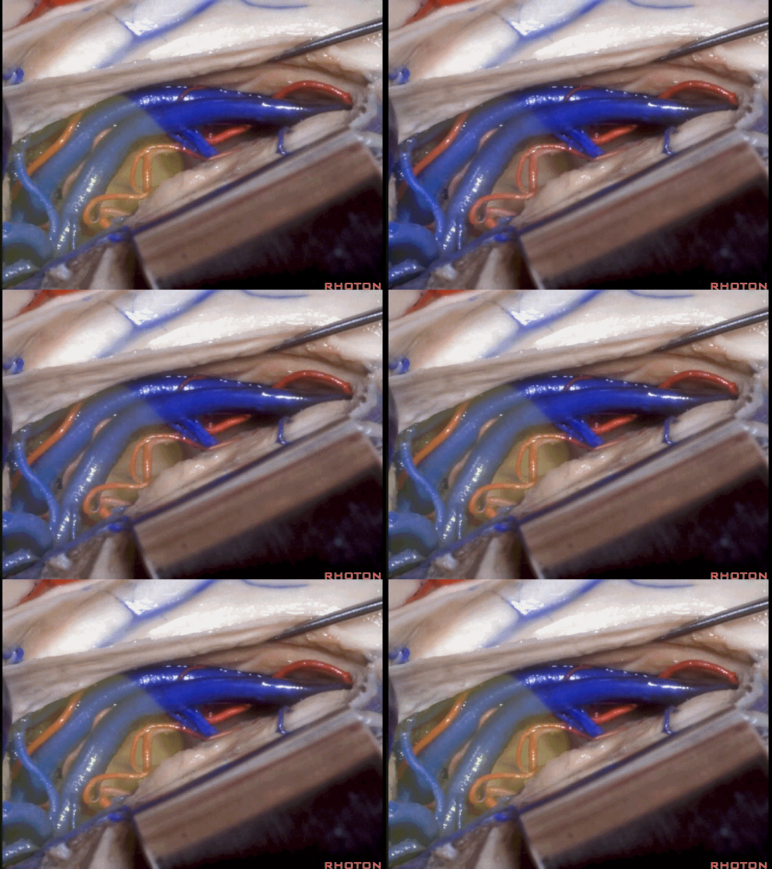
▼我们向后暴露至四叠体池(下图)。
We come back to the quadrigeminal cistern.
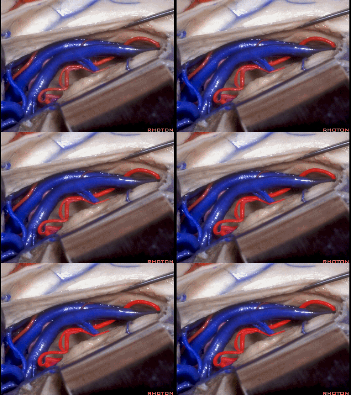
▼这是 脉络膜后内侧动脉
What arteries? Medial posterior choroidal,
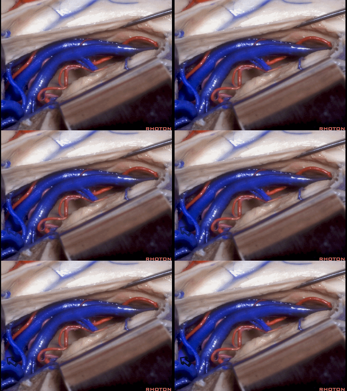
▼这是 Galen静脉(大脑大静脉)
vein of Galen.
▼我们将脉络丛始终留于丘脑与内囊那一侧。沿着脉络裂继续深入内侧,可见基底静脉(下图)
We're looking...we've opened...we let the choroid plexus always go with the thalamus. and internal capsule. And as you work directly medial and come around the fissure, we see the...basal vein
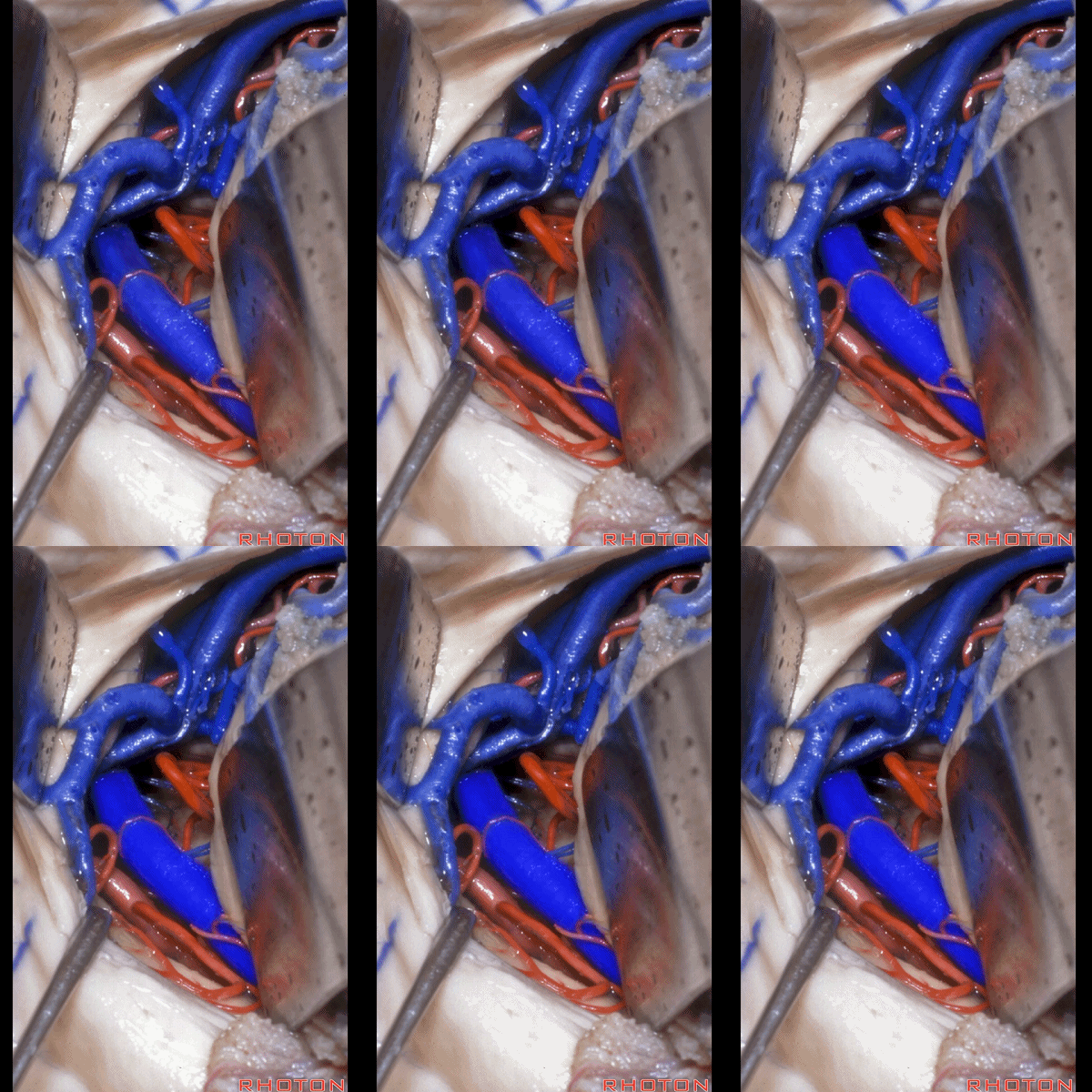
▼这是 大脑内静脉
internal cerebral vein
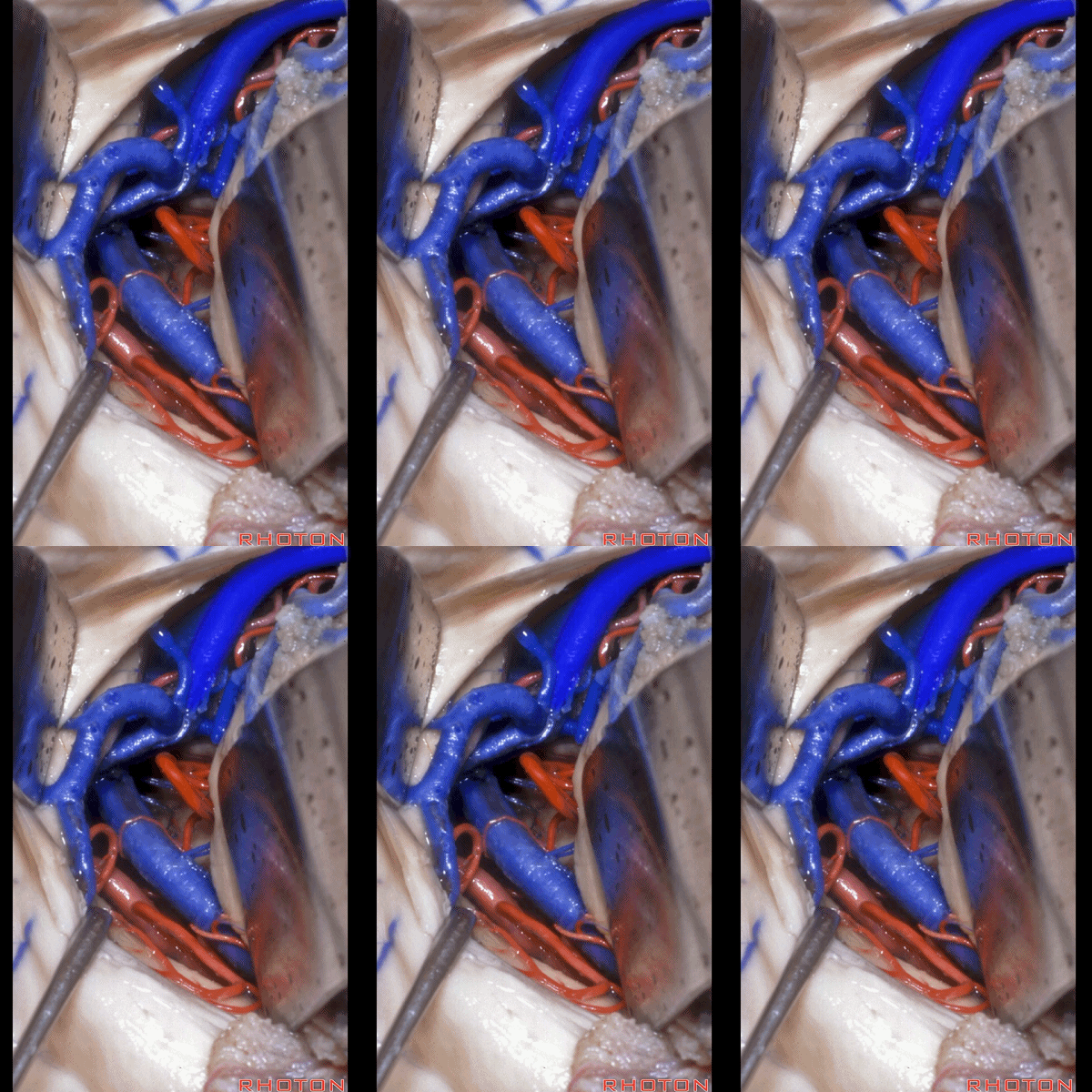
▼这是 Galen静脉
vein of Galen.
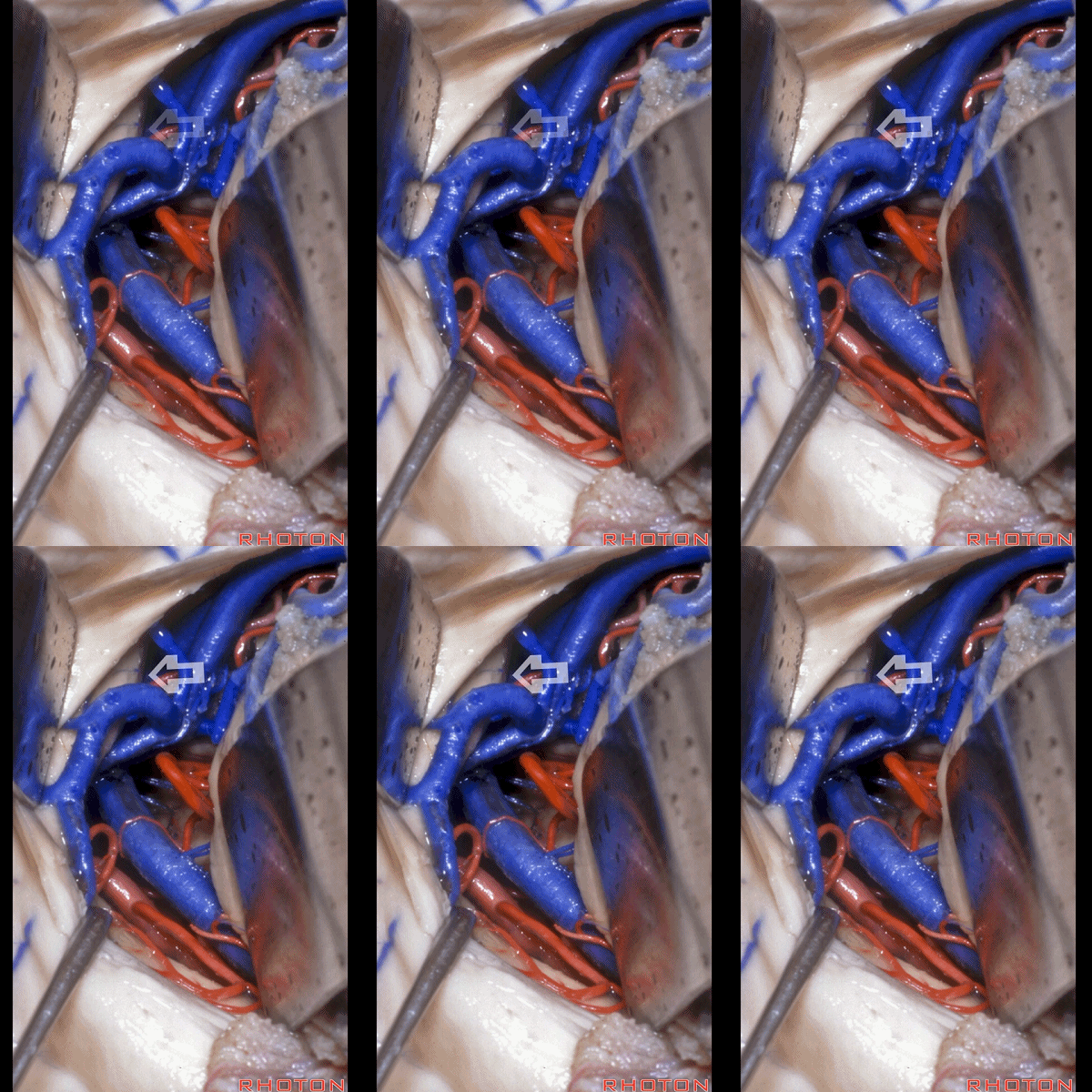
▼而就在此处脉络裂的内侧,可见松果体(下图)。以上就是经脉络裂 至 四叠体池 入路
And right in this area directly medial to the fissure, you see the pineal. So this is an approach through the choroidal fissure,to the quadrigeminal cistern.
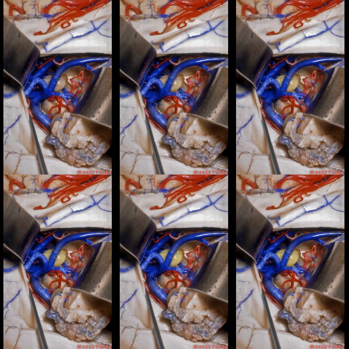
▼现在需要在侧脑室房部用到此入路的情况,是当我们处理侧脑室房部的血管畸形、脑膜瘤,或 脉络丛肿瘤。因此这就是经侧脑室房部 脉络裂入路暴露四叠体池。
The only time that we're coming through this route from the atrium is it for dealing with lesions and the glomus like AVMs or meningeomas in the atrium, or choroid plexus tumors. So, this will be a transchoroidal approach from the atrium to the quadrigeminal cistern.
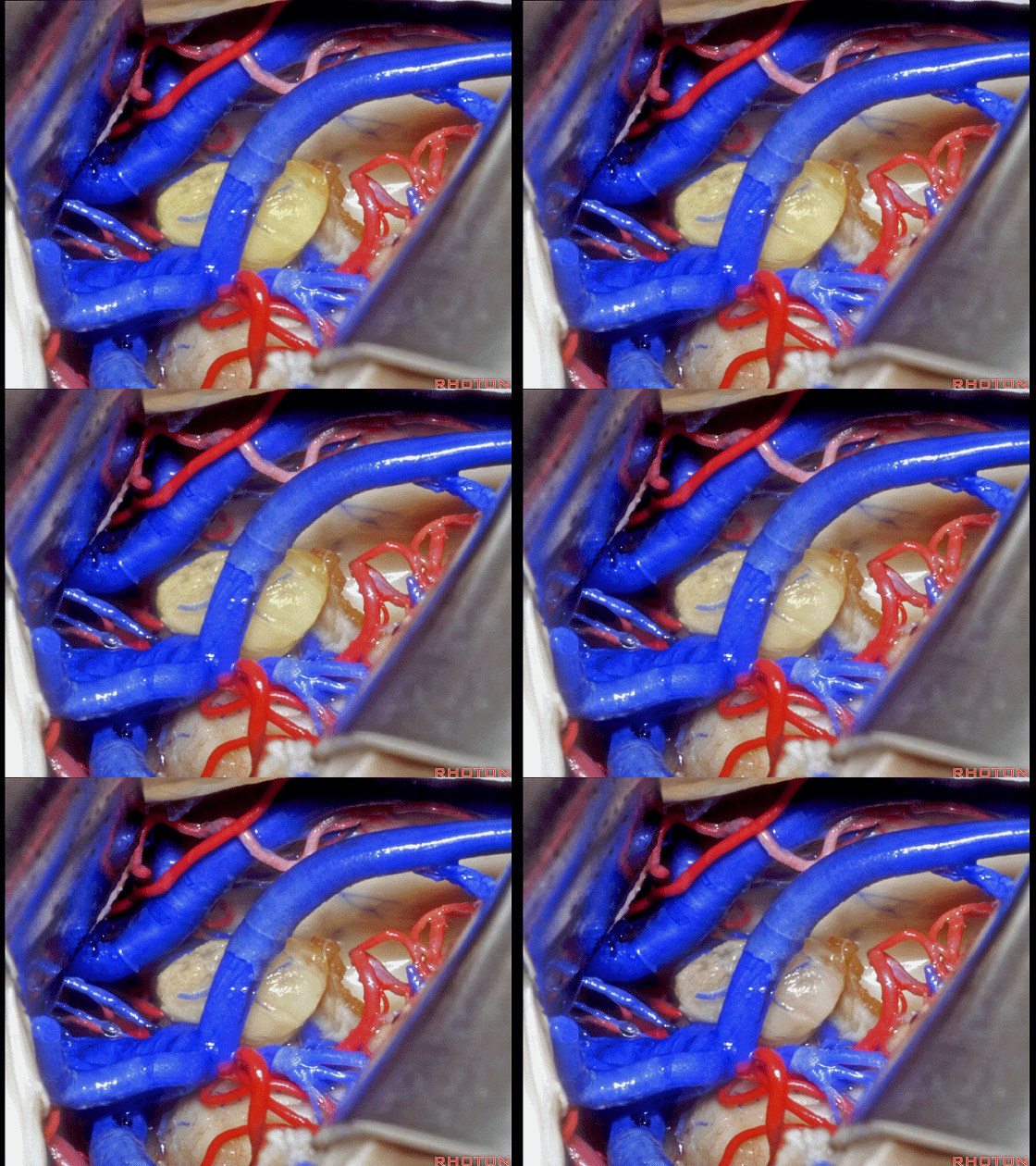
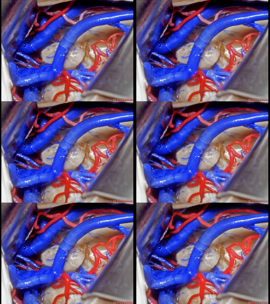
▼脉络丛肿瘤 其由 脉络膜动脉(下图) 供血并引流于 脉络膜静脉。
that are fed by choroidal arteries and drained by choroidal veins.
![]()
▼对于侧脑室房部的暴露,我们也可经由大脑半球的内侧面。这是 距状沟 calcarine sulcus
Usually for atrium, we'll either come in through the medial surface of the hemisphere. And here we see...what sulcus?
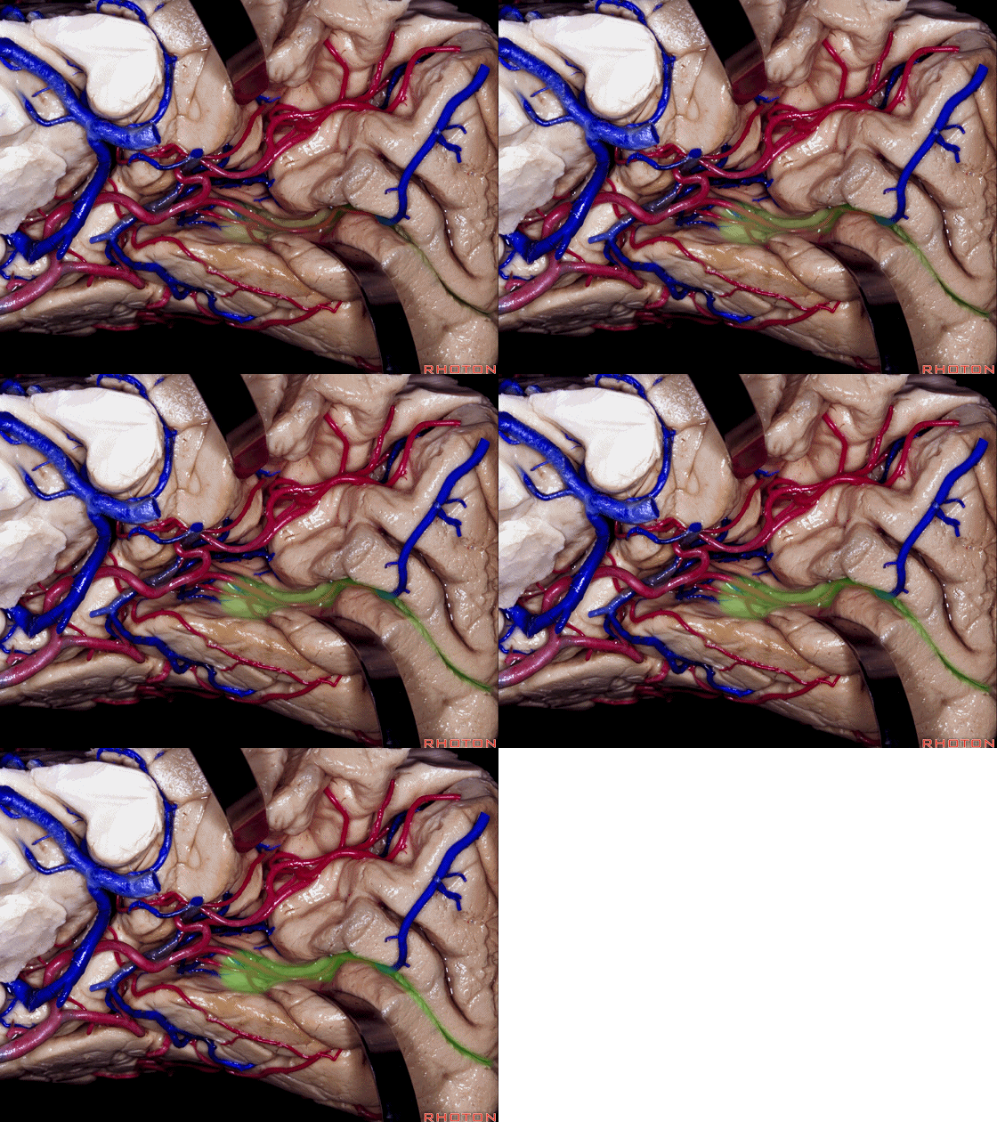
▼这是 顶枕沟。
parieto-occipital.
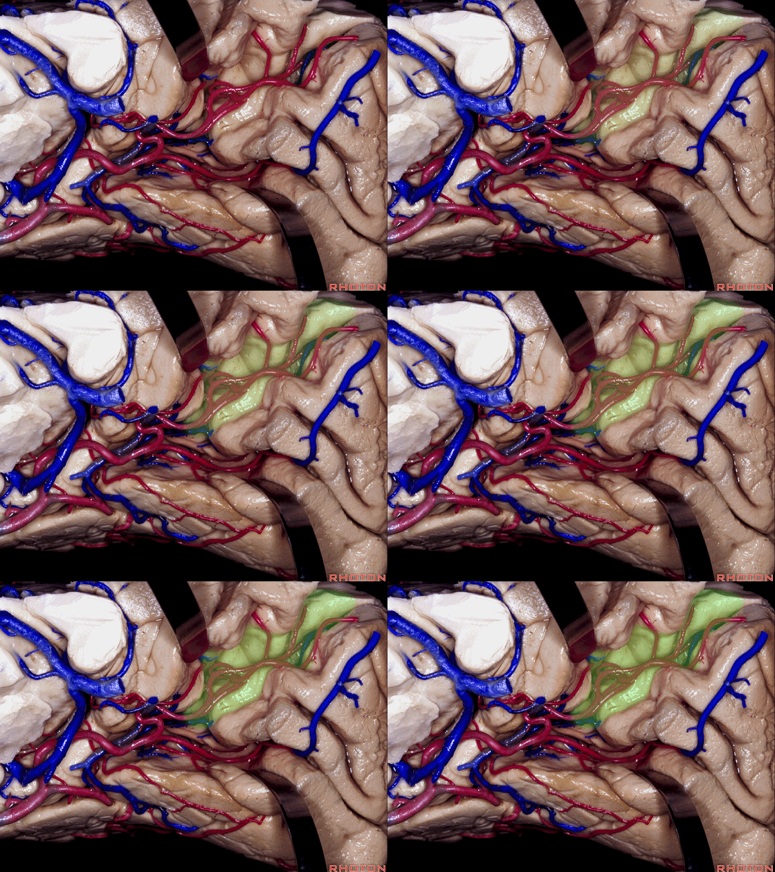
▼这是 舌回
This is lingual gyrus ,

▼位于距状沟和顶枕沟之间的脑叶是 楔叶。这是下象限视野的投射区。
and between calcarine and parieto-occipital sulcus, we call that... cuneus. That's lower visual field.
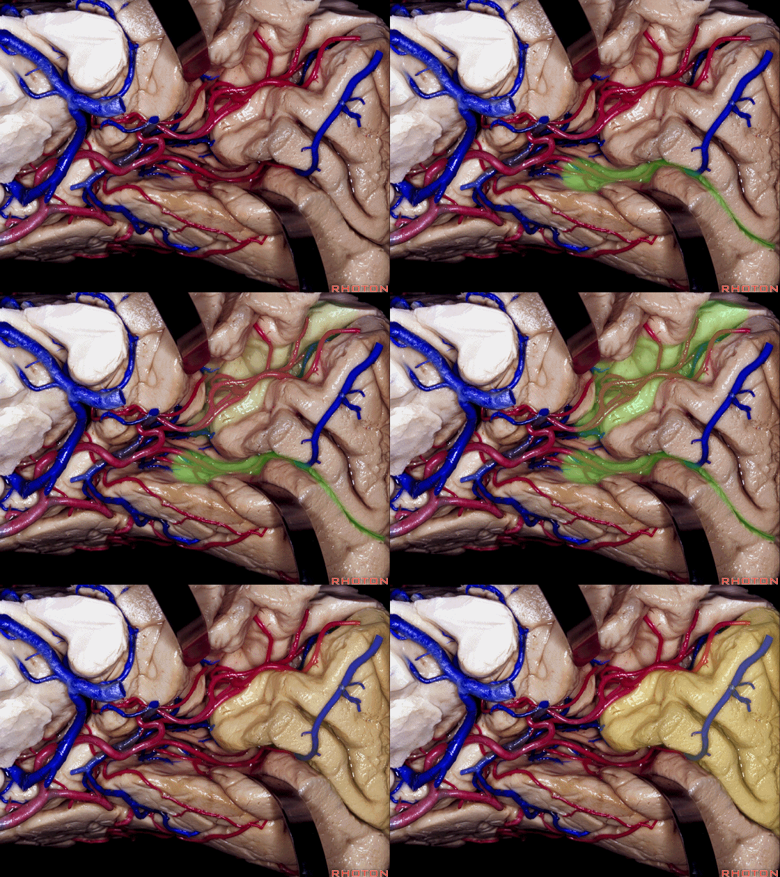
▼位于顶枕沟前方的是 楔前叶。
And then this area in front of parieto-occipital area is precuneus.
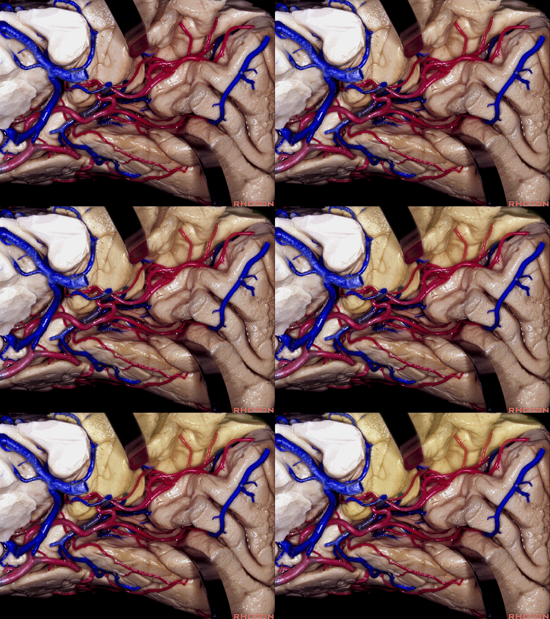
▼再从后方看一下枕叶内侧,这是 距状沟
calcarine sulcus
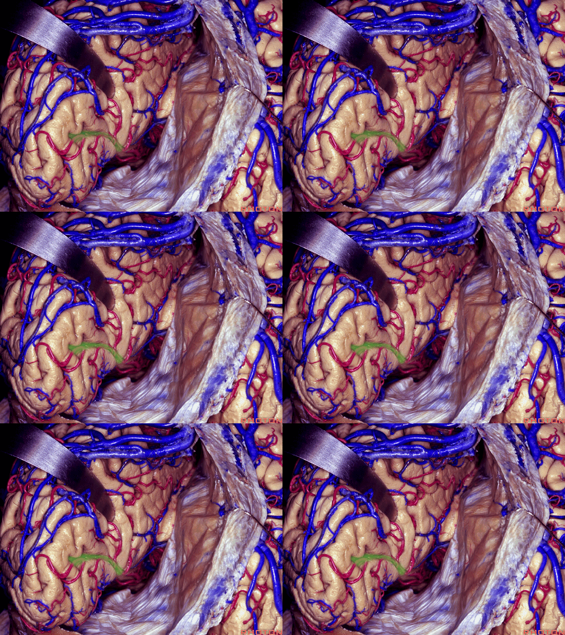
▼这是 顶枕沟
parieto-occipital sulcus
![]()
▼这是 楔前叶,其位于视野投射区的前方,
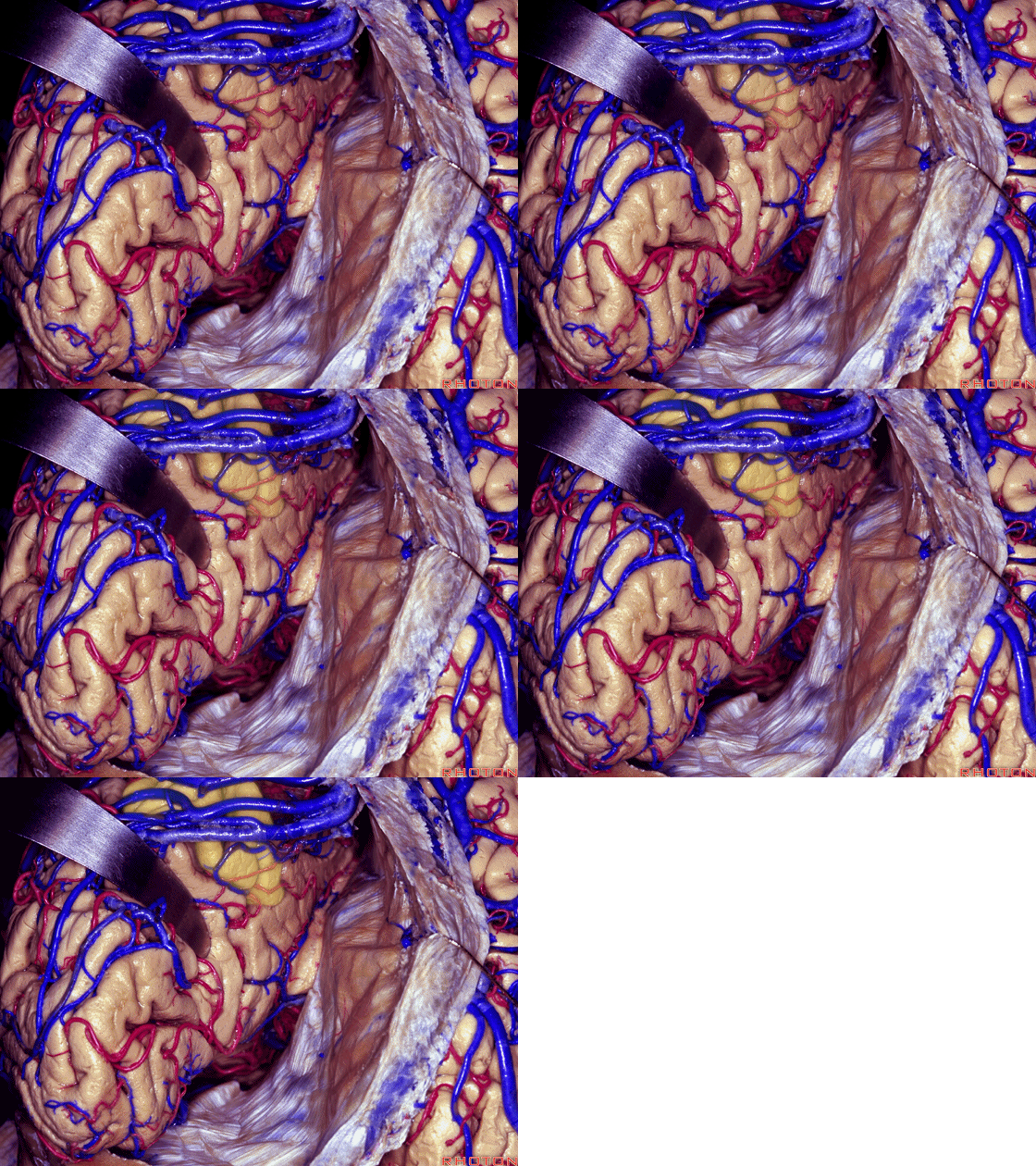
▼Yassagil发明了 楔前叶入路。可经由楔前叶进入 侧脑室房部。
So that Yassagil is giving us this precuneus approach. you can come through precuneus to atrium.

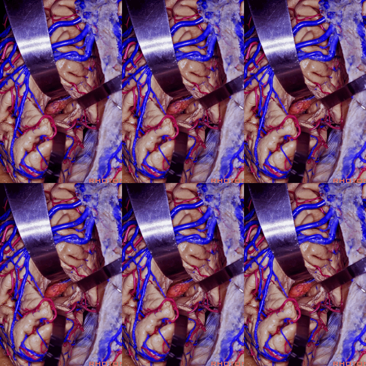
▼顶枕沟也深入到大脑半球内侧面,打开它可进入侧脑室房部。
Also this parieto-occipital sulcus cuts deeply into the medial surface of the hemisphere and you can open it and come...

▼这一侧是 楔叶
this is cuneus side
▼这一侧是 楔前叶
precuneus side
▼我们可打开前方的 楔前叶 进入 侧脑室房部。
You can open through this anterior wall to precuneus side into the atrium,

▼这是侧脑室房部的 脉络球。但我们不能打开 顶枕沟的楔叶侧。
▼或者也可经由此处(下图)的胼胝体外侧界,进入侧脑室房部。
Or you can come through the lateral margin of the corpus callosum here, just in this area to the atrium.
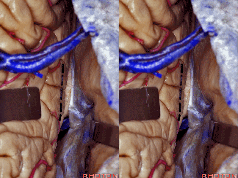
▼这属于经胼胝体入路 至侧脑室房部。
That will be considered a transcallosal approach.
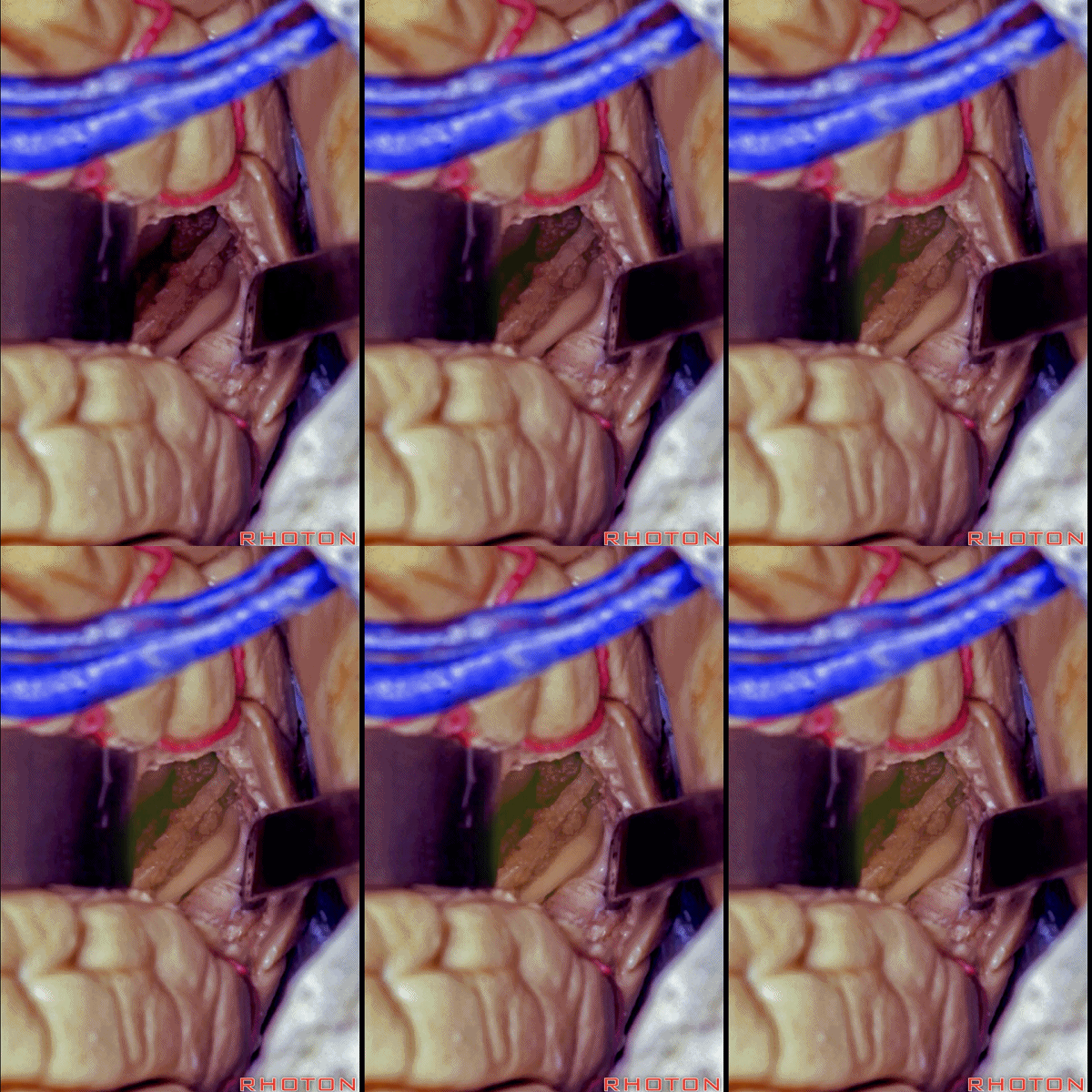
▼然而最常用的入路还是经由 顶上小叶 或顶间沟。下图示 顶上小叶 superior parietal lobule。
But probably the most common approach is to come through the superior parietal lobule or the interparietal sulcus.
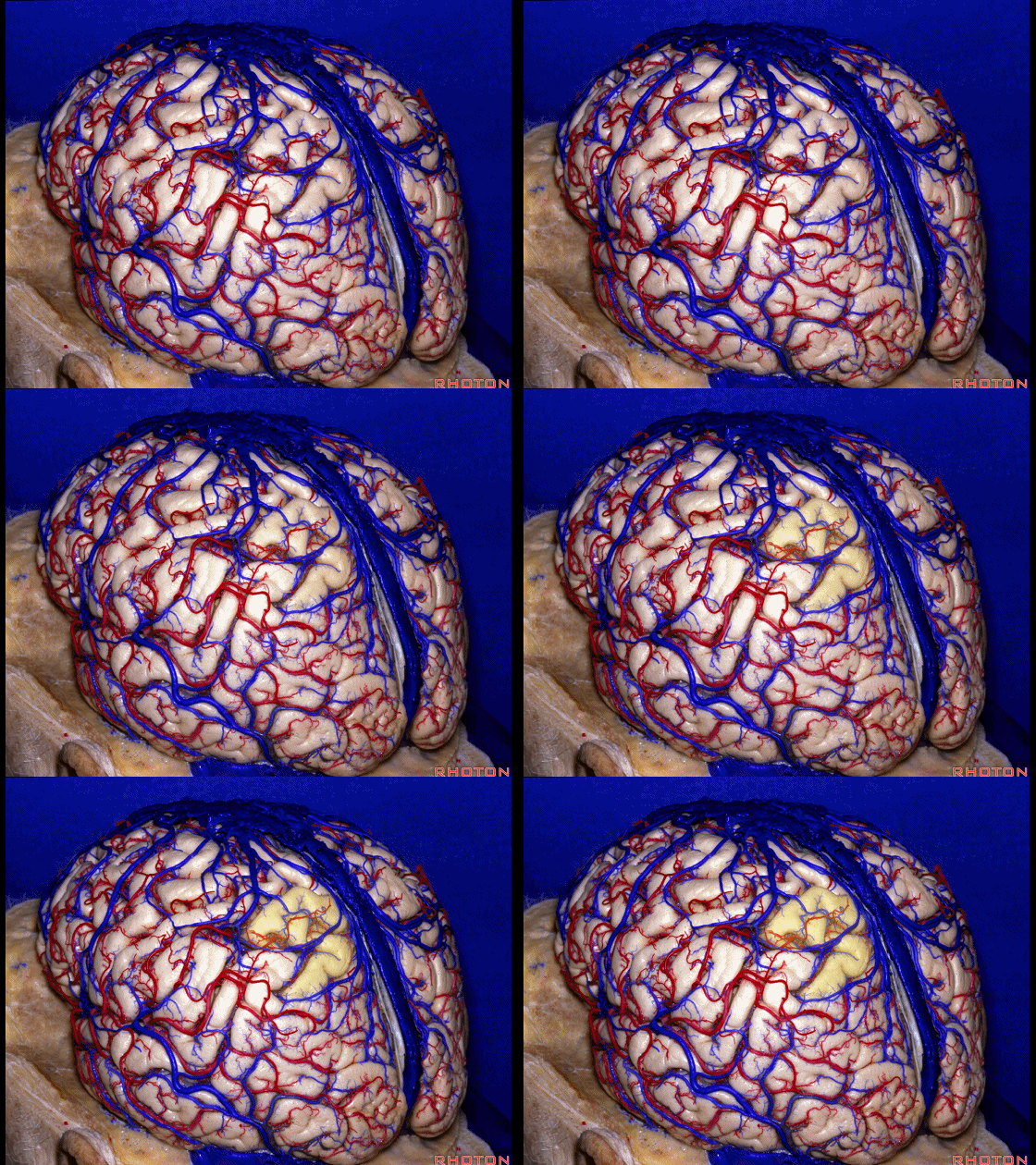
▼下图示 顶间沟
interparietal sulcus
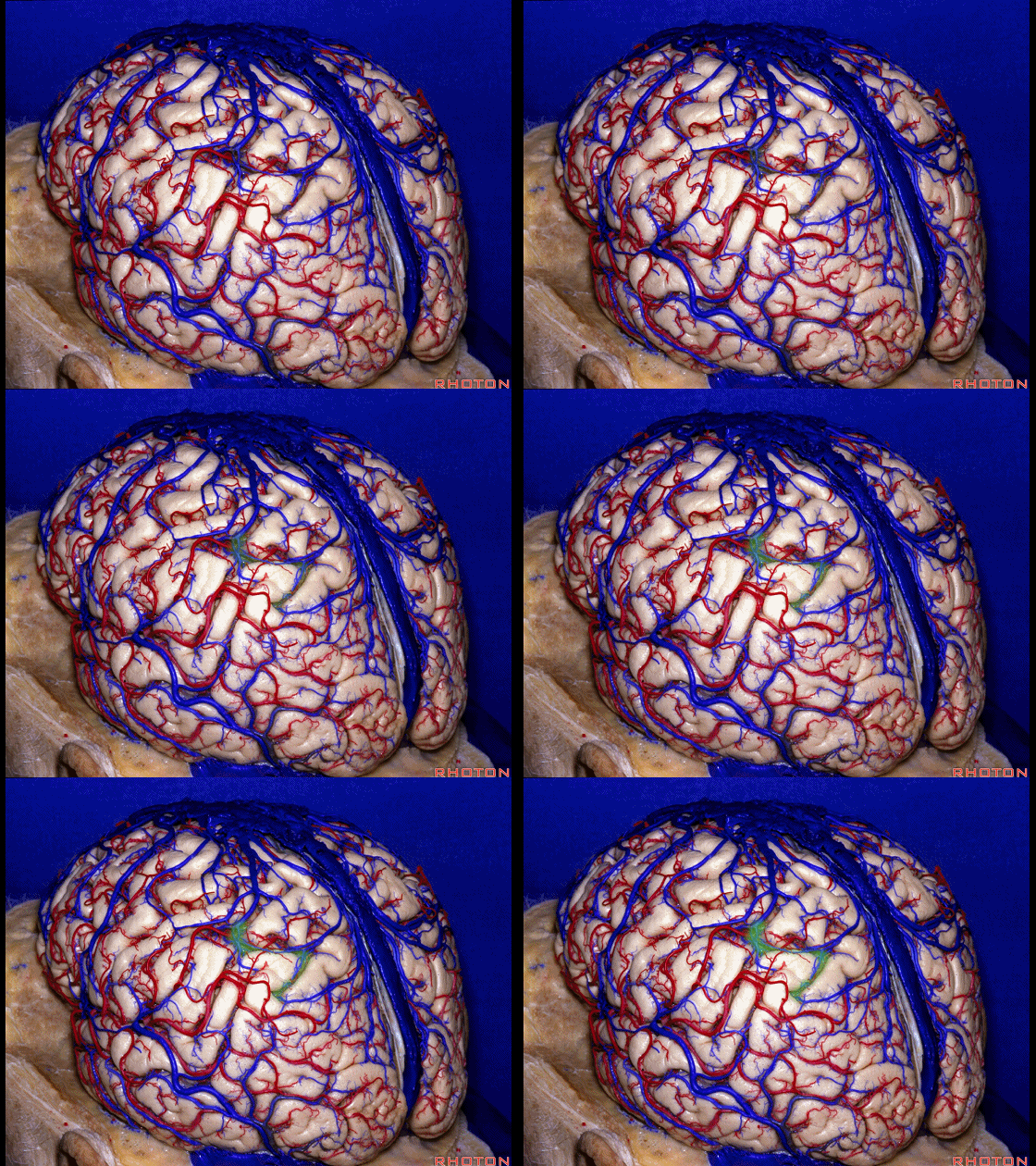
▼顶间沟深入到半球内部。我们可经由它进入侧脑室房部
And that interparietal sulcus cuts deeply into the hemisphere. And you can open through it into the atrium,
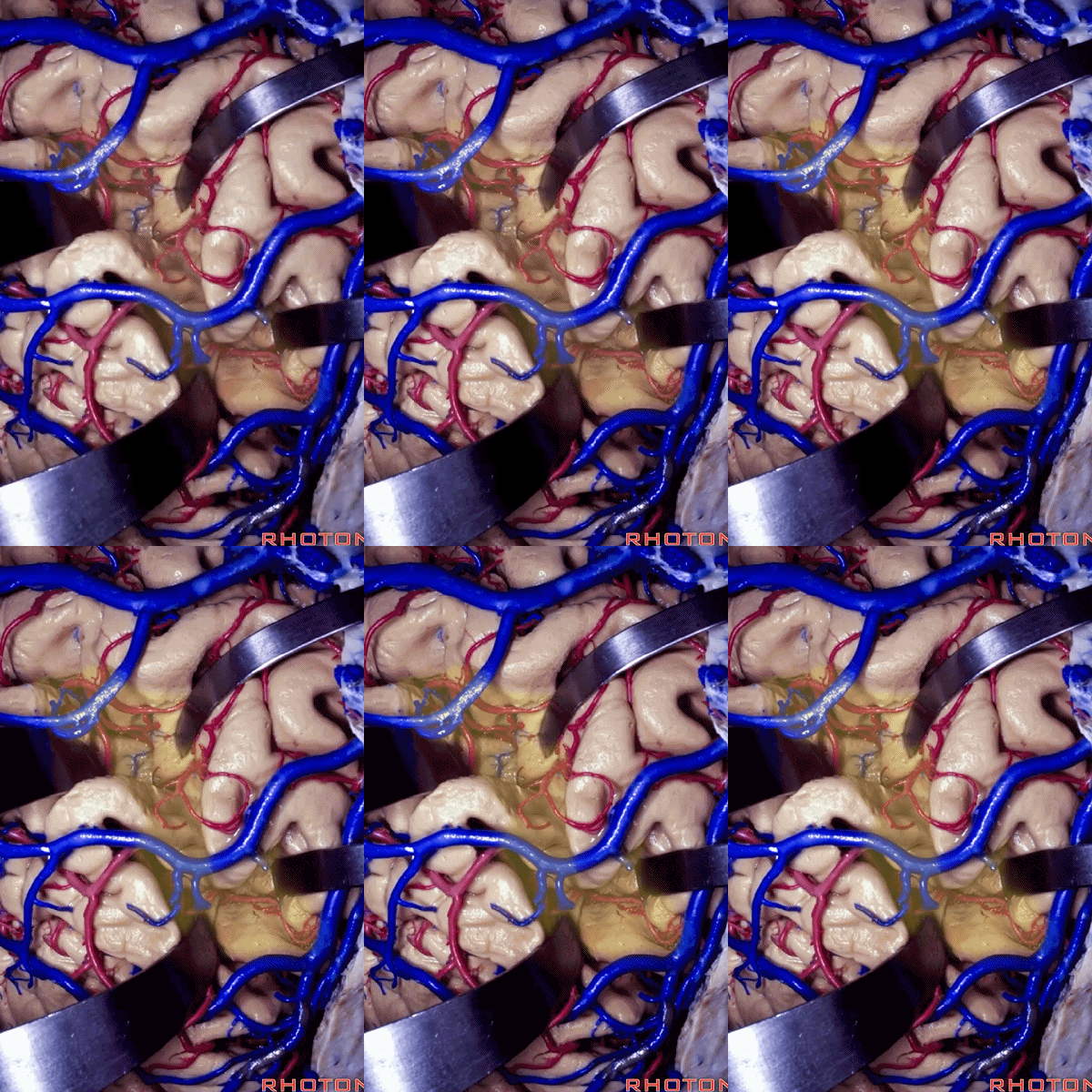
▼下图示 打开后的 侧脑室房部
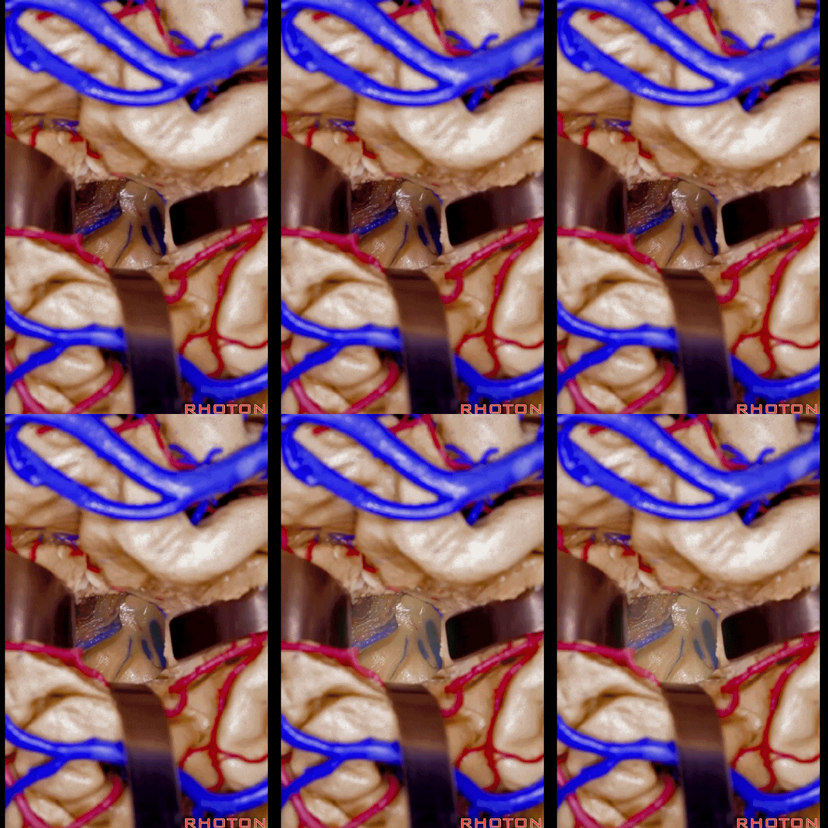
▼尽管我认为更常用的还是经顶上小叶 入路。

![]()
▼现在来看松果体区的入路,下图示 松果体
But for approaches to the pineal region,
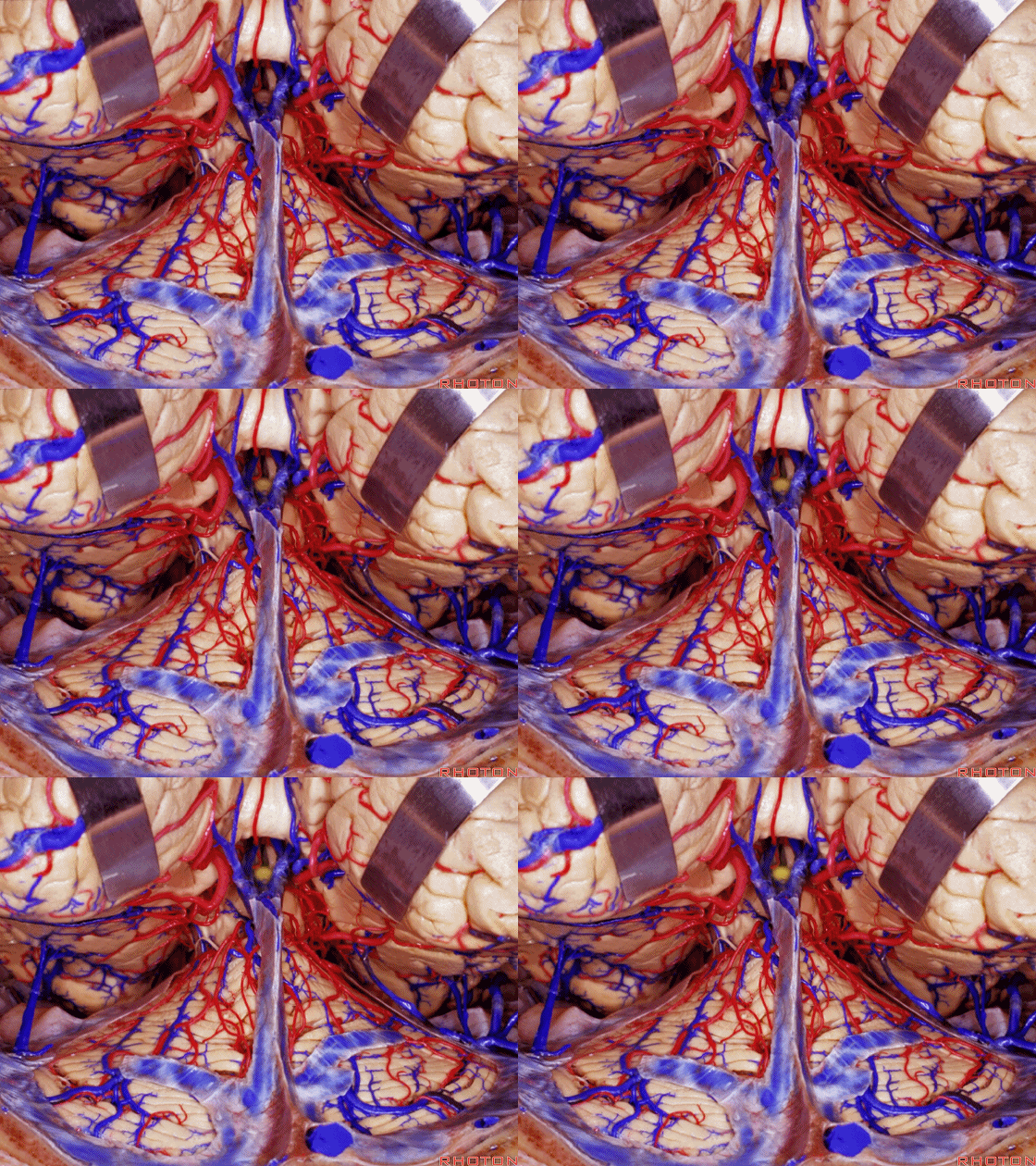
▼我们可从幕上,也可经幕下。大体上,我认为成人神经外科医师偏好幕上入路,而小儿神经外科医师则偏好幕下入路。
we either come above, or below the tentorium. In general, I will say surgeons operating on adults prefer to come above the tent, pediatric neurosurgeons tend to use the infratentorial approaches more commonly.
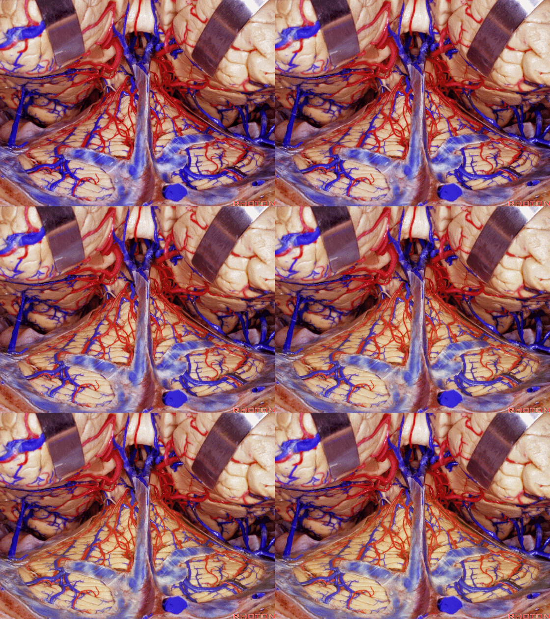
▼来看看松果体区,这是 上丘、下丘。
And here we're looking at pineal region, superior, inferior colliculi, 4th nerve.

▼这是 滑车神经
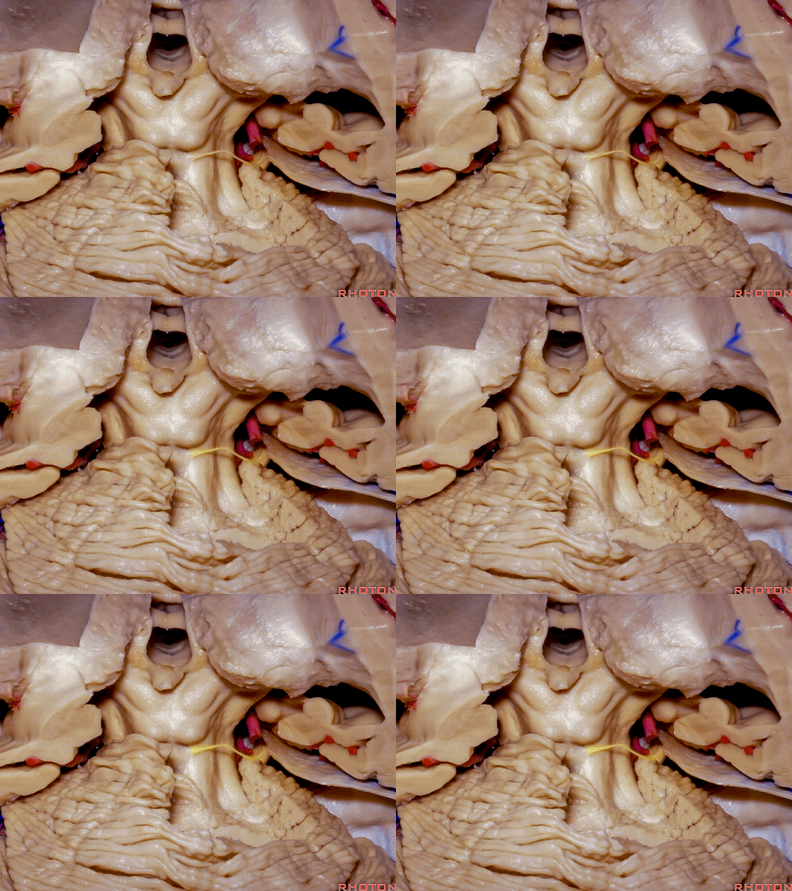
▼如果从小脑蚓尖部的上方进入,很难暴露至下丘的下方,并沿着丘板向下至滑车神经。
And if you come over the apex of the vermis, it's difficult to get down below the inferior colliculus, along the collicular plate down to the 4th nerve.
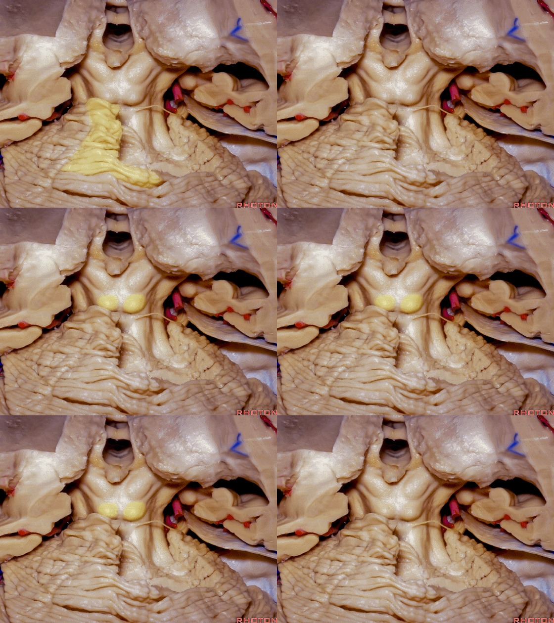
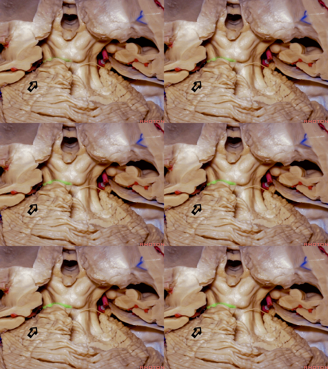
▼但如果采用旁正中入路, 将更轻易地暴露小脑中央前裂,也称为 小脑中脑裂(下图)。
But if you come paramedian off the midline, it's easier to get low in this precentral cerebellar fissure or we call it cerebello-midbrain fissure.
▼这是正中入路。经小脑蚓尖上方的暴露。下图示 蚓上静脉。
So here's the median approach over the apex of the vermis. Superior vermian vein.
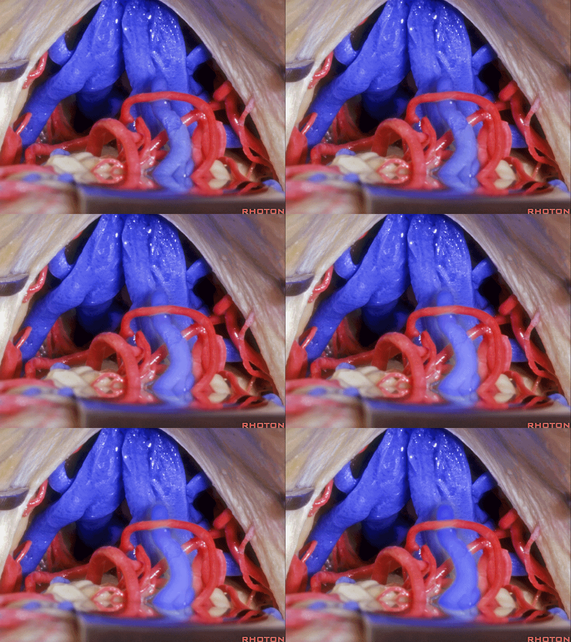
▼这是 基底静脉
Here is basal vein
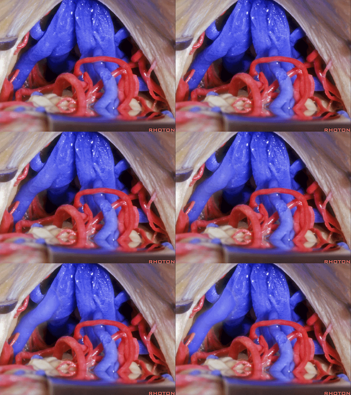
▼这是 大脑内静脉
internal cerebral vein
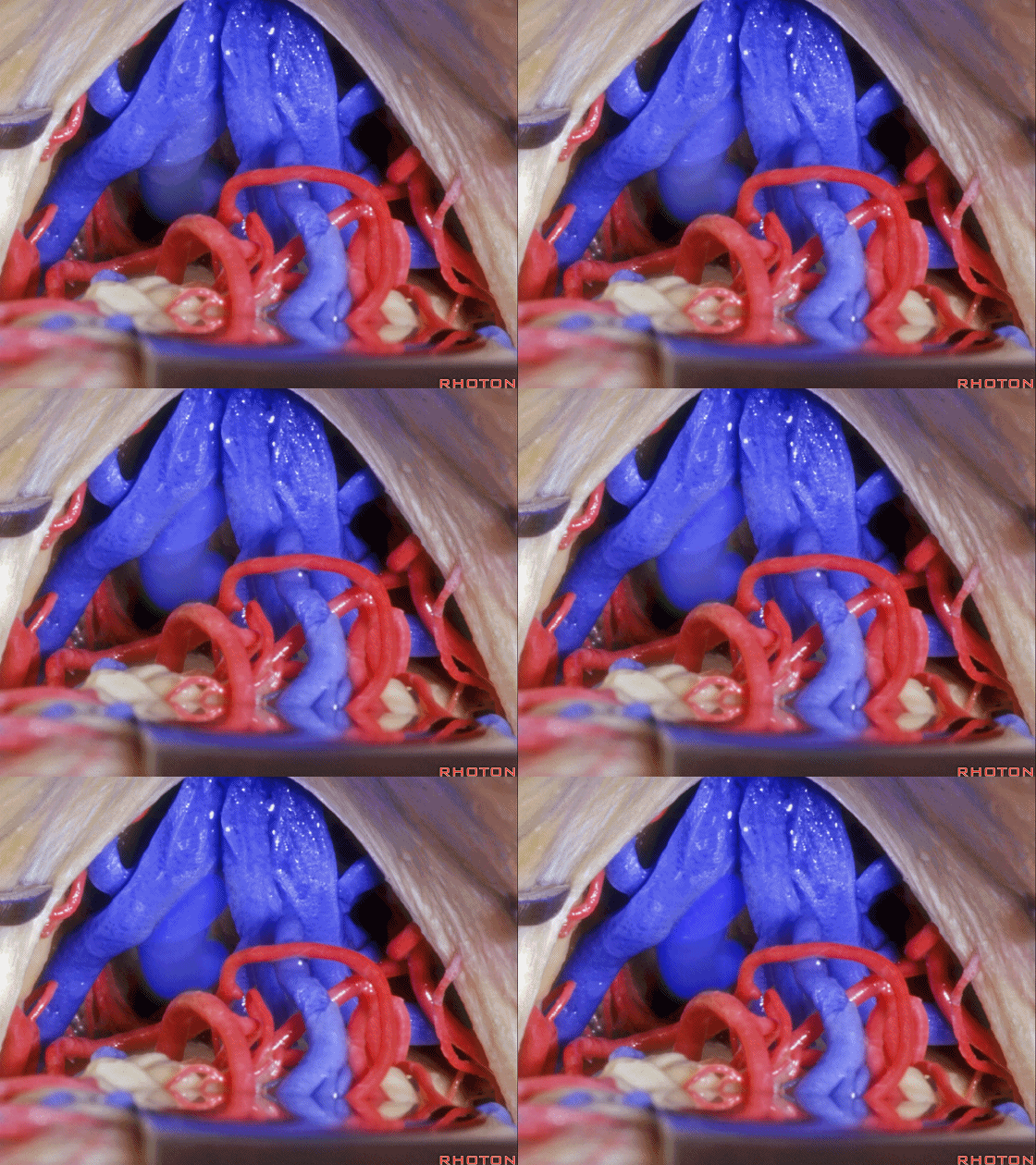
▼这是 小脑上动脉
Superior cerebellar artery
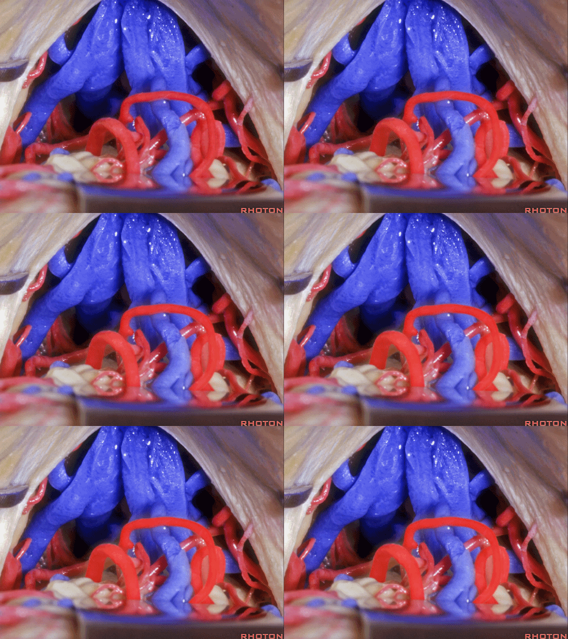
▼这是 大脑后动脉
posterior cerebral artery
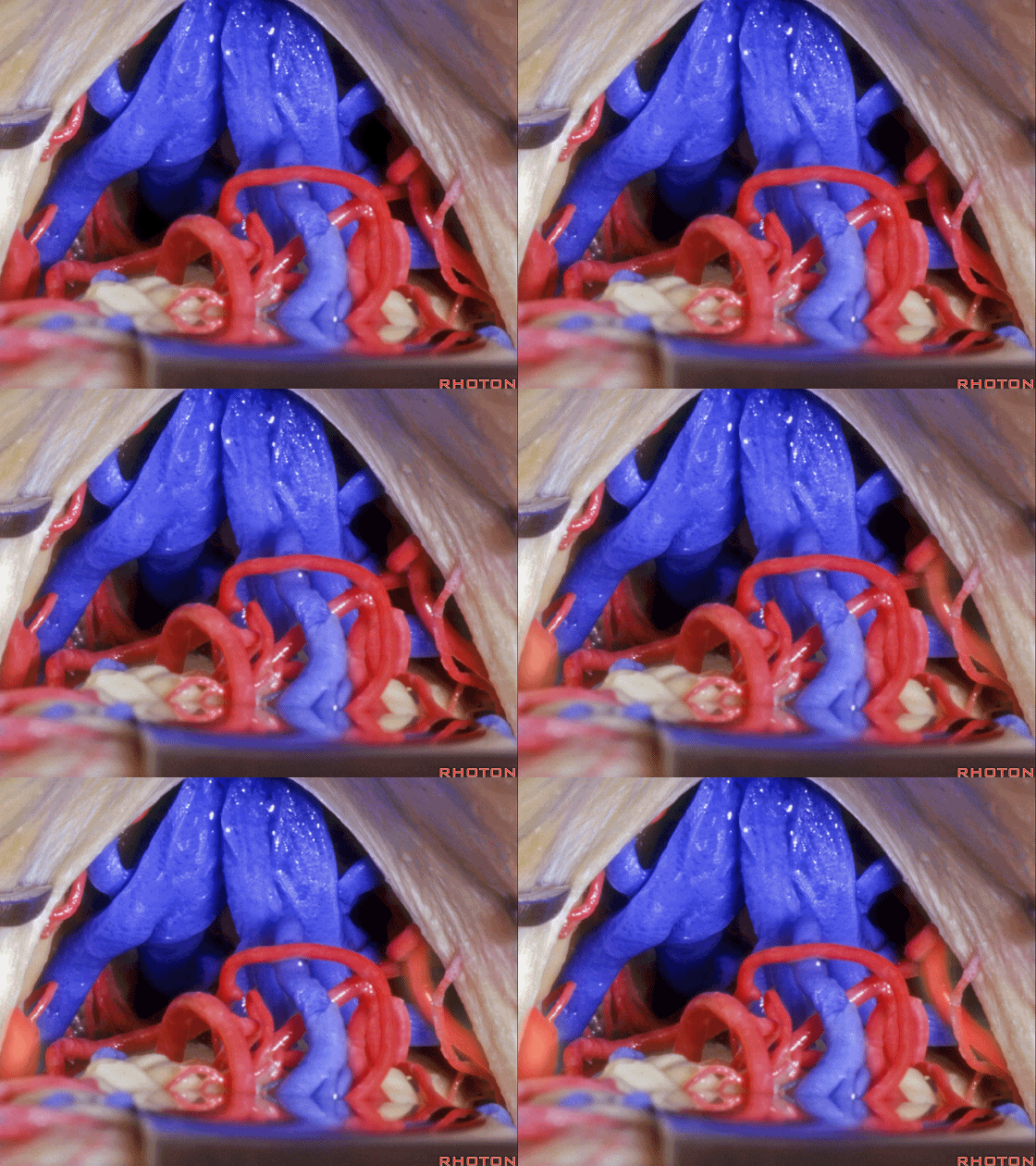
▼经小脑蚓尖上方可显露松果体。
And coming over the apex of the vermis you can see the pineal.
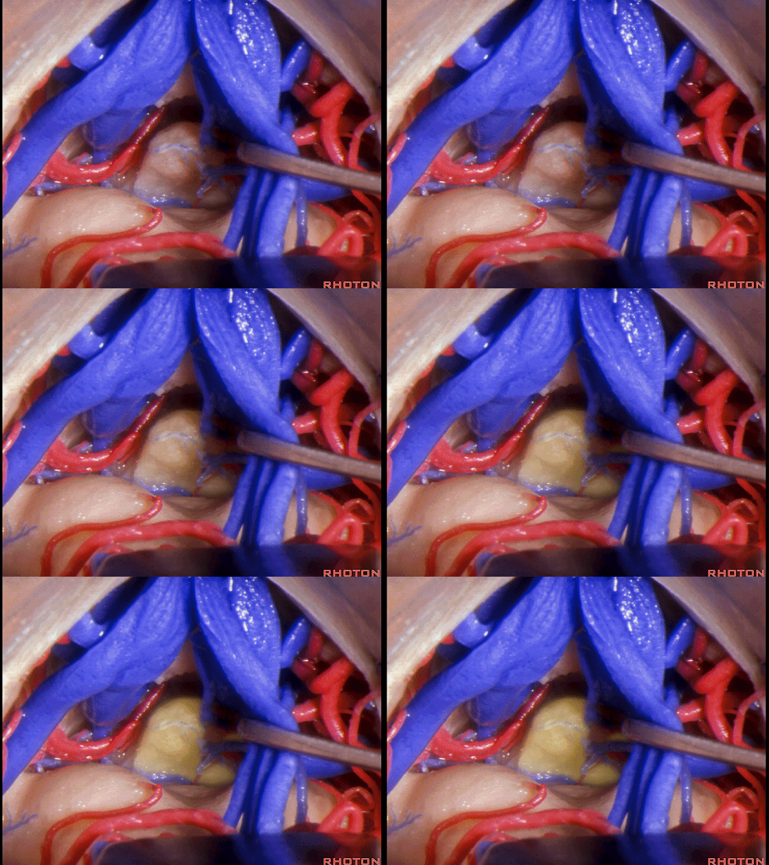
▼这是 脉络膜后内侧动脉
Medial posterior choroidal.
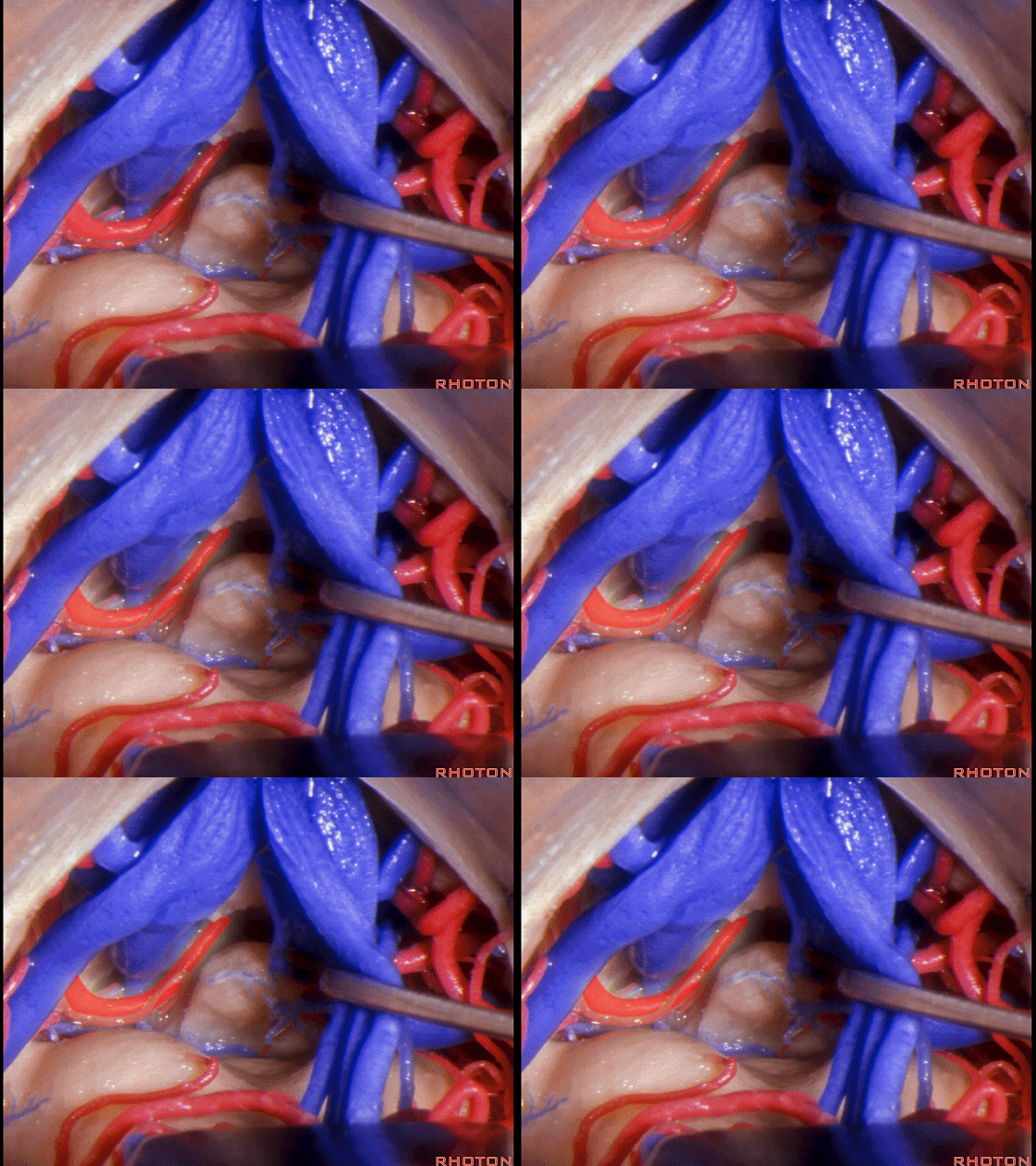
▼可暴露上丘,但往往无法暴露下丘。
You can see this superior but usually not the inferior colliculus.
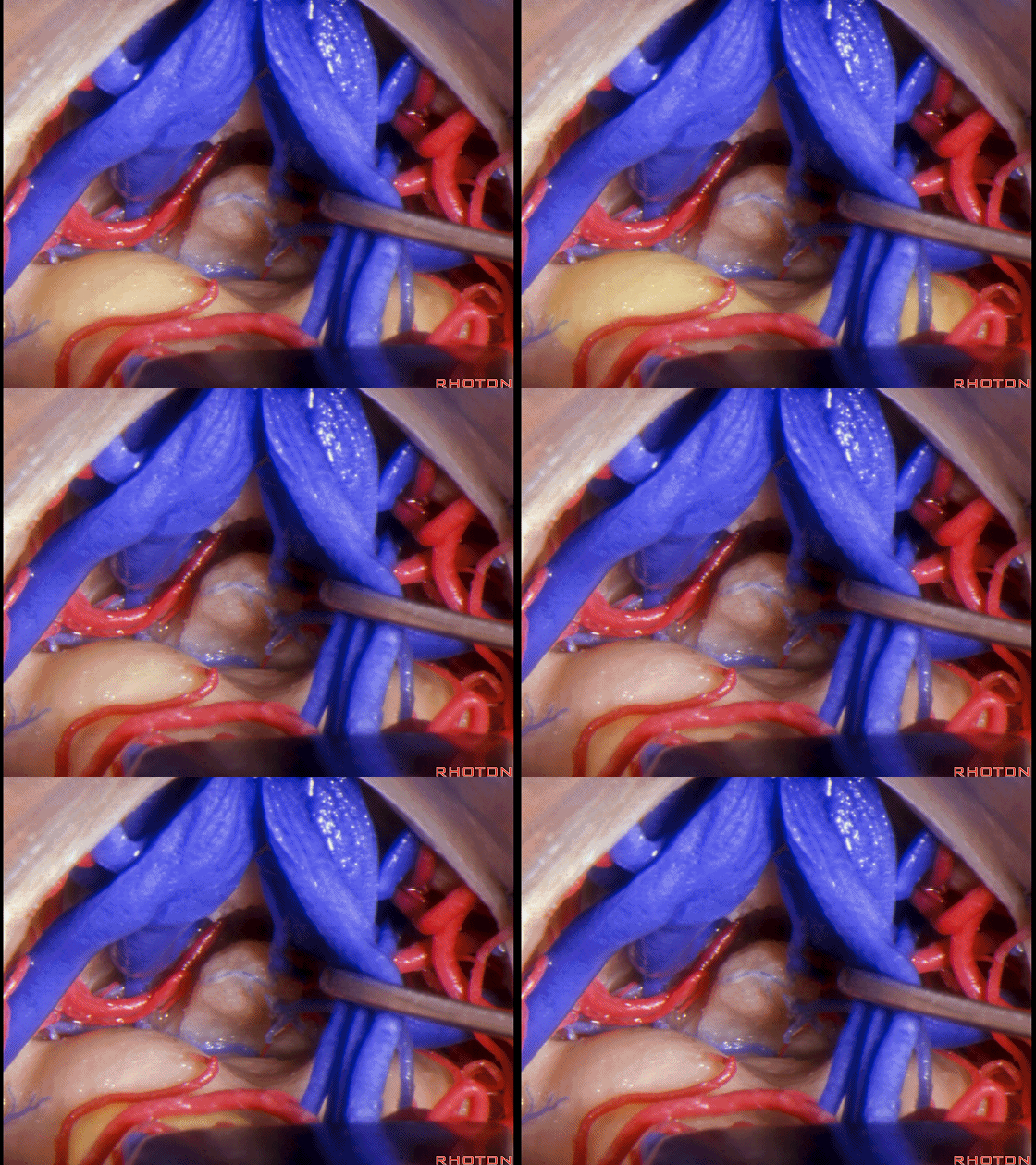
▼通过这个松果体上经中间帆入路,可进一步打开中间帆并从此处向前直至Monro孔,
You can open the velum interpositum and work forward from here all the way to the foramen of Monro using this suprapineal velum interpositum approach.
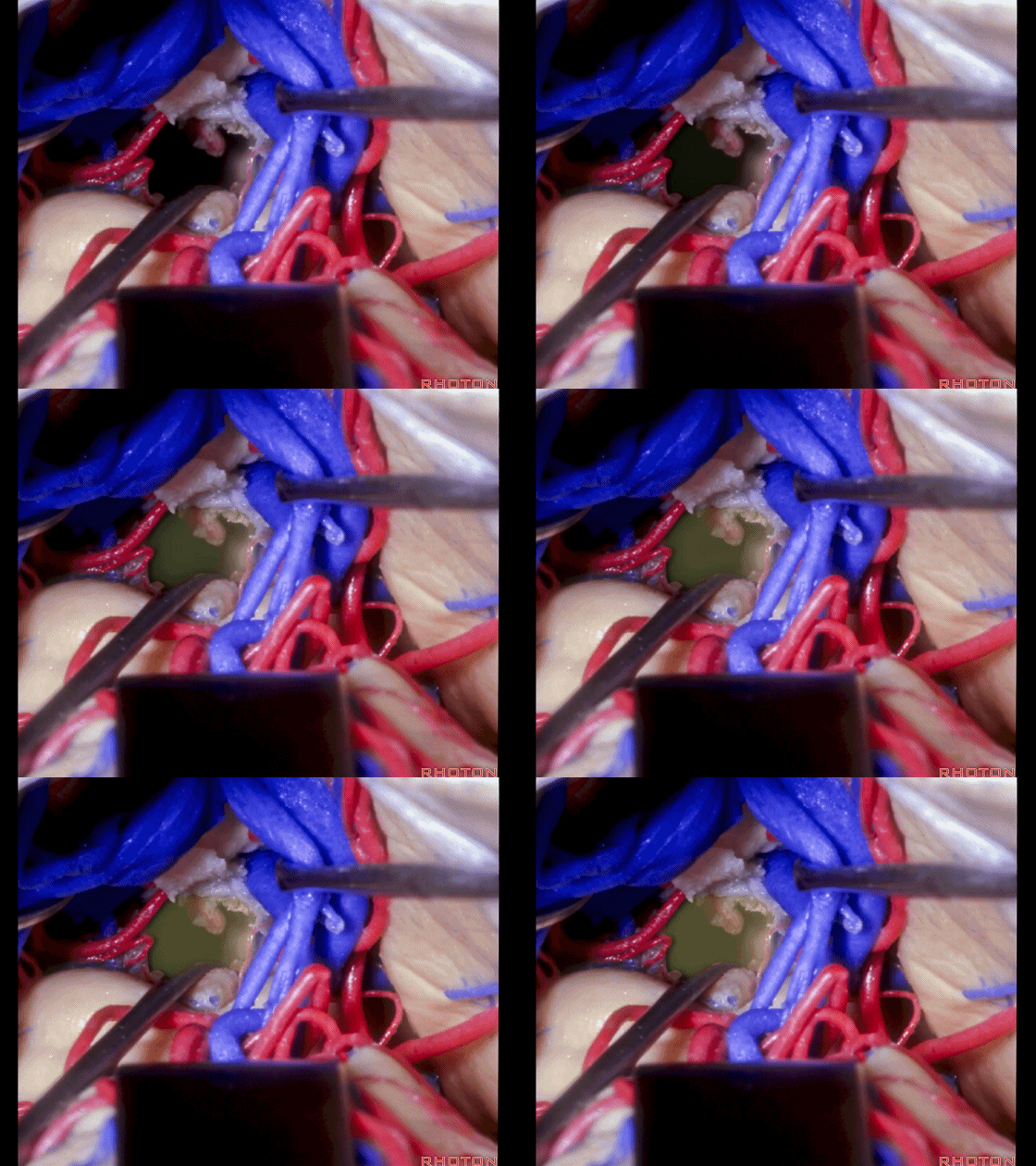
▼但如果想要暴露至中脑下部、下丘下方的滑车神经区域,则需避开小脑蚓尖部
But if you want to come lower on the midbrain below the inferior colliculus to the area of the 4th nerve, then you can come off of the apex of the vermis
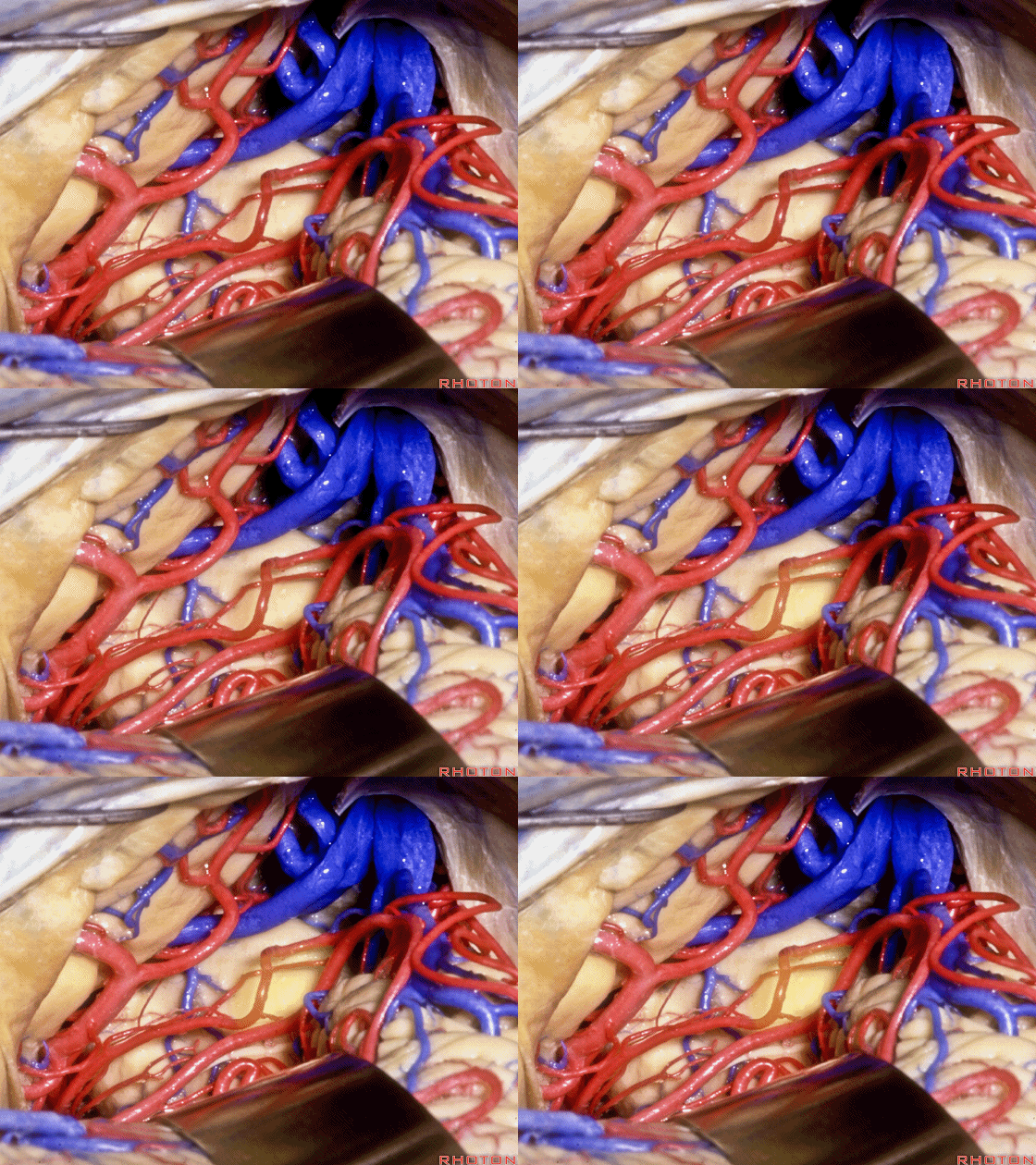
▼下图示 滑车神经
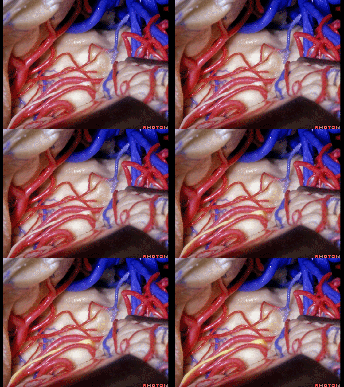
▼下图示 小脑蚓尖部
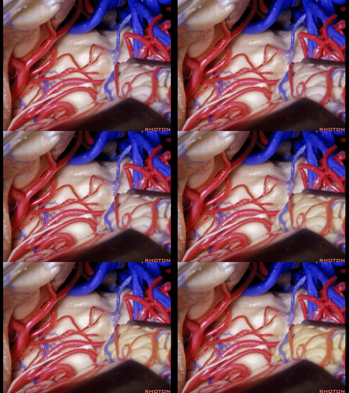
▼经旁正中入路 可暴露 四叠体池、丘板、滑车神经
and do a paramedian approach that gives you quadrigeminal cistern, collicular plate, 4th nerve,
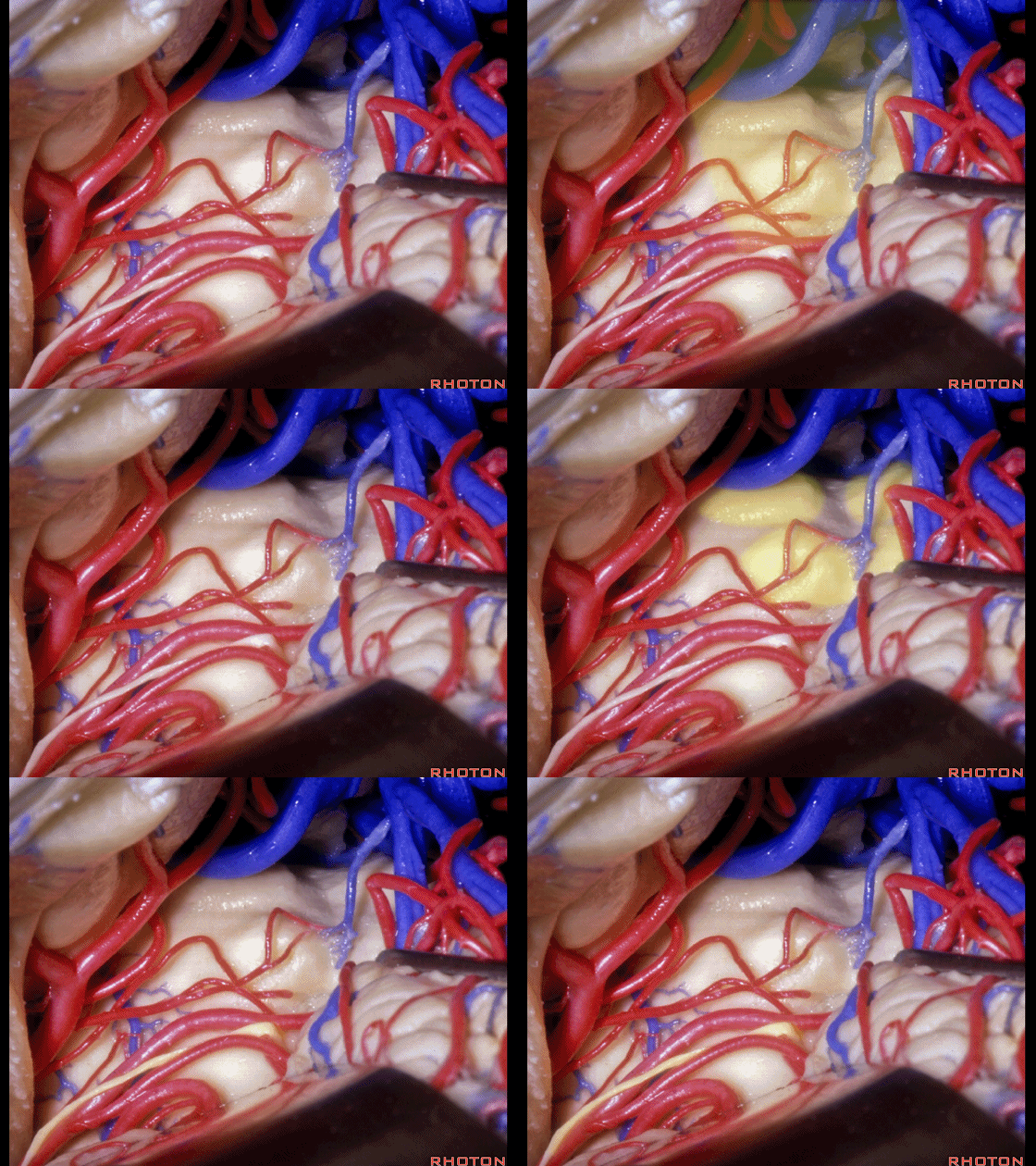
▼这是 大脑脚
here's the cerebral peduncle.
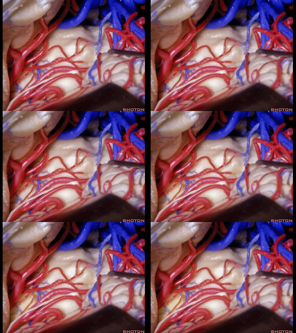
▼这是 脚池后部 以及 环池
this is gonna be...what cistern in here? It gives you back end of the crural and ambient cistern
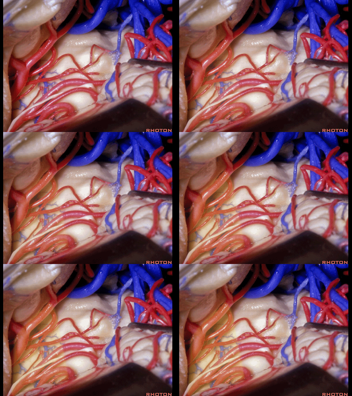
▼经由此幕下入路,可通往 四叠体池
leading back to the quadrigeminal cistern using this infratentorial approach.

▼松果体区的另一入路是枕叶经天幕入路。观察这一区域的上矢状窦,位于人字缝下方,没有桥静脉汇入窦。
The other route to this area is occipital transtentorial. And if you look at the area of the saggital sinus below the lambdoid suture, there are no bridging veins to the sinus.
![]()
▼这些后方的静脉向前汇入矢状窦。上矢状窦后6cm范围内通常没有桥静脉汇入。
The veins posteriorly are directed forward and usually there are no bridging veins to the posterior 6 centimeters of the saggital sinus.
![]()
▼因此我们可牵开枕叶,在天幕上方,可见松果体。
So that we can retract the occipital lobe, here we're on the tent, pineal
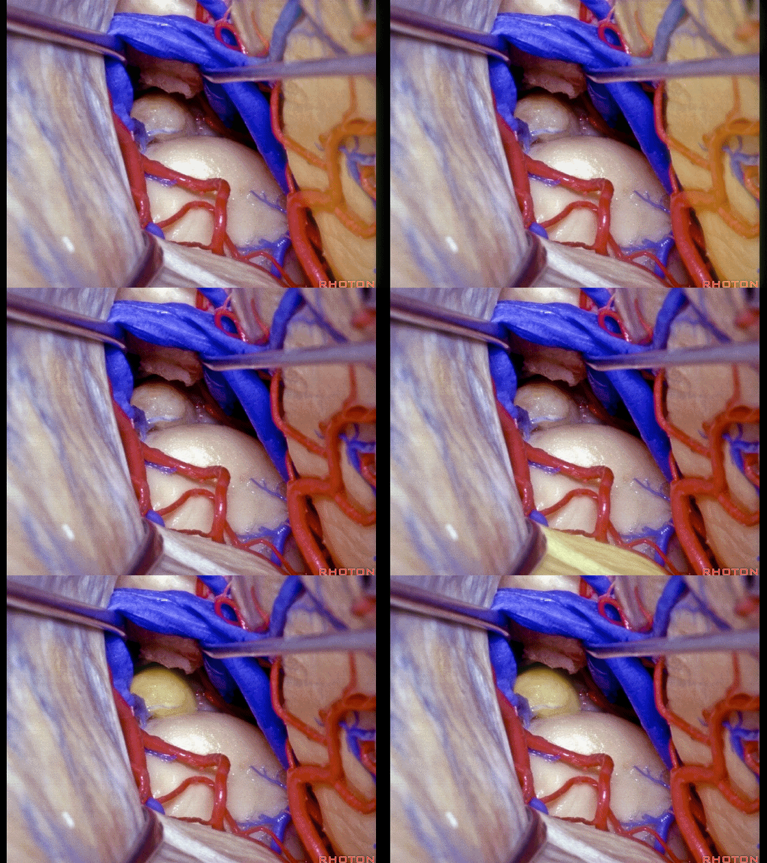
▼随后可贴着直窦 切开天幕
And then we can divide the tent adjacent to straight sinus, and gain this exposure of quadrigeminal cistern, pineal, superior colliculus.
![]()
▼从而暴露 四叠体池
You also have ambient,
![]()
▼暴露松果体、上丘,也可显露环池,以及一小部分脚池。
and a little bit of crural cistern in this area.
![]()
▼也可在Galen静脉上方暴露至胼胝体,均通过此枕叶经天幕入路。
And it also gives you access above the vein of Galen, to corpus callosum using this occipital transtentorial approach
![]()
▼这里我们切开了天幕,枕叶、大脑镰。
And here we've divided the tent, occipital lobe, falx.
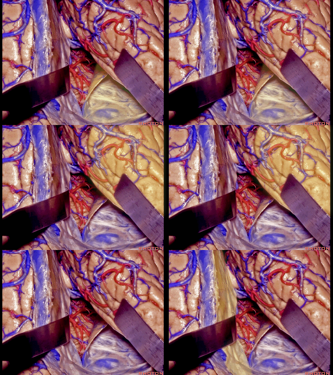
▼针对天幕尖部脑膜瘤的入路,即在切开天幕后,再切开大脑镰后部,从而暴露天幕尖部。
And an approach that I've used for meningeomas of the tentorial apex is after dividing the tent, to divide the posterior falx that gives you access to the tentorial apex.
![]()
▼还可进一步切开对侧的天幕,保留这里的汇入直窦的静脉。可由此处理位于天幕尖区域的肿瘤。
And then you can divide the controlateral half of the tent, persevering the venous connections here into the straight sinus. You can deal with tumors here, in the area of the tentorial apex.
![]()
▼如果这一侧的横窦为非优势侧,还可切断横窦,从而进行联合幕上-幕下的暴露,充分暴露这一区域。
And if you're on the side of a nondominant transverse sinus, you can divide that transverse sinus, and do a combined supra-infratentorial exposure, then, of the area above and below the tent.
![]()


