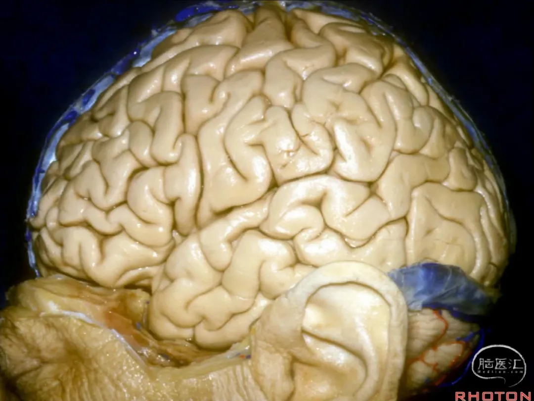
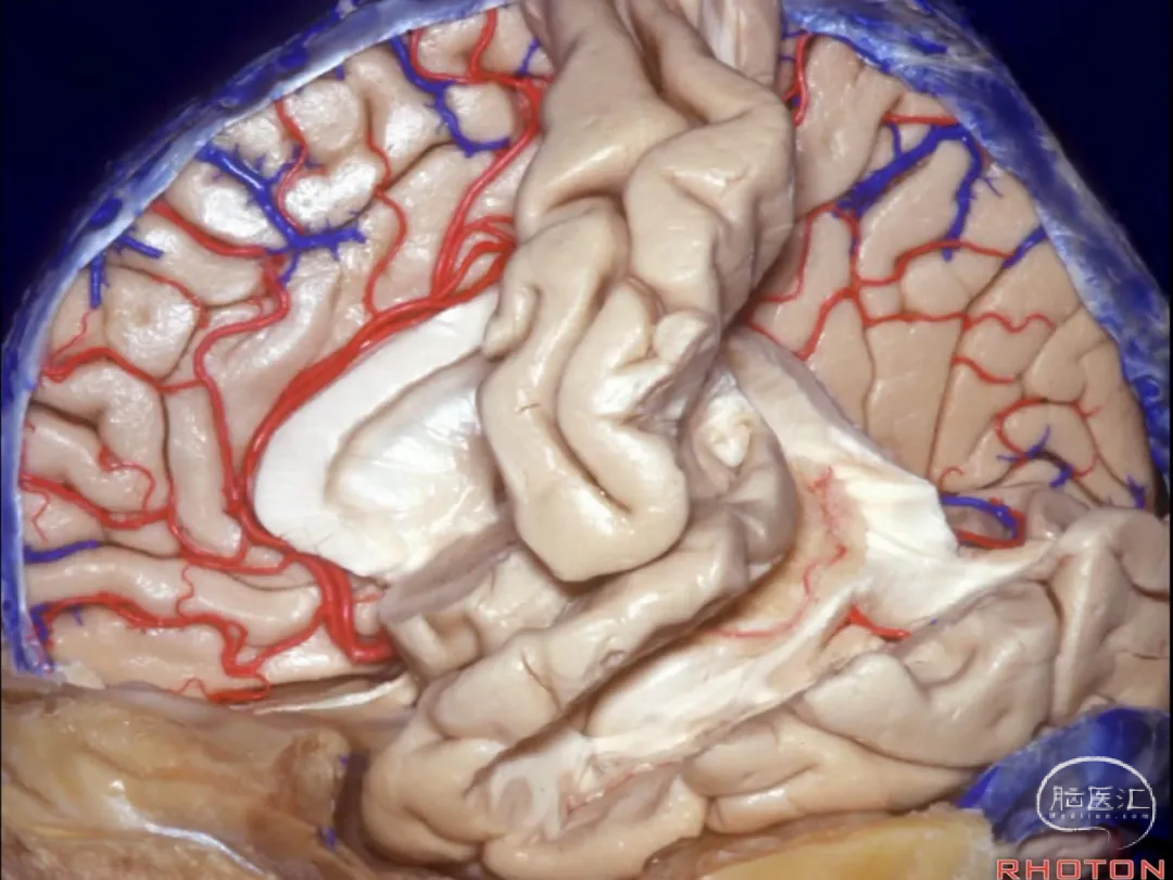
▼下图示岛叶
▼再来看看大脑前动脉,胼周动脉在半球外侧面的投影在C处(下图),该层面约为额下沟水平。
And now where is the anterior cerebral going to be located as you look through the lateral surface, at A, B or C, that pericallosal? Anyone wanna help me? It'll be running at C about the level of the inferior frontal sulcus.
▼胼周动脉沿着胼胝体走行,位于侧脑室额角的上方。
And it's running around the corpus callosum at the upper margin of the frontal horn.
▼再来看看大脑后动脉,其在半球外侧面的投影在B处。 其在外侧面的终末支的实际位置要低于投影的位置。
Now where are you going to find the posterior cerebral artery as you look through from the lateral surface? Anyone for C, B, ok, A?Well it's lower than this lateral surface. It'll be at B.
▼究其原因,让我们从这个视角观察大脑后动脉 在 环池 中的走行
So, and the reason for that is if you look at the course here, in the ambient cistern of posterior cerebral artery,
▼可见小脑幕 呈现一倾斜角度。
the tent and cerebellum are all sloped upward.

▼因此,越往内侧,你会发现大脑后动脉(下图)越是高于 大脑半球外侧面的下界。
So that as you get further medial, the area where you find posterior cerebral artery is significantly higher than that lower margin of the lateral surface.
▼通常, 从上方来看,在环池内,大脑后动脉 位于 海马旁回的上表面,而非下方。
So, and often that posterior cerebral artery...if you look at the upper surface of the parahippocampal gyrus in the ambient cistern, it's going to run on, not below the parahippocampal gyrus,
▼下图示 海马旁回
▼海马旁回 有下表面、内侧面、上表面。
So that parahippocampal gyrus here has a lower surface, a medial surface, and an upper surface.
▼下图示大脑后动脉
▼大脑后动脉 常行于 海马旁回 (下图)的上表面,位于颞角(下图)的内侧。
And posterior cerebral artery often runs on that upper surface of parahippocampal gyrus, medial to the temporal horn.
▼再从侧面来看,画面中加入了脑室。
So, looking through again from laterally, we fit it in the ventricles into this view.
▼下图示 颞上沟 层面
And here we plotted at the level of the superior temporal sulcus,
▼颞上沟层面 代表 岛叶的下界。故岛叶的下界沟即位于该水平。下图示岛叶。
the lower margin of the insula, so the inferior limiting sulcus of the insula is at this level.
▼这是 脉络裂。
And here we see the choroidal fissure
▼穹窿(下图) 构成脉络裂外侧界
which has the fornix on the outer margin,

▼这是 穹窿体部
▼这是 穹窿脚

▼这是 穹窿伞
▼丘脑 为 脉络裂 的内侧界
and thalamus on the inner margin.
▼这是 侧脑室体部
▼侧脑室 房部
▼侧脑室 颞角
▼穹窿 为脉络裂的外侧界。丘脑 为脉络裂的内侧界。
Fornix is on the outer margin.Thalamus is on the inner margin.

▼打开侧脑室体部的脉络裂,即可进入第三脑室。
And if you open the choroidal fissure in the body, you end up in third ventricle.

▼打开侧脑室房部的脉络裂,可暴露松果体。
If you open it in the atrium, you end up beside the pineal.

▼打开颞角 的脉络裂,则可进入环池。
You open in the temporal horn, you open up in ambient cistern.

▼这个位于环池顶壁的结构是外侧膝状体。它是视觉的中继站。属于丘脑的一部分。
Now, what is this structure that hangs down into the roof of the ambient cistern? This is...it's part of thalamus. It's the visual relay. So that's lateral geniculate body.
▼脉络裂终于 海马头(下图)后方
And here the choroidal fissure ends behind the head of the hippocampus,

▼这是杏仁核(amygdala下图),位于颞角前壁
and behind this structure in the anterior wall of the temporal horn which is the amygdala
▼可见 脉络膜前动脉(下图) 进入该区域。
We see the anterior choroid artery coming into this area.
▼下图 我们去除丘脑,保留穹窿的体部、穹窿脚、穹窿伞。
And here we've removed the thalamus, persevere the body, crus, fimbria of fornix.
▼打开脉络裂(下图)时,我们通常在穹窿侧进行切开,
Always when we open the choroidal fissure, we want to open on the forniceal side,
▼沿着穹窿体部、穹窿脚、穹窿伞 打开脉络裂
on the side of the body, crus, fimbria.

▼我们打开颞角的脉络裂(下图),即可见 大脑后动脉(下图)行于环池内。
Here, if we open in the temporal horn, we see the posterior cerebral artery in the ambient cistern.

▼在穹窿脚前方打开脉络裂,即可进入四叠体池,至松果体旁。
If we open in front of the crus of the fornix, we end up in the quadrigeminal cistern beside the pineal.
▼若打开侧脑室体部的脉络裂,则可进入第三脑室,从侧脑室体部进入第三脑室。
And if we open the fissure out of the body of the ventricle,we end up in third ventricle.But coming through from the body to the third
▼打开的这一间隙,称为中间帆(下图 绿色区域),其位于穹窿和丘脑髓纹之间,
we open this space between the fornix and the striae medullaris thalami that we call it velum...what...velum interpositum
▼侧脑室颞角内侧为 脉络膜前动脉,穿行于脉络裂并供血于脉络丛。
So now, what are the arteries that supply and pass through the choroidal fissure to supply the plexus? Well in the temporal horn we have anterior choroidal artery that
▼脉络膜前动脉 发自颈内动脉(下图),绕行于钩回(下图 黄色区域)前段和钩回后段,
anterior choroidal artery arises from carotid, passes around the uncus.and posterior segment of the uncus,
▼钩回分为 前段、后段和尖端。
The uncus has an anterior segment, a posterior segment, and an apex.
▼钩回前段(下图)包含杏仁核。
So we see anterior segment of uncus containing amygdala.
▼钩回后段 包含 海马头。
Posterior segment contains head of hippocampus.
▼钩回尖端 位于 动眼神经外侧。
The apex of the uncus is lateral to the 3rd nerve.
▼脉络膜前动脉 绕过 钩回尖端,其进入 侧脑室颞角(下图)的部位称为 下脉络点(下图 绿色区域)。
And it passes around the apex, and enters the temporal horn at the inferior choroidal point.

▼这里是丘脑区以及环池的下面观。(下图示 环池)
Now, here we're looking at the thalamic area, the ambient cistern from below.

▼这是 大脑后动脉。
And here we see posterior cerebral artery.

▼大脑后动脉P2段 发出 脉络膜后外侧动脉。
And arising from the P2 segment, are the lateral posterior choroidal arteries
▼脉络膜后外侧动脉,其走行于穹窿伞和丘脑枕之间
lateral posterior choroidal arteries pass between the fimbria and the pulvinar,
▼脉络膜后外侧动脉,供血于 侧脑室颞角后部 和 房部的 脉络丛。
to enter choroid plexus in posterior temporal horn and atrium.
▼这里 我们去除了舌回(舌回 是 上象限视野的投射区)
When we look below we also...here have taken off the lingual gyrus,where the upper visual field is located.

▼这是距状裂,和其 深部底端。
Here we see the calcarine sulcus, the deep end of it.
▼距状裂深部底端 拱入侧脑室房部的内侧界,即为禽距的位置。 禽距 是 侧脑室房部内侧壁的一个隆起。侧脑室房部与距状沟的深部底端之间仅相隔这一薄层皮质。
It cuts into the medial margin of the atrium at the level of the calcar avis.And it's separated from the deep end of the calcarine sulcus by only a thin layer of cortex here at the level of the calcar avis which the prominent here in the medial wall of the atrium.
▼下图示 脉络膜后内侧动脉,其发自P1段,行于脚池、环池、及四叠体池
And then arising from P1 passing through the crural and ambient cisterns, and then quadrigeminal cistern is the medial posterior choroidal artery

▼这是 脉络膜后内侧动脉

▼这是 脉络膜后外侧动脉。
Here the lateral posterior choroidal arteries.
▼随后,脉络膜后内侧动脉 在松果体旁向前走行,进入中间帆
And then the medial posterior choroidal arteries turn forward beside the pineal, right in the velum interpositum,

▼脉络膜后内侧动脉 穿过 脉络裂,供血于侧脑室体部的脉络丛。
and pass through the fissure to supply the choroid plexus in the body of the ventricle.

▼这是侧脑室额角、体部的上面观。
So here we're looking into frontal horn, and body from above.
▼如果从此处(下图)切开胼胝体,半数可能进入右侧脑室,半数可能进入左侧脑室。
And if you come through the corpus callosum in this area, half the time, you'll open into the right lateral ventricle, half the time, into the left lateral ventricle.

▼很多情况下,我们并不能看到这些突起和凹陷的脑室壁结构,而且一旦Monro孔(下图)处脑室沟通受阻,你面对的就是一圆形的腔隙。
And often you lose these...prominences and, sort of sulcal areas between the structures in the wall, if the ventricle at the foramen of Monro is occluded,and it becomes a round cavity.

▼因此你在打开胼胝体进入侧脑室处理脑室肿瘤时,难以辨识尾状核和丘脑(下图)。
So that when you look into the ventricle coming through the corpus callosum with the ventricular tumor, it's hard to pick out the caudate and the thalamus.
▼但仍可见丘纹静脉,以及脉络丛。
But you see the thalamostriate vein, the choroid plexus.
▼如果丘纹静脉位于脉络丛的右侧,则说明进入的是右侧的侧脑室。
If the thalamostriate vein is to the right of the choroid plexus, you've opened into the right lateral ventricle.
▼如果丘纹静脉位于脉络丛的左侧,则说明进入的是左侧的侧脑室。
If it's to the left of the choroid plexus, you've opened into the left lateral ventricle.
▼从侧脑室体部进入第三脑室,则需经过 中间帆。
And in coming from the body to the third ventricle, we come through the velum interpositum.
▼因此我们需再经过四层结构进入第三脑室。最上层(第一层)为穹窿。
So that you come through or around four layers of tissue. And the upper layer is the fornix.

▼将穹窿翻向后方,才可暴露 中间帆。
And here we've folded the fornix backwards,and we see the velum interpositum
▼中间帆 上壁为一层脉络膜,其为室管膜组织, 位于穹窿下方。
that it has an upper layer of tela,an ependymal membrane,below the fornix.
▼因此先是穹窿,然后是脉络膜。再打开脉络膜,则进入中间帆(下图),
So we have fornix,and then, tela.And then we open the tela, we're into the velum interpositum
▼其内有大脑内静脉
that contains the internal cerebral veins,
▼这是 脉络膜后内侧动脉。
and what group of arteries, what arteries do you see here? Medial posterior choroidal arteries.

▼随后再打开下层脉络膜,这才进入第三脑室。
And then we open that lower layer of tela, and you're looking into third ventricle.
声明:脑医汇旗下神外资讯、神介资讯、脑医咨询所发表内容之知识产权为脑医汇及主办方、原作者等相关权利人所有。未经许可,禁止进行转载、摘编、复制、裁切、录制等。经许可授权使用,亦须注明来源。欢迎转发、分享。


