


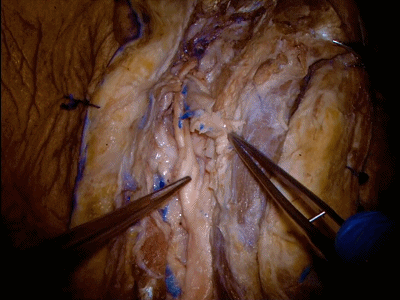
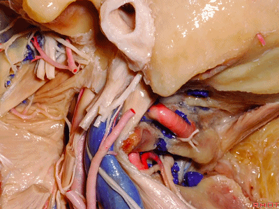
▼通过面神经出茎乳孔处的前方和后方可触及茎突底部。将面神经向前牵开可显示茎突全程。
The styloid base can be palpated anteriorly and posterior to the facial nerve as it enters the stylomastoid foramen.Here the facial nerve is retracted anteriorly to expose the extent of the styloid process.
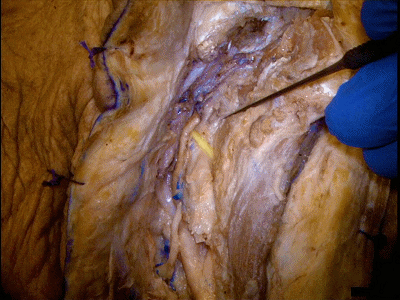
▼下图可见面神经穿入腮腺后部(该部腮腺已部分切除)。腮腺若被切开,则需缝合腮腺包膜,可选用薇乔缝线间断缝合。这可避免术后腮腺涎液漏的发生。
We also see the facial nerve entering the posterior aspect of the parotid gland which has been partially removed for exposure.If the parotid gland is entered, it is important to close the capsule of theparotid,with an interrupted vicryl suture.This will help to spanvent a postoperative seroma.
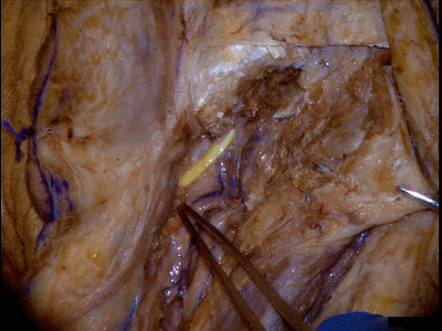
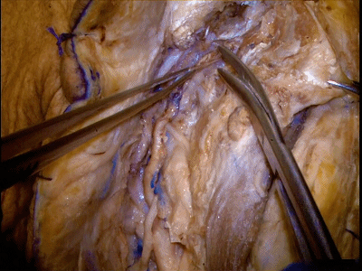
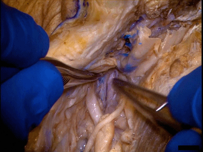
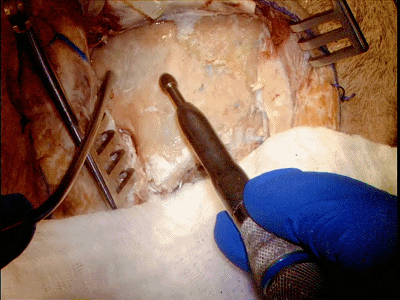
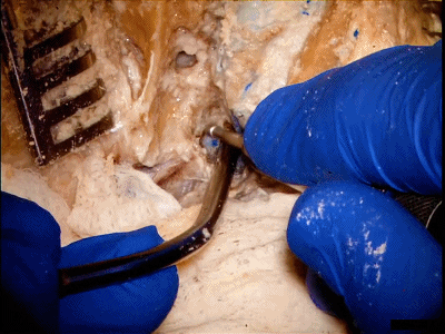
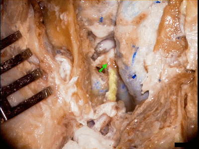
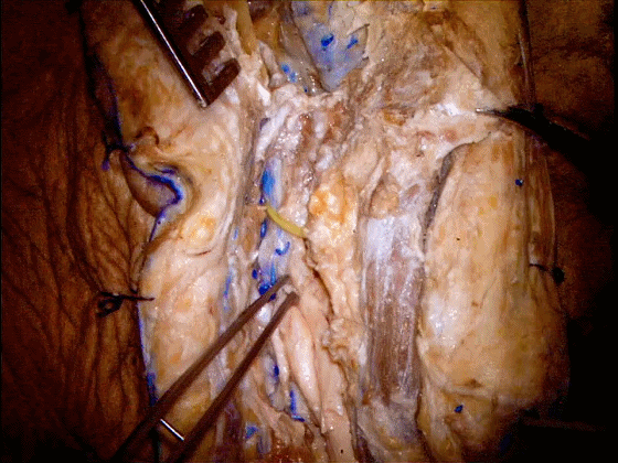
▼这里可见茎乳动脉,通常发自耳后动脉。茎乳动脉需予以保护,以保证面神经乳突段的血供。
Here one sees the stylomastoid artery, which typically arises from the posterior auricular artery. The stylomastoid artery should be spanserved because of its blood supply to themastoid segment of the facial nerve.
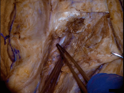
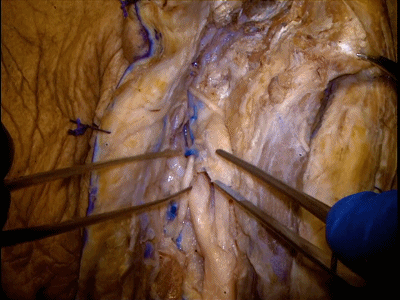
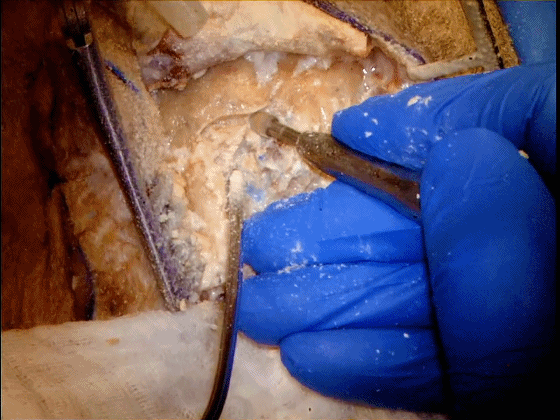
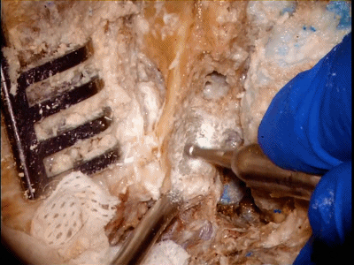
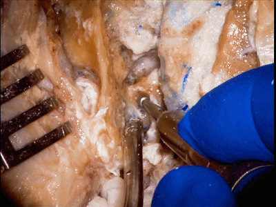
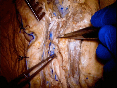
▼切除茎突,方可充分暴露颈内动脉。如肿瘤包裹颈内动脉,或者颈内动脉为肿瘤供血需切除茎突。在本病例中,颈内动脉并未被肿瘤累及。因此上述步骤可以省略。
Removal of the styloid process is necessary to expose the internal carotid artery immediately below the skull base. This should be done when the internal carotid artery is encased by the tumor, or provides arterial blood supply to the tumor. The internal carotid artery was not involved by the tumor in the case we're spanventing today.Therefore the step was not necessary.
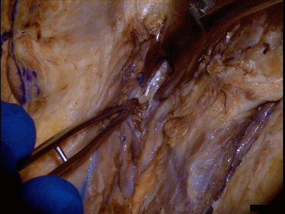
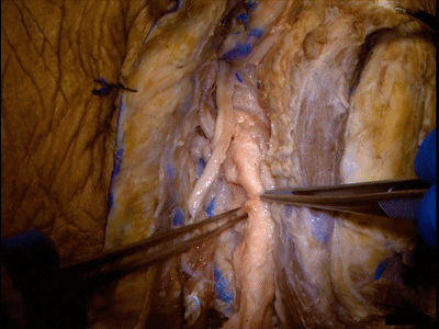
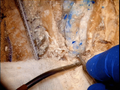
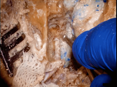
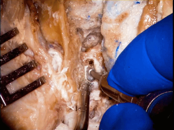
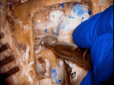
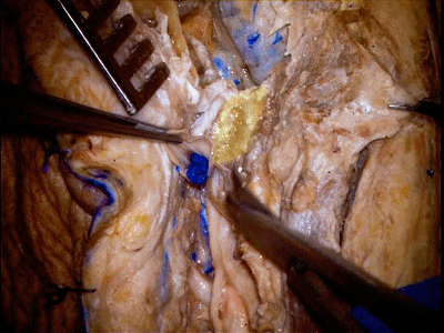
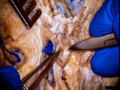
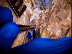
▼剪断与茎突黏连的软组织

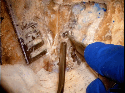
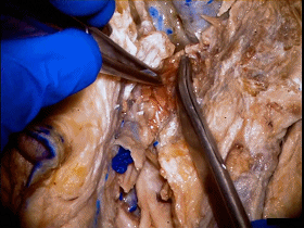
▼咬骨钳咬除茎突。
The styloid process is removed with a rongeur.
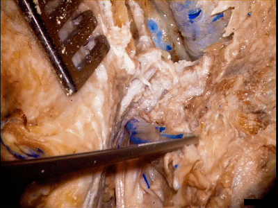
▼接下来将视野转移至下颈部,进行颈内静脉和颈总动脉的暴露。去除面静脉(下图),可暴露颈动脉分叉部。
Attention is turned to the lower neck to expose the jugular vein and the common carotidartery. Removal of the facial vein allows exposure of the carotid bifurcation.
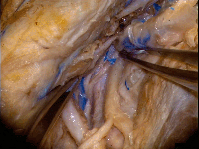
▼舌下神经(下图)通常于颈动脉分叉部水平或稍上方进一步向内进入舌根。
The hypoglossal nerve is identified typically at the level of the bifurcation or immediately above the bifurcation, as it courses medially to the base of the tongue.
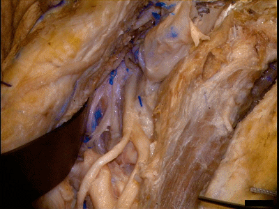
▼副神经(下图)通常位于寰椎横突尖端(下图)水平。
It is noted that the accessory nerve can typically be found at the level of the tip of the transverse process of C1.
▼在此层面,副神经可走行于颈内静脉的前方或后方。
The accessory nerve can pass either anterior or posterior to the jugular vein at this level.

▼枕动脉被游离并切除,以利于上颈部结构的暴露。
The occipital artery is mobilized and removed in order to gain exposure of the upper cervical region.
▼向上翻折颈内静脉,以暴露颈动脉分叉部、迷走神经、舌下神经(下图)。
The jugular vein is mobilized superiorly exposing the carotid bifurcation, the vagal, and the hypoglossal nerves.
▼甲状腺上动脉(下图)位于颈外动脉起始部。
The superior thyroid artery is seen at the base of the external carotid artery.
▼牵开颈外动脉,显露颈内动脉。
The external carotid artery is retracted to allow exposure of the internal carotid artery.
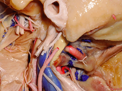
▼颈内动脉的显露范围向上至颅底。在该层面,可见舌咽神经(下图)从颈静脉孔内侧部穿出,跨过颈内动脉前缘。
The internal carotid artery is exposed immediately below the skull base. The 9th cranial nerve can be seen at this level crossing the anterior margin of the internal carotid artery. In this view, one can see the 9th cranial nerve exiting the medial aspect of the jugular foramen.
▼可见舌下神经、迷走神经相吻合(下图)。后组颅神经的这种吻合可见于神经恰穿出颈静脉孔的颅底附近。迷走、副、舌下神经之间均可出现上述吻合。
This view also shows the fusion of the hypoglossal and the 10th cranial nerves as they approach the jugular foramen. The fusion of the lower cranial nerves can occur immediately below the skull base as they approach the jugular foramen. This may be true of any combination of the 10th, 11th, or 12th cranial nerves.
▼下一阶段将进行乳突切除术。此处的解剖标志包括上方的乳突上嵴(下图)
Attention is turned at this stage of procedure to the mastoidectomy. The anatomical landmarks to be identified include the supramastoid crest superiorly,

▼后方的星点(下图),其可作为横窦-乙状窦转角处的标准,以暴露乙状窦;
posteriorly the asterion, which provides localization of the junction of the transverse sigmoid sinus, and exposure of the sigmoid sinus
▼这是前方的Henle嵴(下图),可定位外侧半规管。
anteriorly the spine of Henle should be noted, to provide the location of the lateral semicircular canal.

▼首先磨除三角形皮质骨区域

▼随着乳突的切除,可见该标本一个较小的乳突气房。
As mastoidectomy proceeds, it is noted that there's a small mastoid air cell space in the specimen.
▼要点在于暴露范围需足够大,需从横窦、乙状窦到颈静脉球水平。需要同时暴露乙状窦后方,及前方的硬脑膜。
It's very important that exposure is wide to include the transverse and sigmoid sinus to the level of the jugular bulb. This requires exposure of dura posterior to the sigmoid sinus as well as in the spansigmoid space.
▼下图可见鼓室窦被打开。
The antrum has been exposed in the mastoidectomy.
▼该例标本存在高位颈静脉球,识别该结构的重要性在于,其与前方的面神经、颈内动脉关系密切。
Noted in this specimen is a high jugular bulb, which is important to recognize because of its relationship to the facial nerve, as well as internal carotid artery anteriorly.
▼将面神经乳突段(下图)轮廓化。
The mastoid segment of the facial nerve is skeletonized.
▼此时轮廓化的是后半规管。
Here we're drilling along the posterior semicircular canal.
▼这是鼓索神经(下图)。
The chorda tympani is identified.
▼可见面神经隐窝(下图),其位于乳突段面神经的上段和鼓索神经之间。沿着面神经隐窝可进一步开放中耳腔。
One now sees the facial recess to find between the superior aspect of the mastoid segment of the facial nerve and the chorda tympani. A drilling through the facial recess opens the middle ear space.
▼面神经乳突段的上端沿着外侧半规管下缘(下图箭头)从前向后并转而下行。
The superior aspect of the mastoid segment of the facial nerve can be seen turning below the lateral semicircular canal anteriorly.
▼继续暴露的是上半规管。
The superior semicircular canal is further defined.
▼将颈内静脉结扎后切断,并一直游离至颅底,以完全显露颈静脉孔。
After ligation and division, the jugular vein must be mobilized to the level of the skull base, in order to have full exposure of the jugular foramen.
▼可在寰椎横突尖显露副神经。
The 11th cranial nerve is found at the level of the tip of the transverse process of C1.

▼如果副神经的走行正如该例标本所见,即从颈内静脉前方向后跨行,则需将颈内静脉移至副神经前方以继续向上游离至颈静脉孔水平。
If the 11th cranial nerve passes over the anterior margin of the jugular vein as in this case, the jugular vein must be transposed anteriorly to allow dissection of the jugular vein to the level of the jugular foramen.
▼于颈内静脉后缘松解头外侧直肌(下图)。
The rectus capitis lateralis muscle is dissected from the posterior margin of the jugular vein.
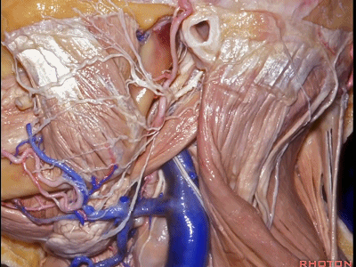
▼从这幅Rhoton解剖图中,可见头外侧直肌(下图),起于寰椎横突上表面(下图),向上附着于颈静脉孔后缘骨质(颈静脉突)。
This Rhoton dissection shows the relationship of the rectus capitis lateralis muscle as it arises from the superior surface of the transverse process of C1, to project superiorly attaching to the posterior bony margin of the jugular foramen.
▼椎动脉恰位于头外侧直肌的后内侧。椎动脉被其周围静脉丛包绕,穿出于横突孔。
The vertebral artery surrounded by the venous plexus exits the foramen transversarium, immediately posteromedial to the rectus capitis lateralis muscle.
▼显露寰椎横突(下图箭头)。从寰椎横突上缘处锐性离断头外侧直肌,将其分块切除直至颈静脉孔后缘。
The transverse process of C1 is defined. This muscle is sharply dissected from thesuperior margin of the transverse process of C1, and then removed in a piecemeal fashionto the posterior margin of the jugular foramen.
▼切除头外侧直肌是暴露颈静脉孔后缘的必要步骤。
Removal of the rectus capitis lateralis muscle is necessary in order to expose the posterior margin of the jugular foramen.
▼在切除头外侧直肌时,需注意避开椎动脉(下图),后者穿出横突孔的位置,恰位于头外侧直肌的后内侧。
As one removes the rectus capitis lateralis muscle, one must be careful to avoid the vertebral artery, which exits the foramen transversarium, posteromedial to the rectus capitis lateralis muscle.
耳后经颞入路至颈静脉孔区---Rhoton解剖视频学习笔记系列(下)
