前篇《Head and Neck Anatomy》(可点击浏览)我们学习了头颈部解剖,本篇在前篇基础上介绍颈静脉孔区 耳后经颞入路,其为处理颈静脉孔区病变最常用的入路。笔者学习后在标本上尝试发现该入路可方便地寻找面神经,降低面神经的损伤几率,并可保留迷路等优点。本文的讲述者是Jon Robertson教授。共118张图片。
笔者水平所限,错误之处请批评指正!

耳后经颞入路至颈静脉孔区
The Postauricular Transtemporal Approach to the Jugular Foramen
▼本集视频将展示颈静脉孔区手术入路,以及一则左侧颈静脉球瘤的病例分享,并同步结合尸头解剖,详解颈静脉孔区手术入路 解剖要点。
This spansentation, to illustrate the approaches to thejugular foramen surgically, includes a case spansentation of a left glomus jugularetumor with surgical footage of the case itself, as well as a cadaver dissection that demonstrates the specific anatomical landmarks inthe approach to the jugular foramen region.
▼颈静脉孔区手术显露困难,缘于其所处位置深在及周围神经血管结构遮挡,包括前方的颈内动脉、侧方的面神经、以及位于颈静脉孔后内侧缘下方的椎动脉。
Surgical accesses to the jugular foramen is difficult because of its deep location and surrounding neurovascular structures blocking its exposure, such as the internal carotid artery anteriorly, the facial nerve laterally, and vertebral artery inferior to the posteromedial marginof the jugular foramen.
▼因此,该区域的手术需做到真正意义上的“解剖性手术”,方可保护该区域错综复杂的神经血管结构。
Therefore skull base lesions of jugular foramen should be approached as a surgical anatomist would to spanserve the complex neurovascular structures of this region.
▼Rhoton将颈静脉孔区的手术入路归结为三组,外侧入路经乳突(耳后经颞入路),后方入路经后颅窝(包括乙状窦后入路,或更广泛的远外侧入路及其经髁扩展),最后一种为经鼓骨的前方入路(耳前颞下-颞下窝入路及其各变型),上述入路均可联合颈部解剖,以根据需要来处理特定病变。
Rhoton has categorized approaches to the jugular foramen into three groups, a lateral group directed through the mastoid bone, this will be the postauricular transtemporal approach, a posterior group directed through the posterior fossa, this will include a retrosigmoid, or a more extensive far lateral or transcondylar variant approach, and finally an anterior group of approachs directed through the tympanic bone, this will be the spanauricular subtemporal infratemporal fossa approach or variations of that. In each of these approaches, a neck dissection can be included as needed to manage the specific pathology that one is dealing with.
▼本集视频重点阐述颈静脉孔区的耳后经颞入路的应用解剖。与颈部解剖相联合,这一入路为处理颈静脉孔区病变最常用的入路。
The goal of this video is to demonstrate the surgical anatomy of the postauricular transtemporal approach to the jugular foramen region. Combined with a neck dissection, this is the most common surgical approach chosen for management of lesions involved in the jugular foramen.
▼迷路通常保留
The labyrinth is usually spanserved in the exposure,
▼但根据病变具体情况,术野可向前扩展,此时需移位面神经,并牺牲中耳结构及外耳道,向内侧扩展时需磨除迷路和耳蜗。
but depending on the pathology, the surgical field may be extended anteriorly by transposing the facial nerve and sacrificing the middle ear structures and external auditory canal, or medially by removal of the labyrinth or cochlea.
▼这是右侧的颅底下表面,我们来看颈静脉孔区。耳后经颞入路联合颈部解剖,可在颅底对颈静脉孔实现270°的暴露。
We're viewing the inferior aspect of the right skull base in the region of the jugular foramen. The postauricular transtemporal approach combined with a neck dissection allows a 270 degree exposure of the jugular foramen at the skull base.
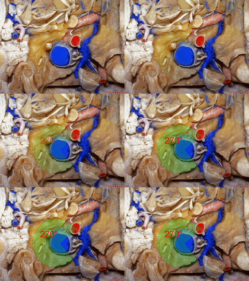
▼这一暴露范围包括前方的颈内动脉,直至颈内动脉管层面
This would include anteriorly exposing the internal carotid artery to the level of the carotid canal,
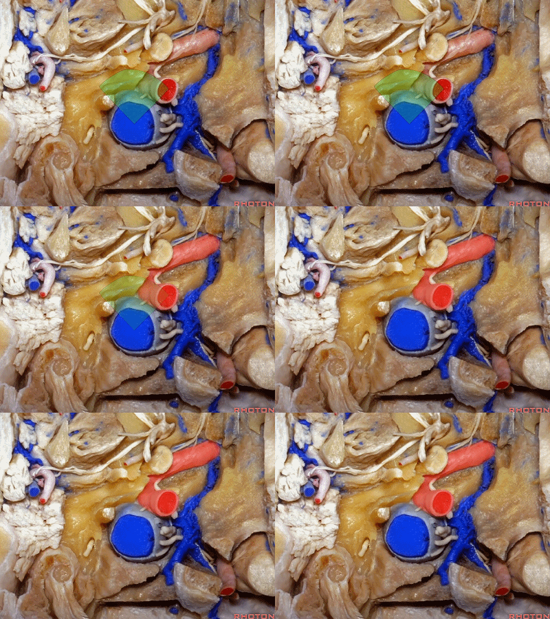
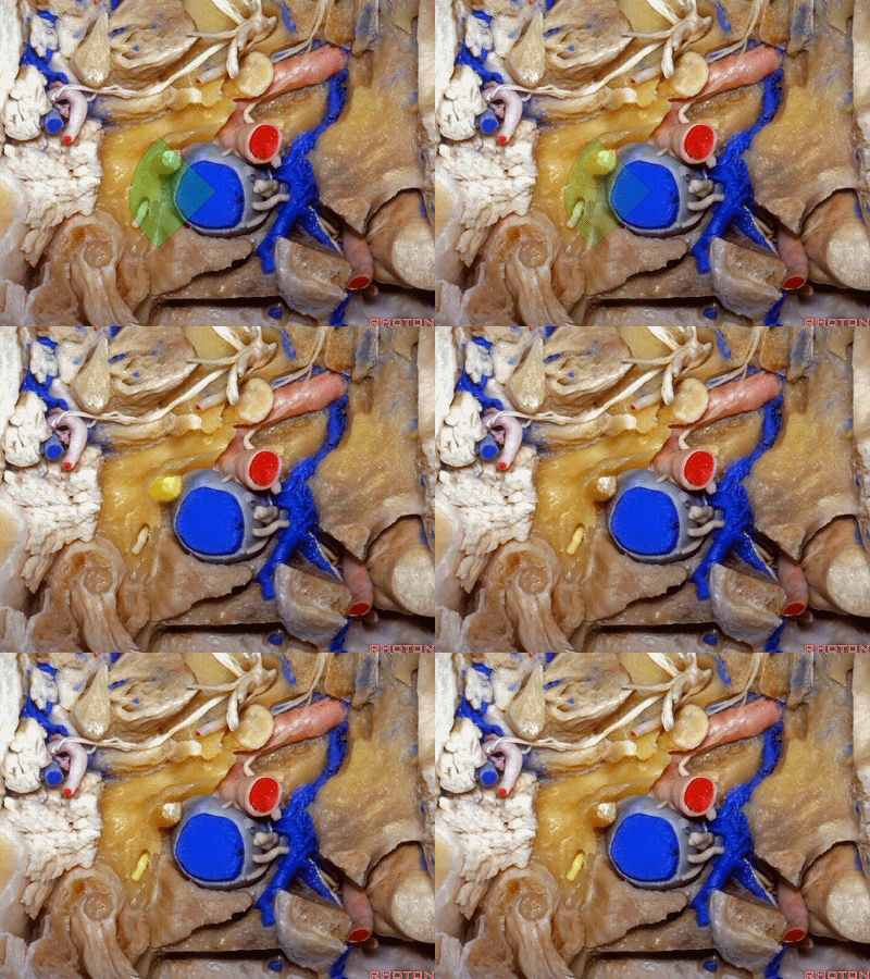
▼侧方的茎突、面神经出茎乳孔处
laterally, the styloid process, and the facial nerve at the stylomastoid foramen,
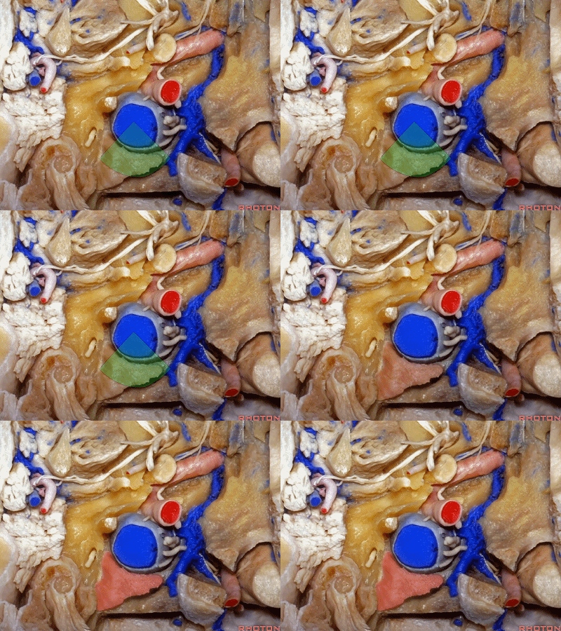
▼后方,则可显露头外侧直肌于颈静脉孔后缘的全部附着范围,附着处即为枕骨的颈静脉突。
and posteriorly, the full extent of the rectus capitis lateralis muscle's attachment to the posterior margin of the jugular foramen, which respansents the jugular process of the occipital bone.
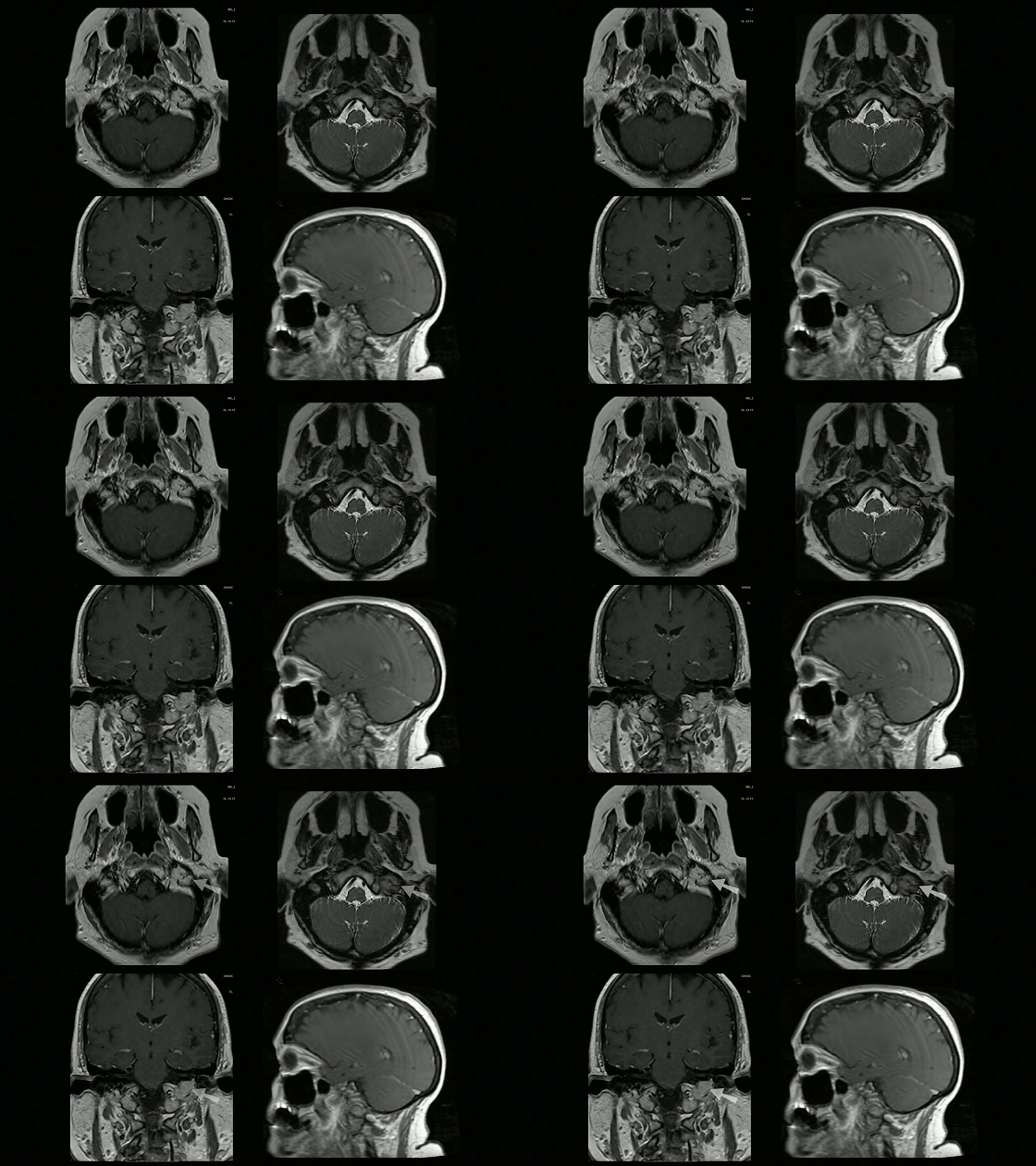
▼我们将结合实际病例来阐述手术技巧:65岁女性,主诉为搏动性耳鸣,左侧听力无减弱。后组颅神经和面神经功能均正常。颅脑MR,提示一3cm大小的颈静脉球瘤。
The case that we're spansenting for illustration of the technique is that of a 65-year-old lady who spansented with pulsatile tinnitus, normal lower cranial nerve and facial nerve function, her hearing was also intact on the left side. Neuro-diagnostic studies included MR scan as well as CT scan revealing a 3-cm glomus jugulare tumor
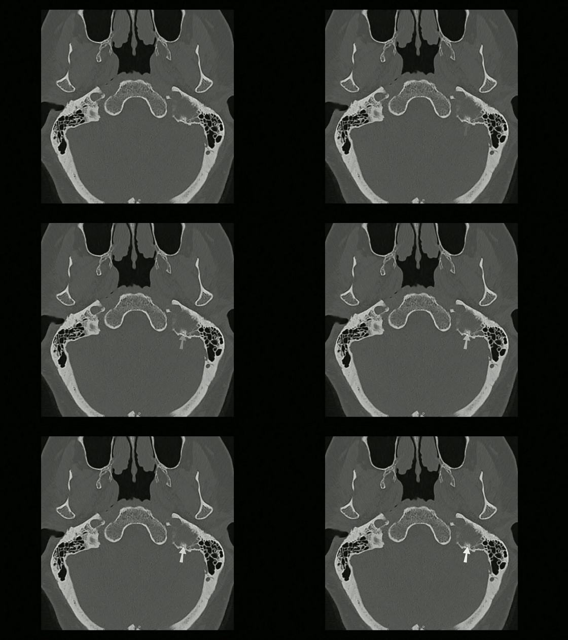
▼颅脑CT扫描主要伴有乳突破坏及侵犯颈静脉球区域。
with destruction of the mastoid bone primarily and the region of the jugular bulb by the glomus jugulare tumor.

▼脑血管造影证实病灶血供丰富,同时由左侧颈外动脉和椎动脉供血,符合颈静脉球瘤典型表现。颅底颈内动脉未参与肿瘤的供血。
The cerebroangiogram demonstrated significant blood supply from both the left external and vertebral arteries, typical of a glomus jugulare tumor. There was no blood supply to the tumor from the internal carotid artery at the skull base.
▼术前1天,该患者进行了血管内介入栓塞治疗。
Preoperatively the patient underwent embolization the day prior to surgery.
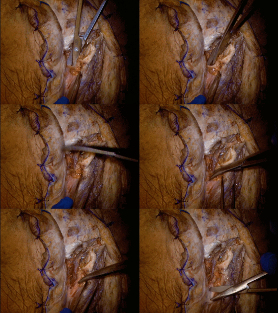
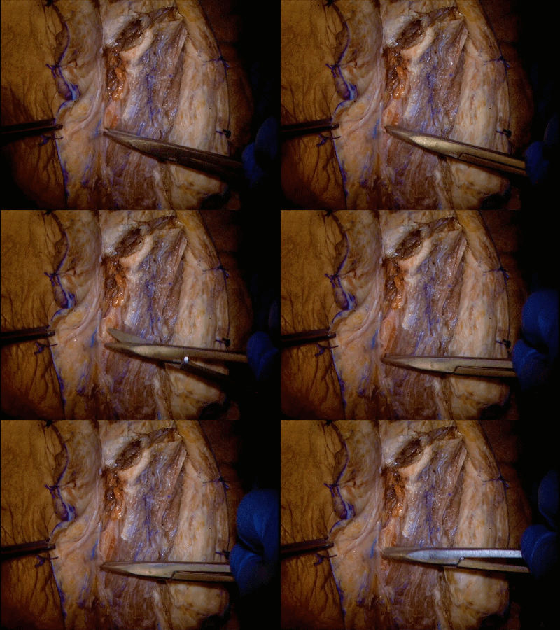
▼手术步骤:患者采取平卧位,同侧肩膀垫高,头转向对侧。耳后部沿C形切开。
The patient was placed on supine position with an ipsilateral shoulder roll, and the head turned away from the surgeon. The postauricular area is exposed along a C-shaped incision.
▼标本解剖首先进行胸锁乳突肌的锐性切开,从乳突尖离断。将胸锁乳突肌小心牵向后方。
Our cadaver dissection begins with the sharp dissection of the sternocleidomastoid muscle from the mastoid tip. The sternocleidomastoid muscle is carefully mobilized posteriorly.
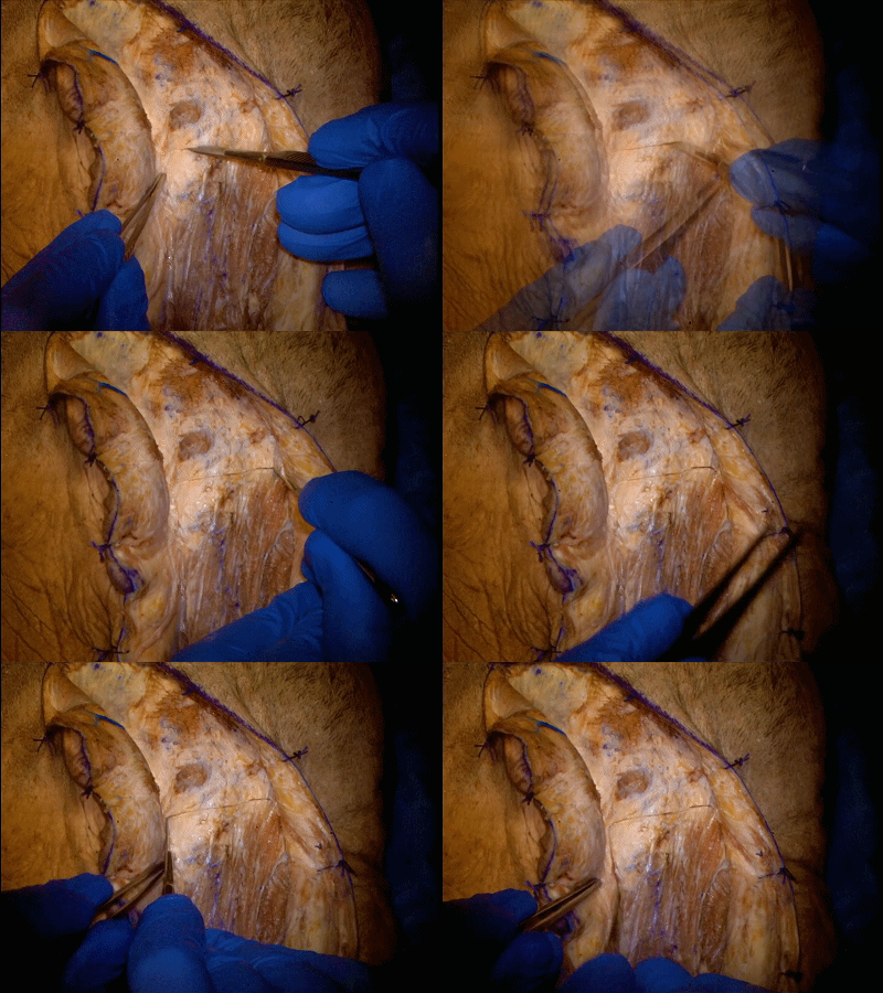
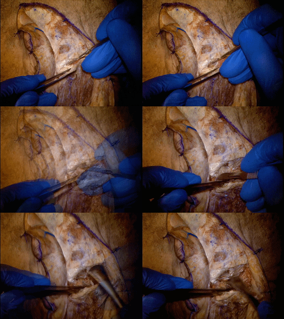
▼注意保护副神经,其穿入胸锁乳突肌上部的后内侧,层面平寰椎横突尖。
Care is taken to spanserve the accessory cranial nerve, which enters the posteromedial aspect of the superior sternocleidomastoid muscle at the level of the tip of the C1 transverse process.
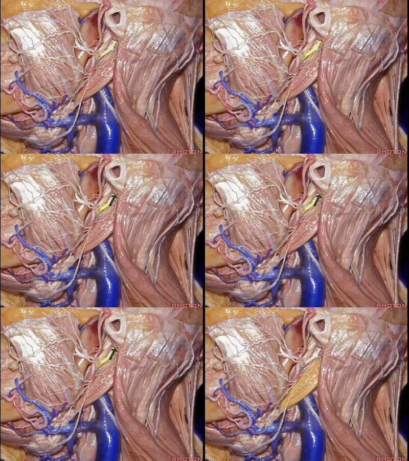
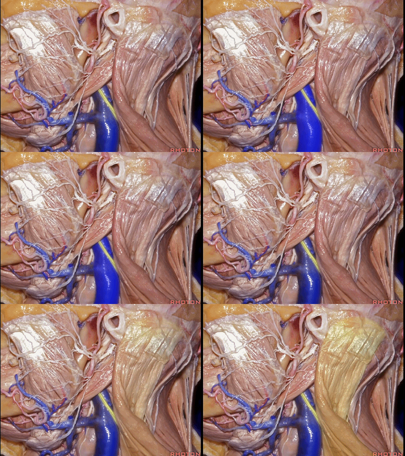
▼下图展示了各解剖结构的关系,包括胸锁乳突肌、二腹肌后腹、面神经、茎突。移除腮腺使得上述解剖结构能如此清晰的显露。
This Rhoton dissection shows the relationship of the anatomy of the sternocleidomastoid muscle to the posterior belly of digastric, facial nerve, and styloid process. These anatomical structures are viewed clearly in this particular dissection because the parotid gland has been removed.
▼这里特别需要注意的结构是从茎乳孔(下图箭头)穿出的面神经,其恰位于二腹肌后腹的前方,二腹肌附着于二腹肌沟。
Aspecific anatomy to be noted in this view is the facial nerve exiting the stylomastoid foramen immediately anterior to the posterior belly of the digastric attached to the digastricgroove.
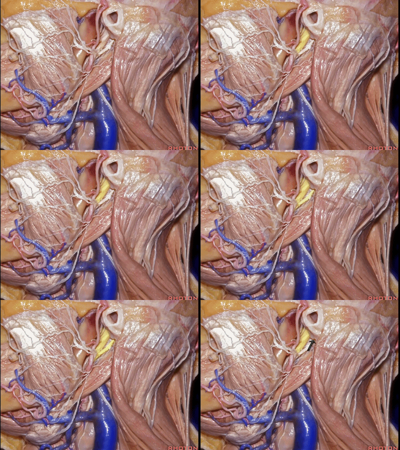
▼茎突(下图)也已清晰显露,面神经沿茎突的后外侧缘穿出于茎乳孔(下图箭头)。
Also note the styloid process can be seen clearly, and the facial nerve exiting the stylomastoid foramen along the posterolateral margin of the styloid process.
▼副神经(下图)从颈内静脉(下图)前缘跨越,进入胸锁乳突肌(下图)的后内侧部。
Note also in this view the accessory nerve crossing the anterior margin of the jugular vein to enter the posteromedial aspect of the sternocleidomastoid muscle.
▼二腹肌后腹已呈90°移位,并用线绳游离。
The posterior belly of the digastric has been mobilized with a right angle, and isolated with application of a vascular loop.
▼锐性分离二腹肌后腹,将其从乳突尖的二腹肌沟上离断,并向下翻折。
The posterior belly of the digastric muscle is sharply dissected from the digastric groove of the mastoid tip and mobilized inferiorly.
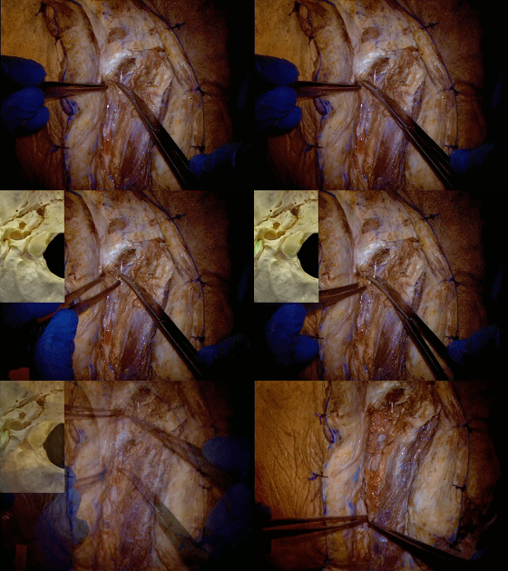
▼从二腹肌沟离断二腹肌后腹,即可显露位于茎乳孔的面神经。
Removal of the posterior belly of the digastric muscle from the digastric groove allows the exposure of the facial never at the stylomastoid foramen.
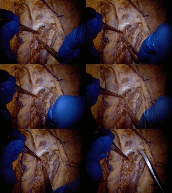
▼同时也可触及 茎突及三块附于其上的茎突肌群。
This also allows palpation of the styloid process and three small muscles attached to the process.
▼茎乳孔(下图箭头)位于茎突底部的后外侧缘。
Stylomastoid foramen is found at the posterolateral margin of the styloid process base.
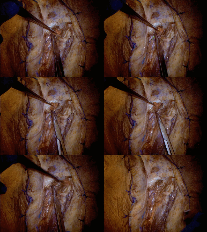
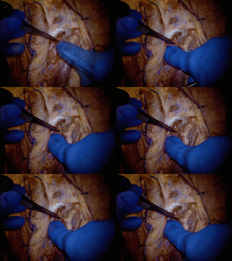
▼需将腮腺(下图箭头)向前游离,直到暴露位于茎乳孔的面神经。
The parotid gland must be mobilized anteriorly, until expose the facial nerve at the stylomastoid foramen.
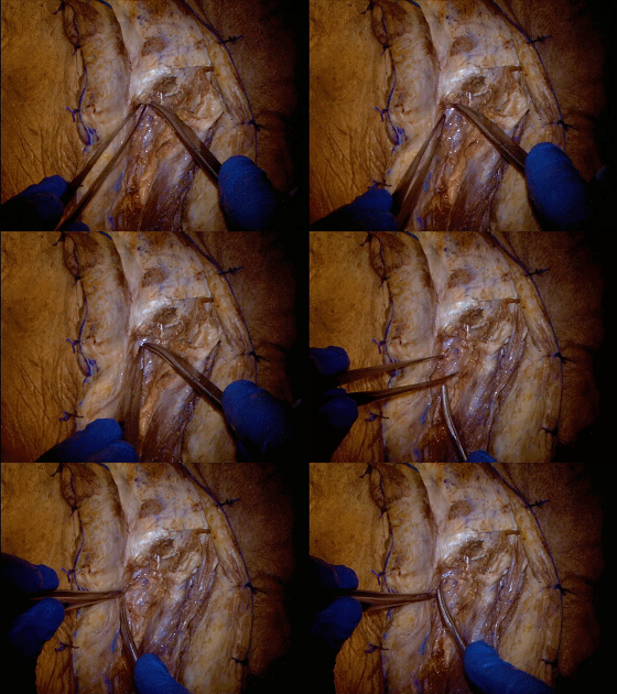
▼这里,茎突舌骨肌已被牵开以显露茎突。
In this view, the stylohyoid muscle has been retracted to expose the styloid process.
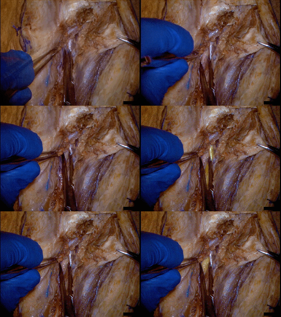
▼锐性分离茎突肌群。茎突舌骨肌已被沿着二腹肌后腹向下翻折。
The muscles attached to the styloid process are sharply dissected. Here the stylohyoid muscle has been reflected inferiorly along with the posteriorbelly of the digastric muscle.
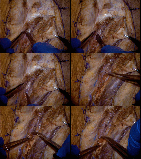
▼沿着茎突底部的后外侧缘仔细松解软组织,以暴露位于茎乳孔的面神经。
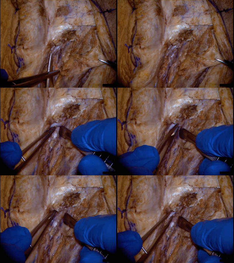
The soft tissue is carefully dissected to expose the facial nerve as it enters the stylomastoid foramen, along the posterolateral aspect of the styloid base.
耳后经颞入路至颈静脉孔区---Rhoton解剖视频学习笔记系列(下)


