

上接“前颅底(2)与鼻腔解剖---Rhoton解剖视频学习笔记系列(中)”,欢迎阅读。
▼在此基础上还可联合额颞开颅,从而显露海绵窦(下图)
And then you combine that with a craniotomy of frontotemporal that you can gain access to the cavernous sinus.

▼可循着三叉神经眼支(下图)至眼眶
It gives access along V1 to the orbit,

▼可循着三叉神经上颌支至上颌窦、眶底
V2 along the roof of the maxillary sinus, floor of orbit.

▼或者循着三叉神经下颌支至 颞下窝。
or along V3 down to the infratemporal fossa.
▼通过下部上颌骨切开入路,可切除斜坡脊索瘤,一些鼻咽纤维血管瘤。
This approach...here, the lower maxillotomy you can do for chordomas of the clivus, some of these juvenile angiofibromas.
▼联合上述两个上颌骨切开入路,以及经颅暴露海绵窦的入路,可处理一些沿着颅神经生长并累及中颅窝以外侵犯颅底的肿瘤。
This approach, combining the upper maxillotomy and the craniotomy exposure of cavernous sinus can be used for some of the tumors that infiltrate along the nerves coming out of middle fossa and going into the skull base.


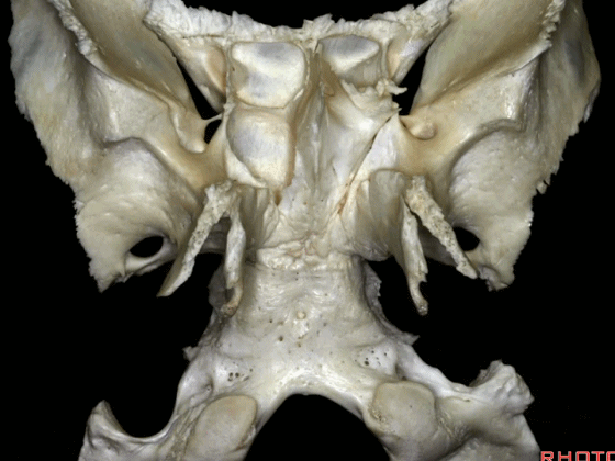
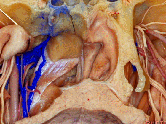
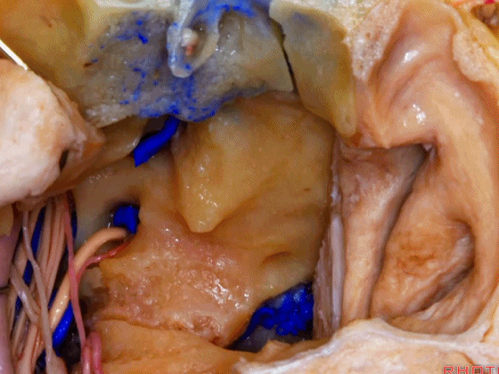
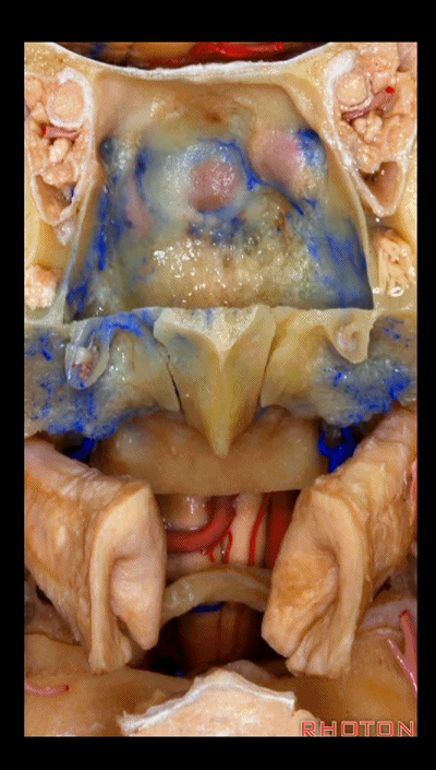
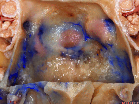
▼接下来我们讨论斜坡相关入路,斜坡病变也越来越多地通过经鼻入路来处理。这是上斜坡、中斜坡、下斜坡。但看一下斜坡,其底部宽于顶部。
Let me just run down the clivus, quickly from upper,middle,and lower third. Now, another situation is going to the clivus and clival lesions are being increasingly exposed transnasally today. But as you look at the clivus, clivus is wider at the bottom than it is at the top.
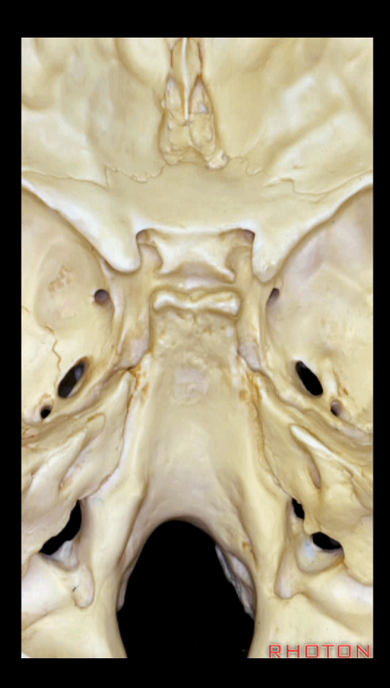
▼处理斜坡病变时,我们难以使用局限化的入路。上斜坡位于蝶窦后方。
So that, upper clivus is behind the sphenoid sinus.

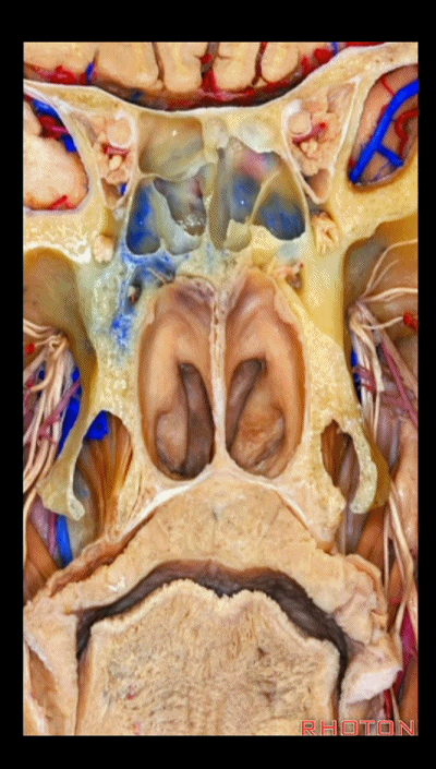
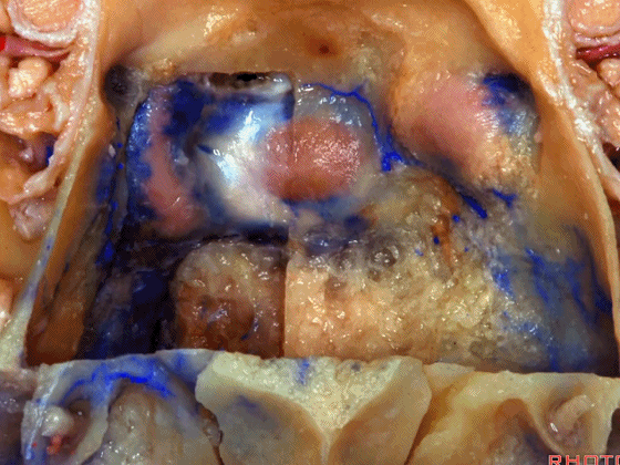
▼这是硬腭
Here is the palate.
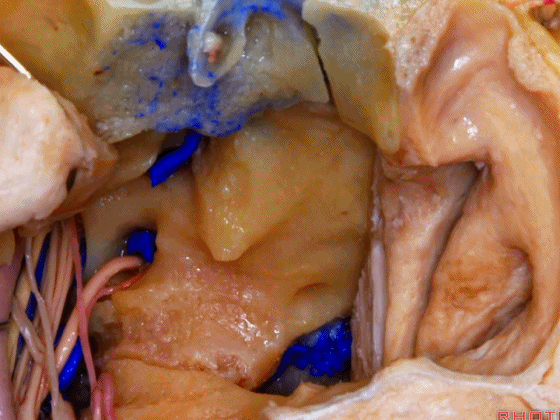
▼首先让我们从下斜坡开始,下斜坡位于寰枕关节上方。
dealing with clival lesions you can't do very focal approaches. And we'll start out, here,lower clivus, just above the atlanto-occipital joint.
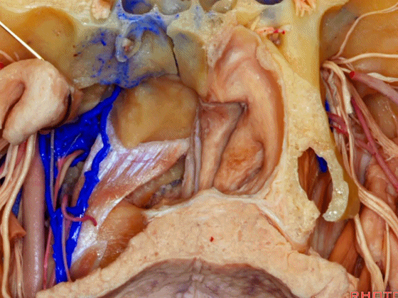
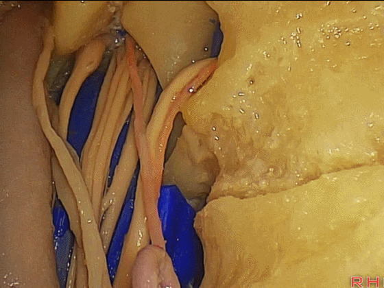
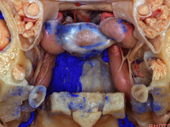
▼下斜坡位于咽鼓管(下图)的后方。咽鼓管位于内侧翼板及下鼻甲后方,是鼻咽部经鼻入路的外侧界限。
Lower clivus will be here,behind the...what is this that sits behind the medial pterygoid plate...what is that...we're in back of the turbinates now,and the lateral limit of the transnasal approach, back in the nasopharynx is the...eustachian tubes.

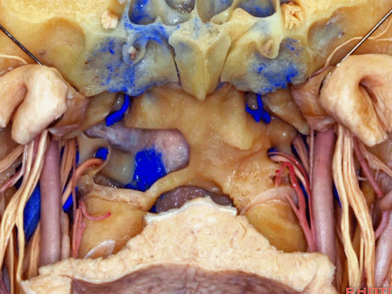
▼从前方来看,这一下垂的结构阻挡了下2/3的斜坡,这是翼突。斜坡的外侧界被其遮挡了。
And if you look at it from the front, what is this sticking down that blocks access to approaches to the lower 2/3 of the clivus? What...you don't see the lateral edge of the clivus. It's hidden behind the pterygoid process and medial pterygoid plate.
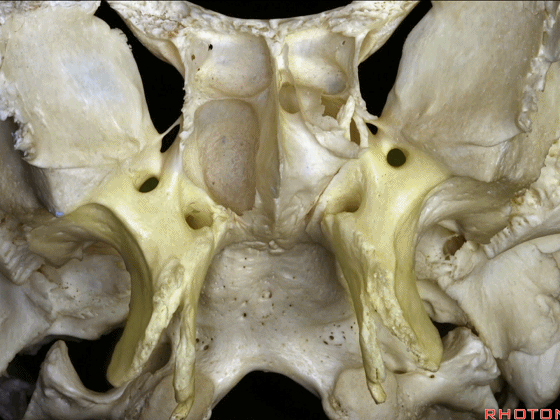
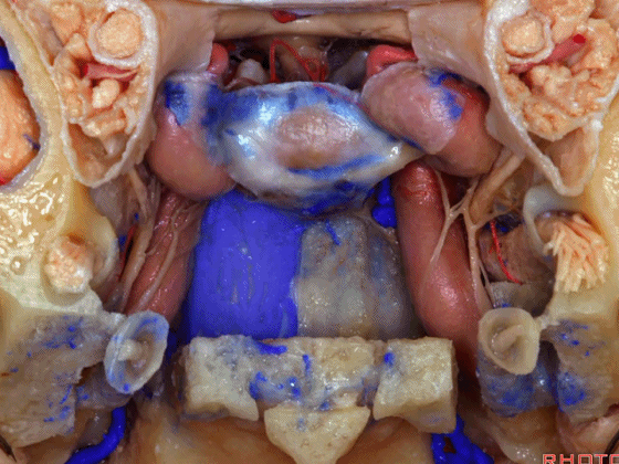
▼这是内侧翼板(下图),因此,想要到达斜坡外侧界,必须知道如何扩大视野。
So, to get to lateral edge of clivus,you have to understand how to widen that opening.
▼这是咽鼓管隆突。
And here is the tubal prominence.

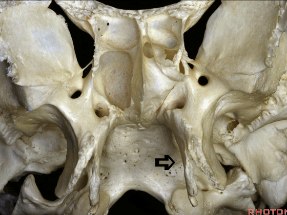
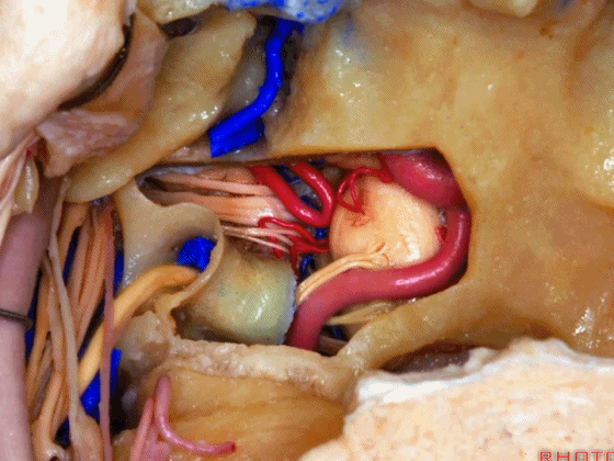
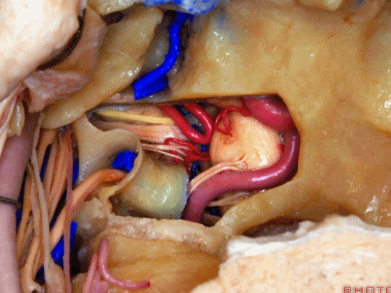
▼切除一块内侧翼板,则可将咽鼓管推向一侧。即可暴露下三分之一斜坡。
If you remove a medial plate then you can retract them laterally. And here we see lower third of clivus, clivus here.
▼下图示 下三分之一斜坡
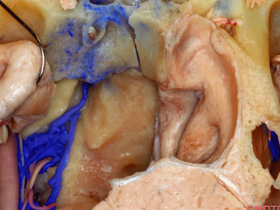
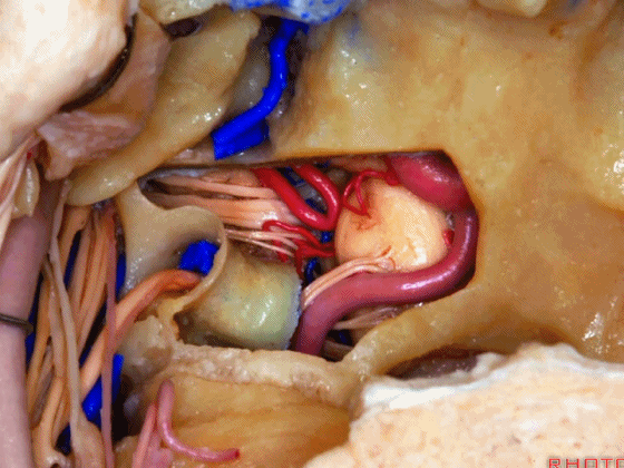
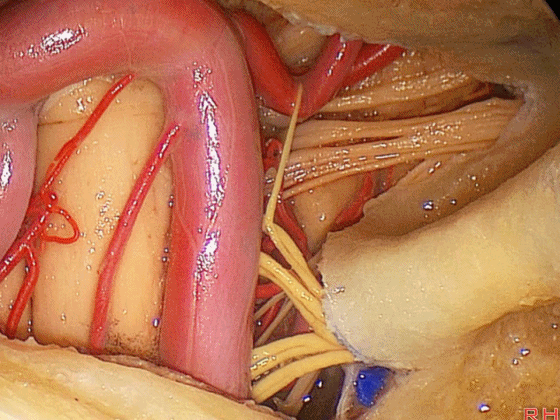
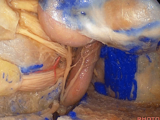
▼这是头前直肌(Rectus capitis anterior 下图),该肌肉从斜坡发出,沿着颈静脉突,跨过寰枕关节,连于寰椎,在其前方为头长肌,需将其牵向外侧。
And a cup of coffee for anyone that can name this muscle that comes off the clivus, along this tubercle, spends the atlanto-occipital joint, attaches to C1, what muscle is it? Rectus capitis anterior,in front of it is the longus capitis that you have to pull laterally.
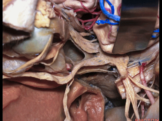
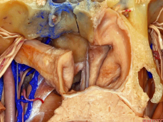
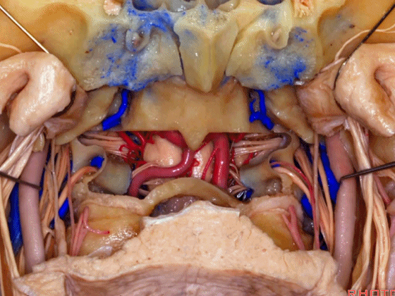
▼这里我们切除了头前直肌。岩斜裂内有岩下窦(下图),但在此裂表面常有静脉丛向下汇入颈静脉孔。
But here we've detached the rectus capitis anterior.We are now...on the inside of the petroclival fissure is the inferior petrosal sinus, but also there are the plexus of veins here on the external surface that runs down the fissure to the area of the jugular foramen.
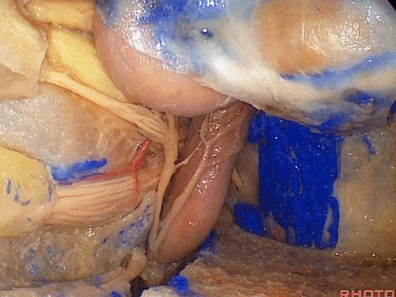
▼这是寰椎关节。
Here's the atlanto-occipital joint.
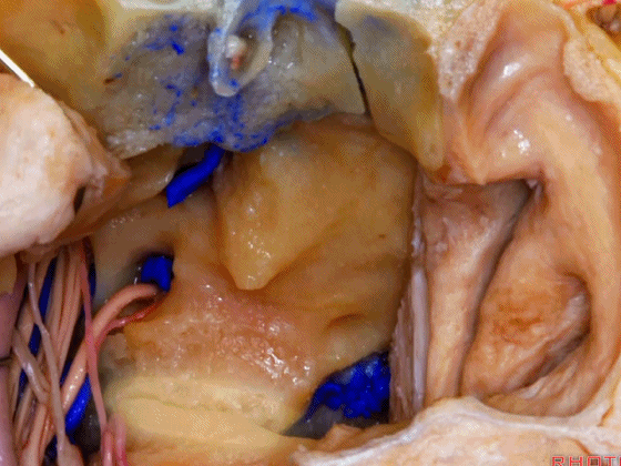
▼在枕髁(下图)上方穿行出颅的是舌下神经(下图)。
And this nerve coming through above the occipital condyle is the...what nerve? Hypoglossal.
▼这是舌咽、迷走、副神经,舌下神经与它们相汇合。
Here's IX, X, XI,and it's joined...XII joins them.
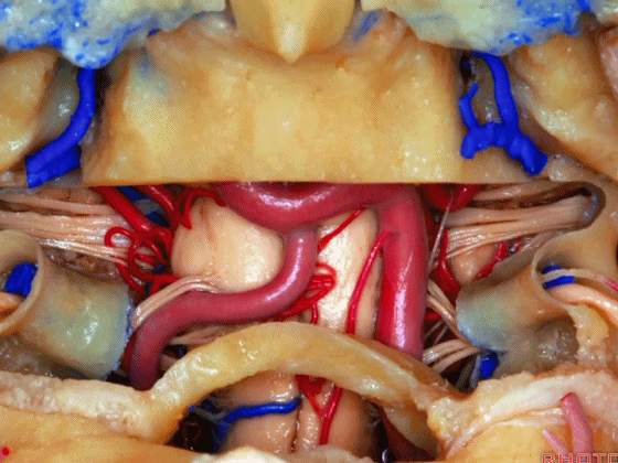
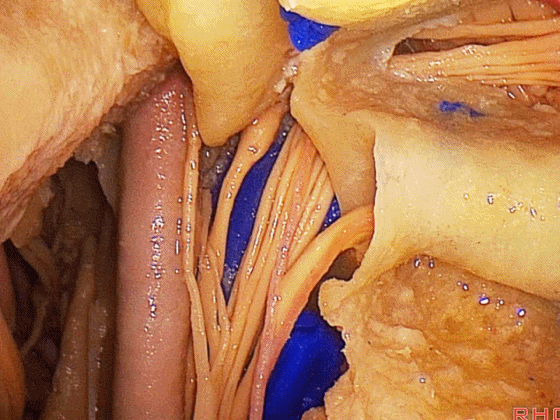
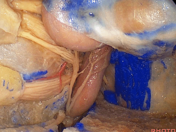
▼我们可打开下三分之一斜坡,这部分操作是我们希望学员们能掌握的
You can open up that lower third of clivus, you know, laterally...this is the kinda work we expect our fellows to do that come with us in the lab after their training,
▼这里可见舌下神经管的暴露。
but you see the drilling of the hypoglossal canal.
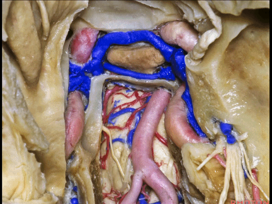
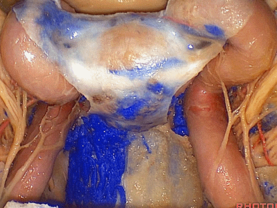
▼在侧方,可见椎动脉(下图)、舌下神经、舌咽、迷走、副神经的硬膜内部分。
You can do a, you know, lateral approach, vertebral, hypoglossal nerves, IX , X , XI intradual.
▼这是舌下神经
▼这是舌咽、迷走、副神经
▼接下来可做双侧暴露,这是双侧的舌下神经管
And then you can extend our approach bilaterally, the pairs of hypoglossal canals.
▼硬膜下部分的舌咽、迷走、副神经,均可经下斜坡暴露。
Here on each side intradually, IX,X,XI,the approach through the lower clivus.
▼这一视野可见舌下神经管(下图)
Here's a view over to the hypoglossal canal here,

▼这是颈静脉孔
jugular foramen
▼这是岩斜裂的下端。
at the lower end of the petroclival fissure.

▼这是用成角内镜下的术野,采用硬膜下入路(下面三图逐级放大)
The view with the endoscope angle laterally, endoscopic view, an intradural approach,
▼这是左侧结构。舌咽、迷走、副神经
and then the opposite side,IX, X, XI

▼舌下神经穿行于舌下神经管
and then the 12th nerve coming through the canal,
▼这是右侧 颈静脉孔下方走行的神经。
the view of the nerves just below the jugular foramen,on both sides.
![]()
▼接下来我们看上三分之一斜坡,要暴露此处,需打开蝶窦后壁(下图)。
And then we go to the upper third of the clivus, and to get there, you have to remove the posterior wall of sphenoid sinus.
▼这是 垂体、颈内动脉海绵窦段、视神经颈内动脉隐窝LOCR。
But here we see pituitary, cavernous carotid, opticocarotid recess.
▼打开蝶鞍,打开右半侧硬膜。
We've opened up the sella, half of it...dura...expose.
▼可见海绵窦前内侧部
We see anteromedial cavernous sinus here,
▼可见垂体上动脉、视交叉。
superior hypophyseal arteries,chiasm.

▼这是上斜坡硬膜的基底静脉丛。
We see the basilar plexus in the dura of the upper clivus.
▼内镜下,这是右侧 海绵窦前内侧部
Here, anteromedial cavernous sinus,
▼这是动眼神经
3rd nerve,

▼这是 三叉神经眼支V1
▼外展神经穿出硬膜,向上行于三叉神经眼支内侧.
6th nerve coming through the dura, running then upward medial to V1

▼这是 三叉神经上颌支V2
▼这是沿颈内动脉上行的交感神经。
sympathetic ascending on the carotid.
▼这是左侧海绵窦前内侧部,这是眼动脉(下图)。该入路也可处理眼动脉的动脉瘤。可进行近端阻断。
and left anteromedial cavernous sinus, and this is ophthalmic. It's not a bad approach for ophthalmic aneurysm. You have proximal control, down here, just a thought.

▼这是左侧 外展神经穿出硬膜,行于颈内动脉斜坡旁段外侧、三叉神经眼支内侧。
Here we see 6th nerve coming through the dura,running medial to V1.running lateral to the terminal petrous carotid.

▼这是左侧 脑膜背动脉,发自脑膜垂体干。
Here's a dorsal meningeal artery coming off the meningohypophyseal trunk.

▼下面我们打开蛛网膜,外展神经穿出硬膜处的硬膜予以保留。
The arachnid has been opened. The dura around the dural exit of the 6th nerve spanserved.

▼这里我们打开了颈内动脉 远环。
Here we open the distal dural ring around the carotid.

▼这是McConnell's被囊动脉,是颈内动脉海绵窦段发出至垂体前表面的分支。它们并不常见
And what is this artery, what do we call these branches as they go from carotid to anterior surface of pituitary? They are not very common. They are...the McConnell's capsular arteries.

▼这是 垂体上动脉。
Here's just the superior hypophyseal arteries.

▼这里我们打开远环内侧。这是鞍膈下方。
Here We've open that distal dural ring medially. We're under the diaphragm.

▼沿着蝶鞍的硬膜分为两层,贴着颅底骨的是骨膜层,贴着垂体和脑组织的是脑膜层。
And here we see along the sella there are two layers of dura a periosteal layer that faces the bone, a meningeal layer that faces the gland and brain,

▼两层之间即为静脉窦(下图)。
and the sinus runs between those two layers.

▼然后我们可沿着垂体的右侧 向后贴着鞍背至垂体后叶。
And then we can work along the side of the gland back to posterior lobe along the edge of the dorsum.

▼随后可显露鞍背。
And then you can get into the dorsum.

▼这是垂体后叶。
Here's posterior lobe.

▼磨除鞍背,即可经上三分之一斜坡暴露至基底动脉尖(下图)
Remove the dorsum, and you have the exposure through the upper third of clivus to basilar apex,

▼这是Liliequist's膜
to Liliequist's membrane, to...

▼这是后交通动脉
posterior communicating

▼这是大脑后动脉P1段

▼这是大脑后动脉
posrterio cerebral

▼这是乳头体
mammillary bodies

▼接下来我们进行中三分之一斜坡的暴露
And then you can expose middle third of clivus,

▼这里是左侧、右侧视野
here one side, the opposite side.

▼这是翼管神经
Anyone, what is this? What do you think? Vidian nerve, again.

▼早期人们都主张可牺牲翼管神经,但其结果是导致患者干眼症。但我相信未来的趋势将是尽可能地显露并保护该神经。它们行于翼管(下图)内,正如舌下神经行于舌下神经管(下图)内一样。
All of the early descriptions of vidian nerve surgery said sacrifice it, that can lead to a dry eye. But it's possible I think...we'll see it become more common in the future to not sacrifice it but actually to drill it out and spanserve it. Here it is in its canal just like the hypoglossal nerves are spanserved in their canal.

▼这是外展神经,于此处(下图)穿出硬膜。
But we see 6th nerves hang on in the dura here.

▼我们可磨除此处的岩尖。
You can drill off...this is petrous apex here in this area.

▼随后利用成角内镜,可见内听道近端的面听神经
You can get in with angled endoscope. And,work laterally to VII and VIII.


▼通过成角内镜亦可向上暴露至三叉神经进入脑桥中部,因此这也不失为一种微创入路,对脑干旁的面神经或三叉神经进行微血管减压术。
You can look above with the angled endoscope and see V entering the mid pons,VII and VIII proximal to the meatus, but not a bad route if you can do a very focal approach to VII or V right at the brainstem for doing a vascular decomspanssion of these nerves.


▼这是三叉神经后根、面听神经。
here we see posterior root of V,VII and VIII.

▼这是位于内听道口的面听神经
VII and VIII over at the porus.

▼经斜坡,我们还可显露海绵窦前内侧部的颈内动脉、三叉神经眼支、上颌支、下颌支。
through the clivus, and here we come now to anteromedial cavernous sinus on the lateral side of the carotid. V1,V2,V3,

▼这是翼管神经,其可定位 颈内动脉岩骨段末端。
and...vidian nerve, again,coming to the terminal petrous carotid.

▼这里保留了外展神经周围的骨质。
Some bone spanserved around VI.

▼外展神经位于三叉神经眼支内侧。
And, here's VI on the medial side of V1.

▼我们可磨除岩尖(下图),可获得三叉神经下方的视野。
You can drill out petrous apex and get this exposure under the trigeminal nerve.

▼这是岩尖磨除后,三叉神经(下图)

▼这是岩上静脉的属支。
Here's one of the superior petrosal veins.

▼因此,向外侧可暴露三叉神经、面听神经、海绵窦前内侧部、以及外展神经。
So, working laterally you can have an exposure of V, VII and VIII,anteromedial cavernous sinus, 6th nerve in this area.

▼内听道放大观。

▼我们查阅了很多相关解剖并加入到这一解剖合集中,以供广大的神经外科医师免费下载学习。这一切最终都是为了提供最好的技术给患者,让他们都能得到最正确、最完美的医疗服务。
We review a lot of this anatomy in the book. One of our aims is to get this nose for neurosurgeon into this collection that's downloaded at no charge.But all of this has been about making what is a delicate fate for, awesome experience for a patient, more accurate, gentle and safe.
点击继续阅读
