上接“前颅底(2)与鼻腔解剖---Rhoton解剖视频学习笔记系列(上)”,欢迎阅读。
▼这是 下鼻甲
inferior turbinate
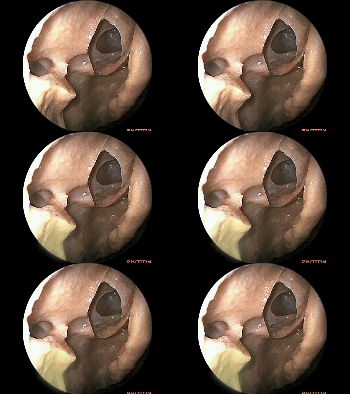
▼中鼻甲(下图)已牵向中线,这些都位于中鼻道内。
the middle turbinate is pulled medially, so this is all middle meatus.
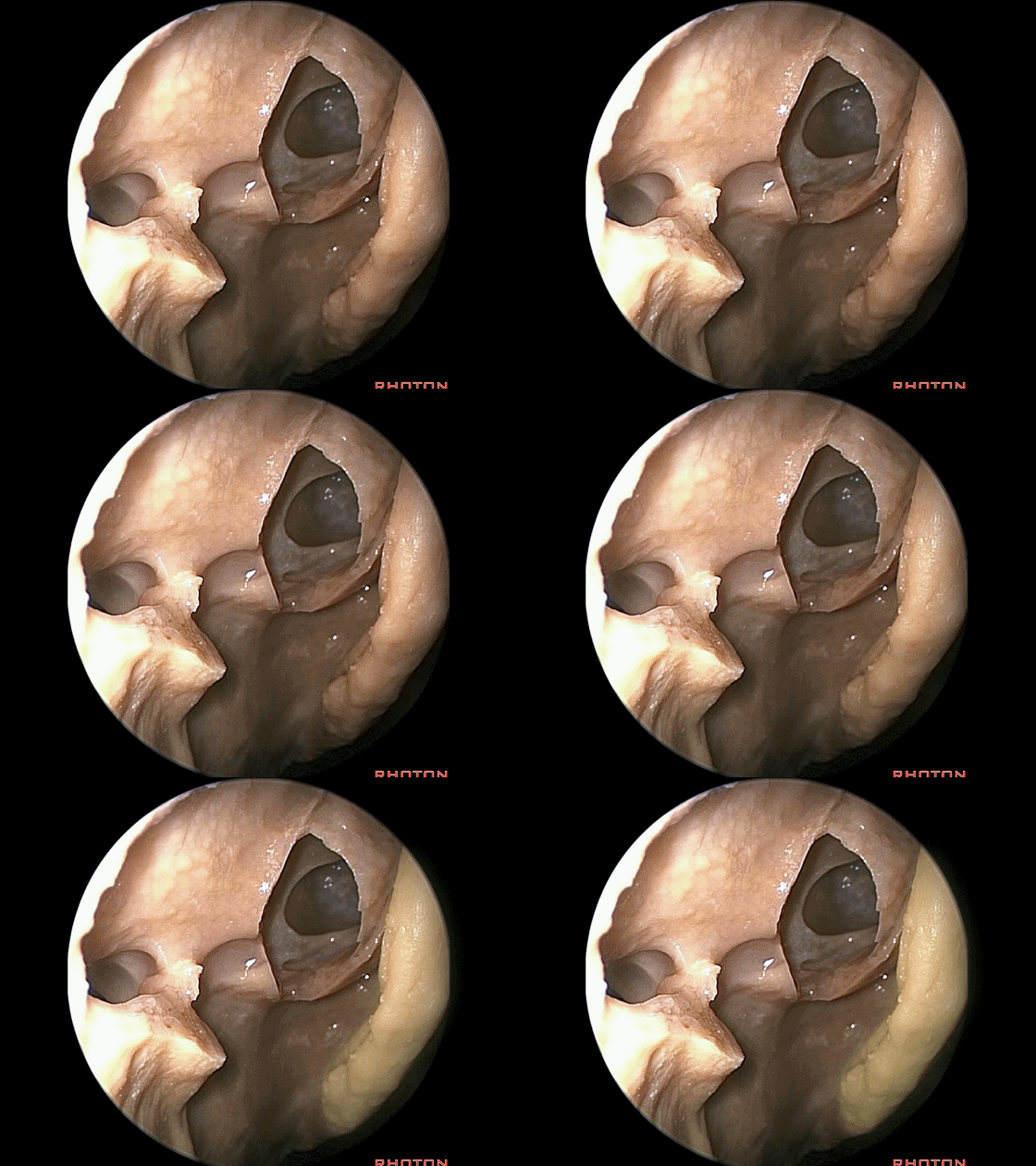
▼那么,切除筛房并打开额窦后,可切除该片区域的骨质。
So coming through this area after the ethmoidectomy and drilling out the frontal sinus,you can do an osteotomy around this area.
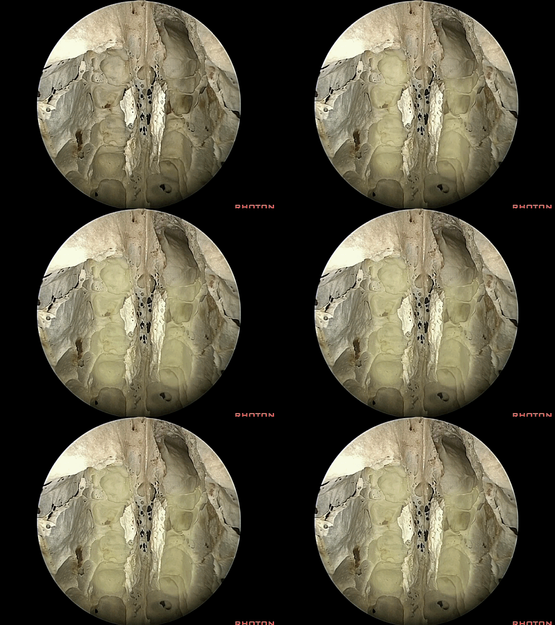
▼这是筛板区域,下图示嗅黏膜,位于筛板下方。
And here we see the cribriform plate area,You can do an osteotomy around this area. Here's the olfactory mucosa sitting just below the cribriform plate.
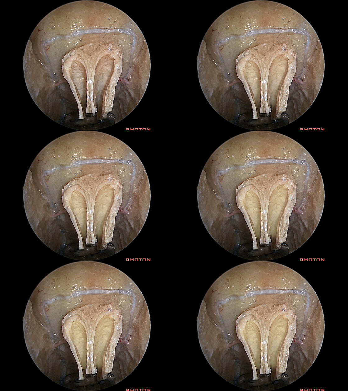
▼这里我们将嗅黏膜予以保留。
Here's the olfactory mucosa spanserved.
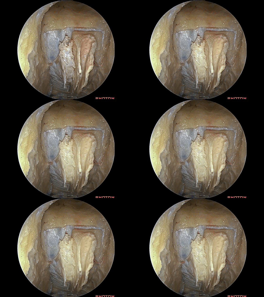
▼这是筛前动脉。如果要牺牲这对血管,则尽量向内侧靠近嗅黏膜处切断之,因为曾经在一些病例中,靠近外侧切断该血管后,其断端回缩入眶内引起严重的眶内血肿。
The anterior ethmoidal arteries.you wanna take those if you're gonna sacrifice some a little bit medial toward the olfactory mucosa, because there're some cases where they were divided laterally and retracted into the orbit and cause significant orbital hematomas.
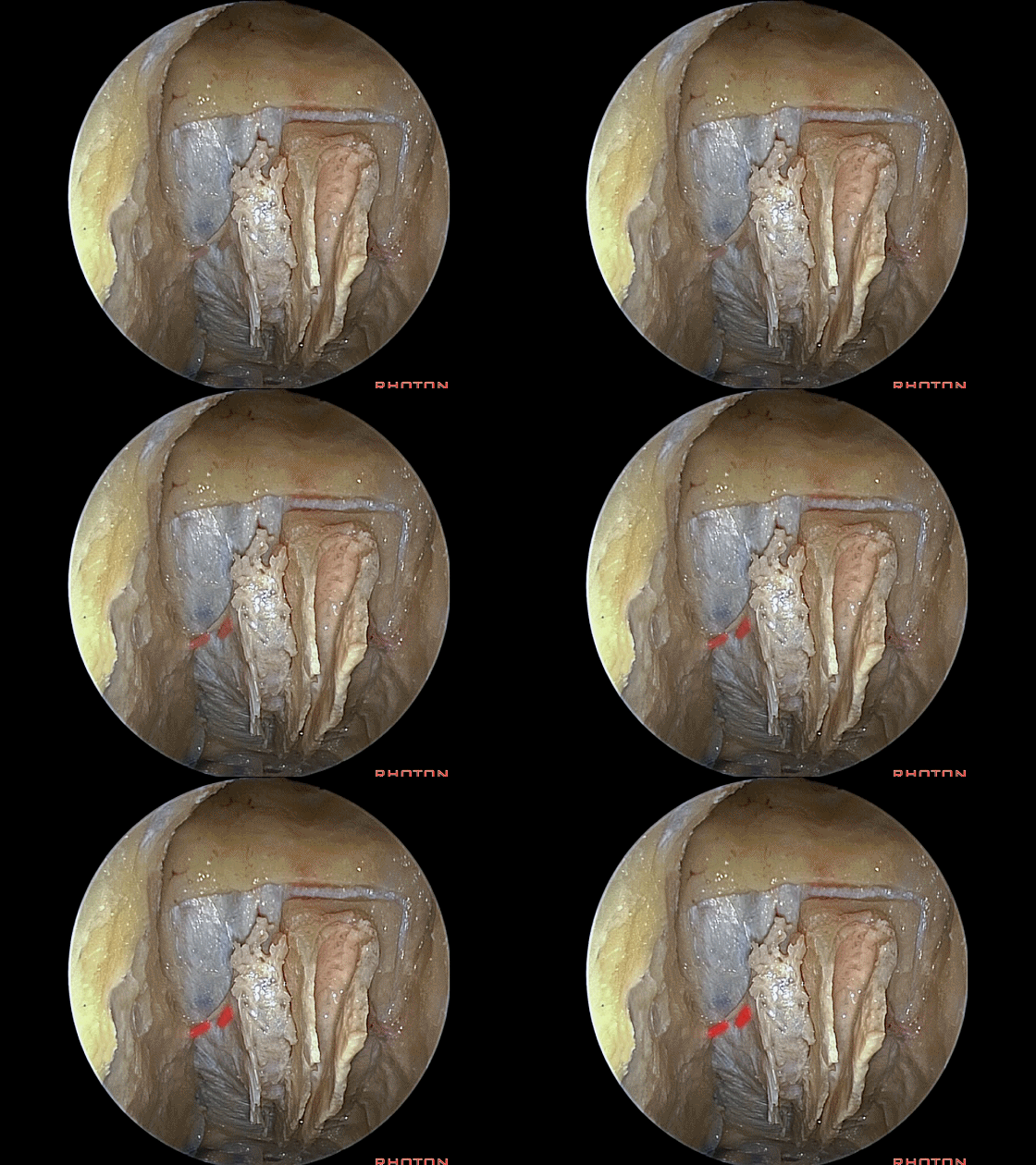
Here are the ethmoidal arteries and nerves. And always that companies the ethmoidal arteries there,these little ethmoidal nerves to the nose.
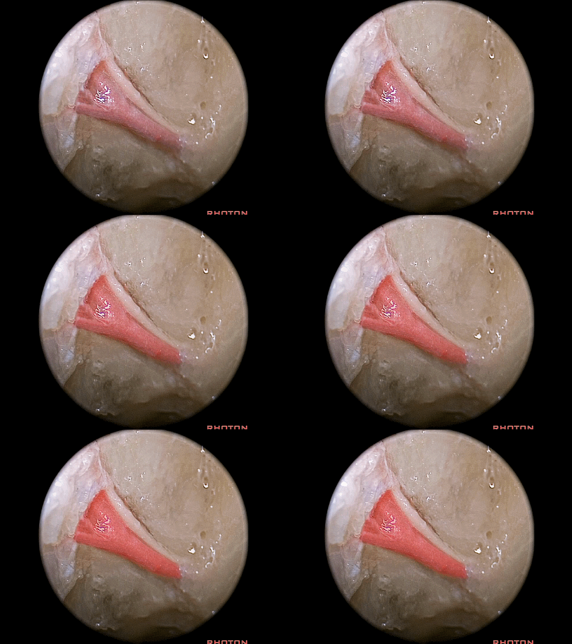
▼切除一侧骨板后,可见嗅球。
And here we've completed the dissection on one side. The olfactory bulbs.
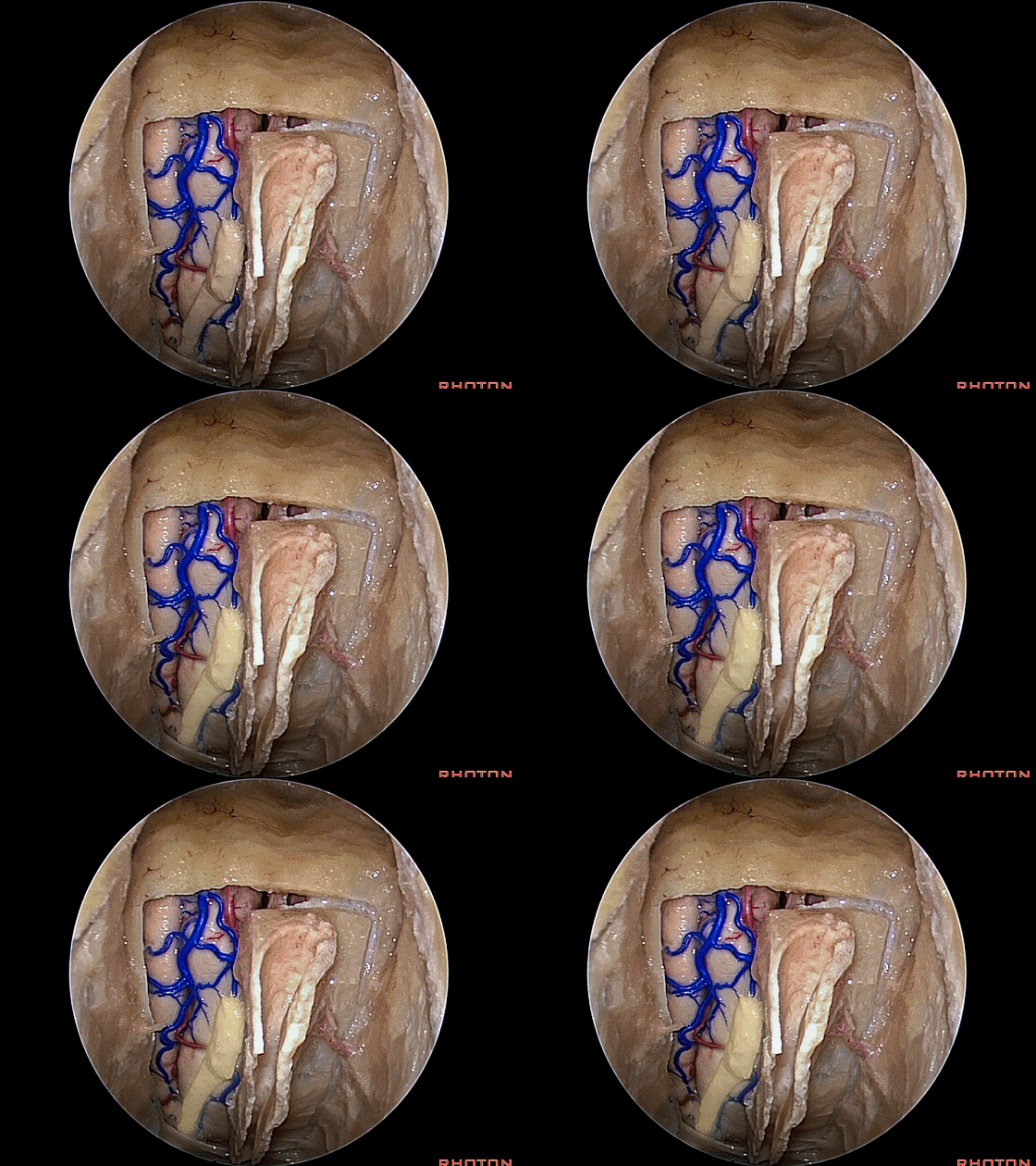
▼暴露上方区域需进一步切开大脑镰。
You have to divide the falx if you're going to open this area above.
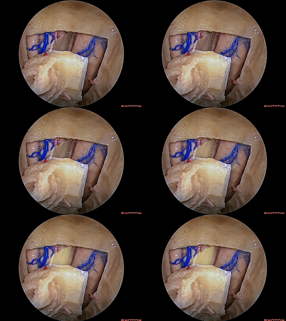
▼这里我们已切开大脑镰
So, the falx is divided,
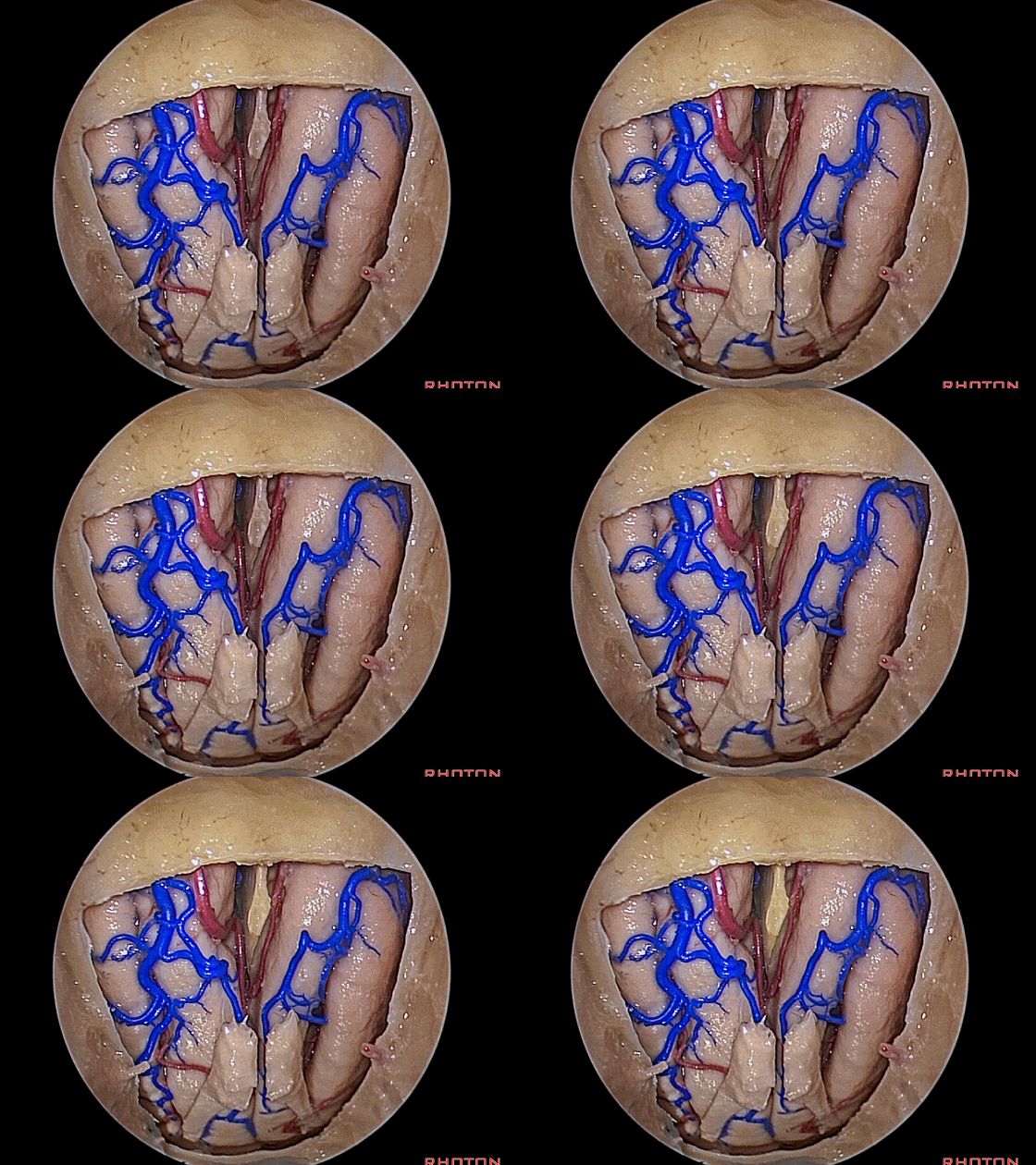
▼以上即为经鼻腔进入前颅窝的入路。皆得益于筛窦迷路的磨除。向后可进入蝶鞍(下图)。
so this is the approach to anterior fossa.aided by the removal of that ethmoidal labyrinth.You can work back to the area of the sella.
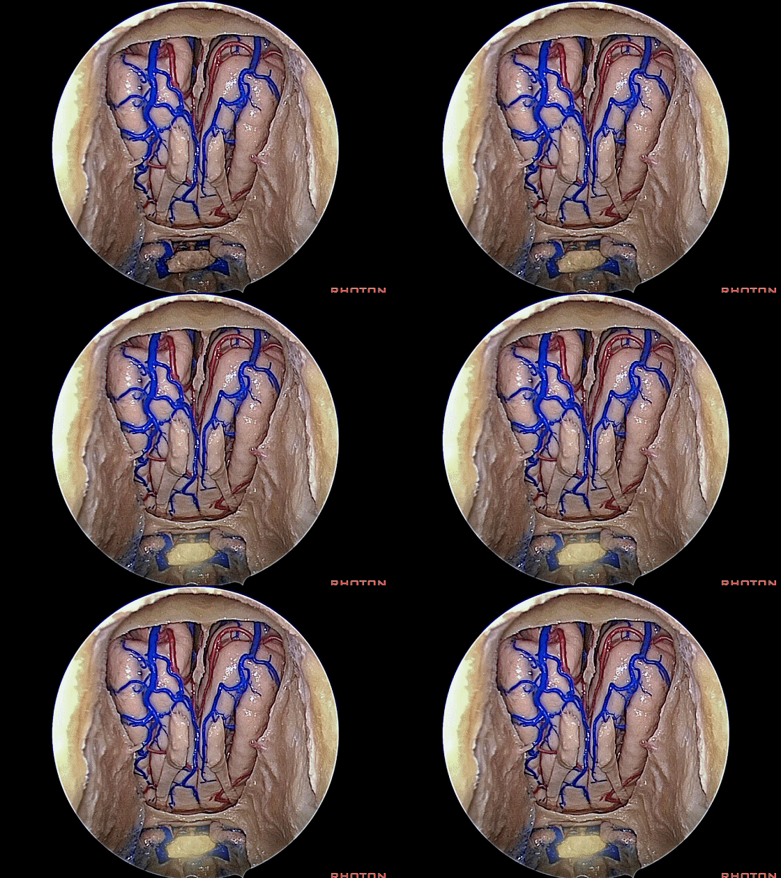
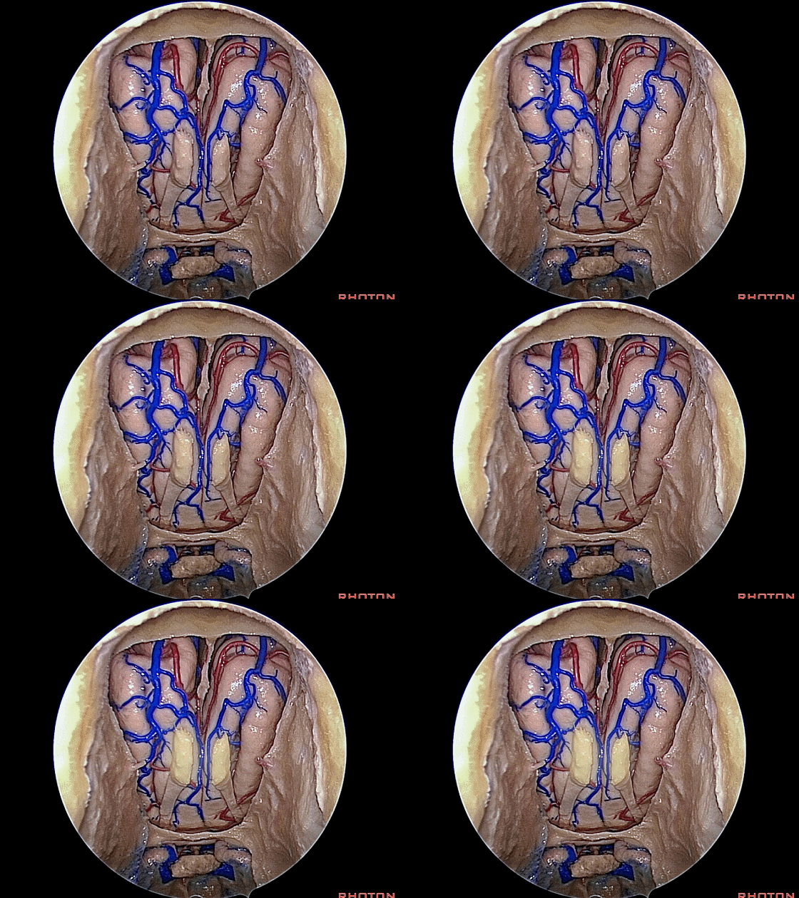
▼鲜有案例报道,可将嗅黏膜保留于嗅球上,在切除病灶时将嗅束垂下,随后复位。这样,嗅黏膜可持续附着于嗅球,保留在鼻腔顶壁,从而保护了嗅觉。但这难度很大。
There're few cases now where the olfactory mucosa has been spanserved attached to the bulbs and folded downward during removal of the pathology and then the closure is done. So that, this mucosa is still in the roof of the nasal cavity attached to the bulbs and olfaction has been spanserved. But I think that's difficult.
![]()
▼接下来我们将讨论经蝶入路。我最初是通过唇下切口进行经蝶入路的,切口在唇下,后来我改变了切口方式,沿着鼻中隔,在黏膜下与鼻中隔之间进入。随后我意识到,在显微镜下,透过鼻孔,即可在鼻甲的内侧、鼻中隔的外侧直接看到蝶窦的前壁,于是我改为直接的经鼻入路,而无需破坏鼻中隔。
We're going to talk about transsphenoidal approach. And I started doing transsphenoidal approach coming sub-labial, under the lip, and then I switched the approach to coming along the nasal septum under the mucosa, between the septum. And then I realized, as I looked up the nostril with a microscope, I can see, medial to the turbinates, lateral to the septum, directly to the face of the sphenoid, and I started doing this direct endonasal approach, where we don't disturb any of the septum.
▼我在进行这一直接经鼻入路时,直接穿过鼻孔,并不剥离鼻中隔黏膜,而是通过扩张器直接到达蝶窦前壁。
Well, when I started doing this direct endonasal approach, we just...we came through the nostril. We didn't strip up any mucosa on the side of the septum and we went with a speculum directly to the face of the sphenoid.
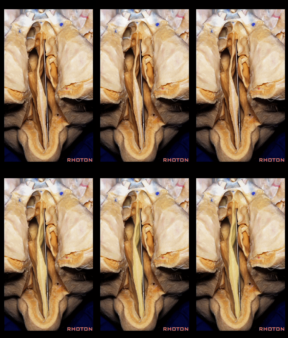
▼透过筛板从上方来看这一径路。
So that, if you look from above through the cribriform plate, this is the septum.
▼我们在鼻中隔一侧放入扩张器。随后离断鼻中隔,将其与蝶窦表面分离。
We place the speculum along one side of the septum. And then, we just crack some of the septum, the packed part, off of the face of the sphenoid,
▼恰位于蝶窦开口下方。离断部分仅仅约有1.5厘米的高度。
just below the sphenoid ostia. And the part that's separated is only about a centimeter and a half in height
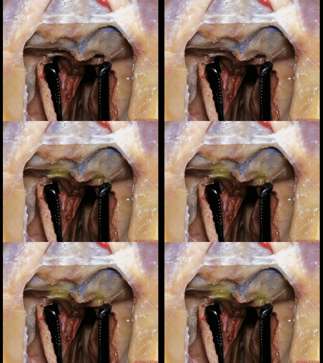
▼随后打开蝶窦,中线的最佳参照物即为蝶窦前壁的这一骨嵴。
And then, we open the sphenoid sinus and the best guide to the midline is this crest on the face of the sphenoid.
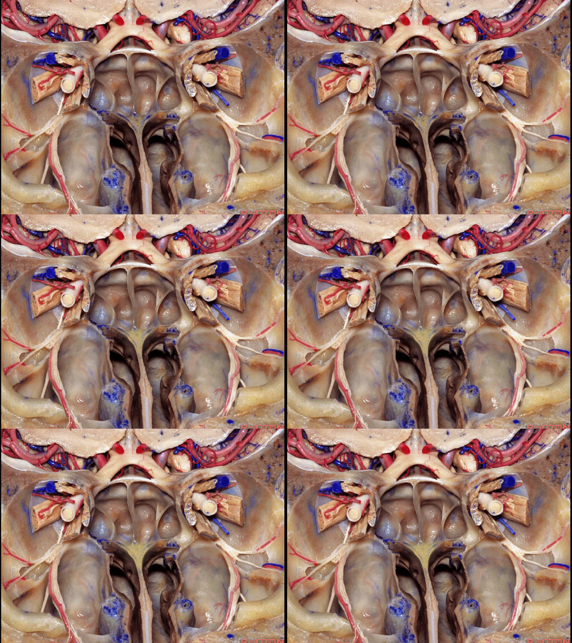
▼放入的这个扩张器,打开后可分开3~4cm能满足我们需要用到的宽度。两侧视神经管之间的距离仅为2.5cm(下图)。在这个区域,我们需要分开的宽度不要超过2cm,否则可能损伤视神经管(下图)。
And these specula, when you put them in, well, they open actually...the blades can separate as much as 3 or 4 cm,but you only want to open them wide enough. Here, in this area, the optic canals are only separated by about 2.5 cm. So that, you don't wanna open them much wider than 2 cm, or else you can get fractures into the optic canal,
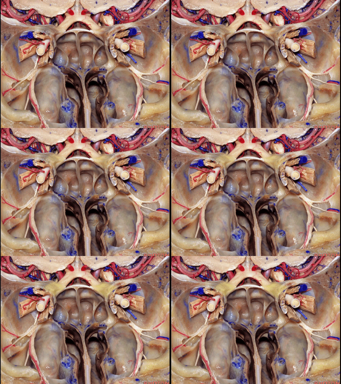
▼如今我们会使用影像导航系统,然而在实际手术开始后,如果影像导航系统提示这个位于蝶窦前壁、蝶窦开口下方的骨嵴偏离了中线,那通常是影像系统出了问题。中线的最佳参照物总是这一骨嵴,此处是筛板垂直板以及犁骨附着在蝶窦前壁的部位。 它是定位蝶窦中线的最佳标志。
Today we often use image guidance,but by the time you get the drapes and the fiducials on, if you look at your image guidance, and it says to you, this spike on the face of the sphenoid, below the sphenoid ostia, if it says that's off the midline, it's usually your image guidance that is off. This is just virtually always directly in the midline. It's the best guide to the midline of the sphenoid sinus. in this area here at the orbital apex. And the best guide to the midline is always this crest medial to the maxillary sinuses where the perpendicular plate of the ethmoid and the vomer attach to the front of the sphenoid
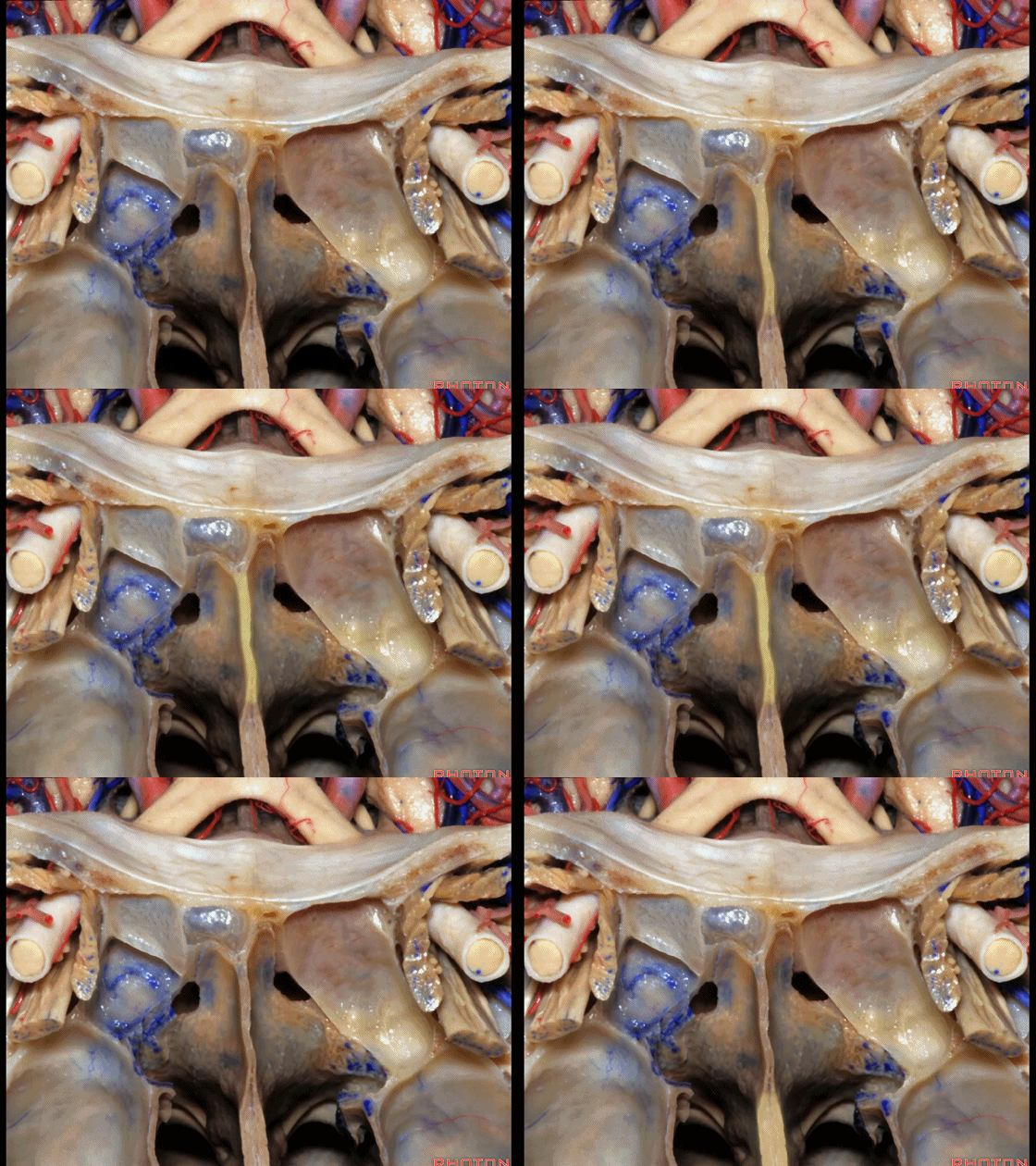
▼直视鼻腔时,最大的阻挡通向蝶窦的障碍物,是靠近中线的中鼻甲后部。
And as you look into the nasal cavity, the biggest obstacle to getting into the sphenoid sinus near the midline is the posterior part of the middle turbinate.
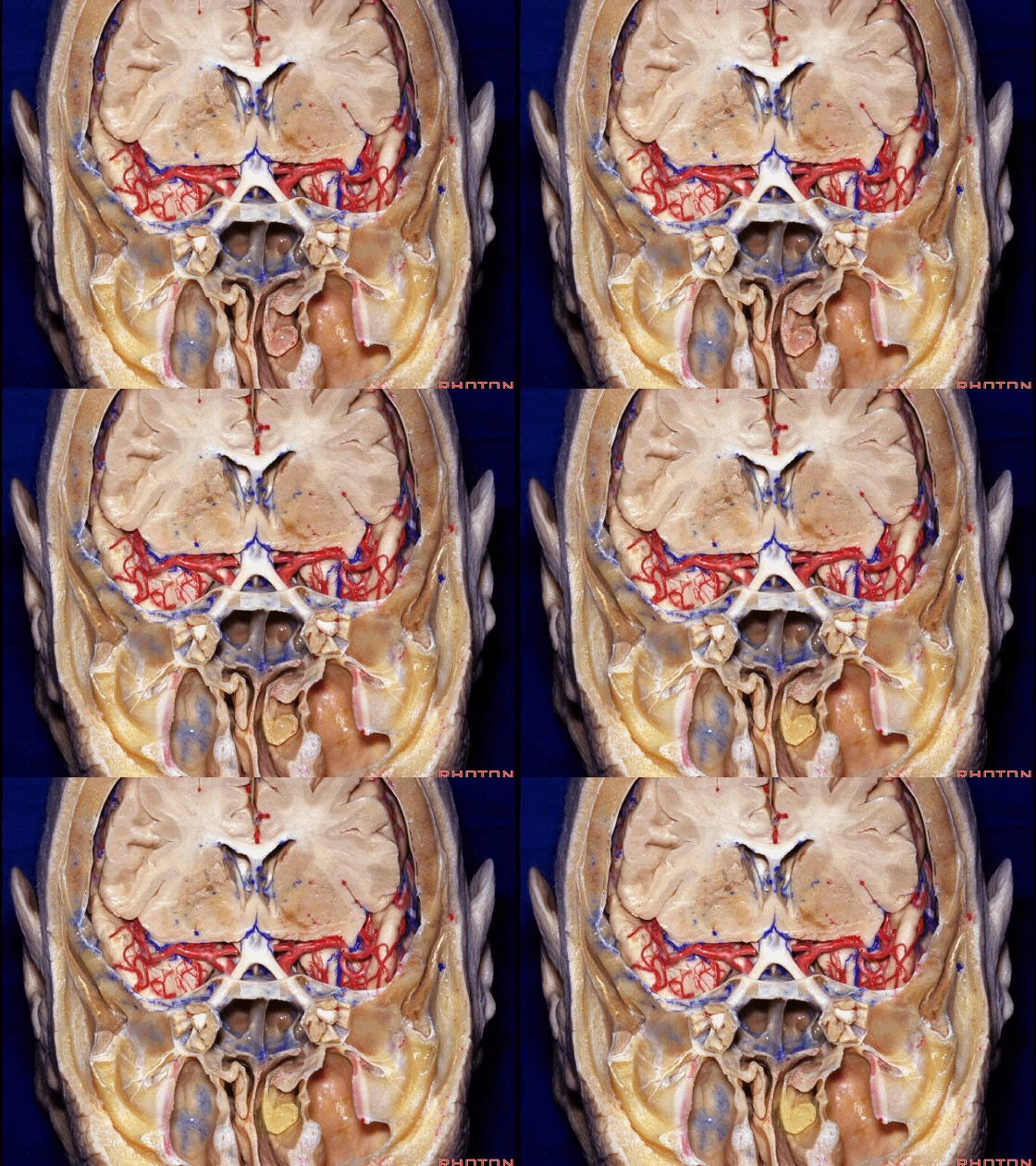
▼中鼻甲向上拱起,并推挤扩张器至对侧。假如扩张器刚好顶在蝶窦前壁的骨嵴上,那么叶片打开后可能会损伤这一侧的视神经和视神经管。因此需要将中鼻甲的后部向外侧推开。一些术者会将其切除,但我从未在经蝶入路中这么做。但我们需尽量推开侧方的鼻甲,使得蝶窦骨嵴位于术野的中线部位。
It bulges over, and it tends to deflect the speculum to the opposite side, and if you get caught on this crest on the face of the sphenoid and open the speculum, it's little more than a centimeter over to that optic nerve and the optic canal. So you always want to push this posterior part of the middle turbinate laterally. Some surgeons resect it, but I've never resected that in a transsphenoidal approach. But you want to get the blades of the speculum positioned in these lateral gutters on each side with the crest on the face of the sphenoid directly in the midline.
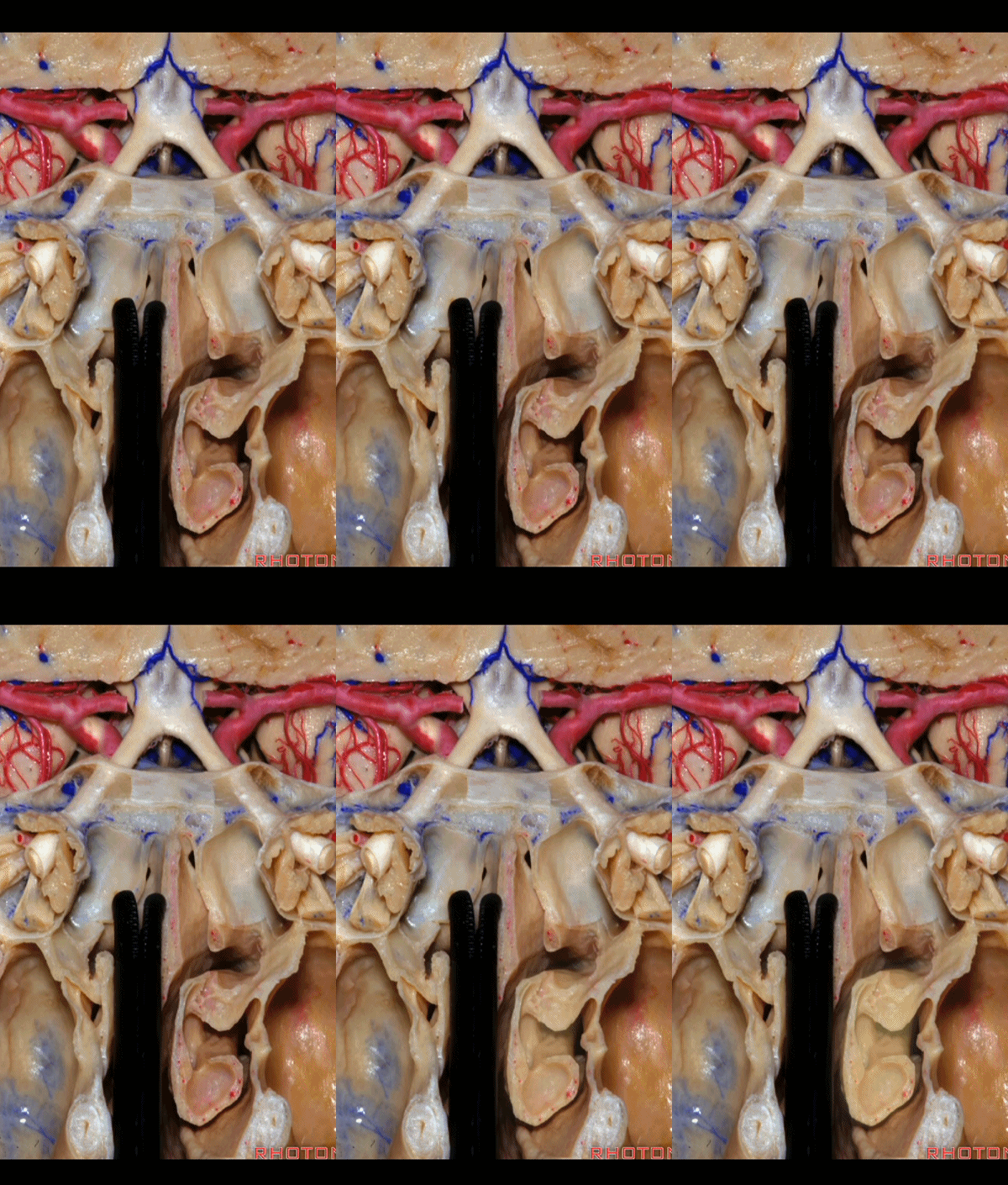
▼进入蝶窦后,多年来我都看到很多人会切除蝶窦内的黏膜,但一旦切除黏膜,就会使得硬膜直接与蝶窦腔,或骨质直接与蝶窦腔相接触。而通常情况下,我们在经蝶手术中,切除肿瘤,关闭蝶鞍前壁,保持蝶窦腔与鼻腔相通以引流。
And then, once you come to the sphenoid sinus, for years I've watched people opened the sinus, take out the mucosa in the sinus, but if you remove that mucosa, you have dura next to the air raided sinus, or bone directly facing the sinus.Usually when we do a transsphenoidal procedure, we take out the tumor, close the anterior sella wall, and leave the sinus open to drain into the nose.
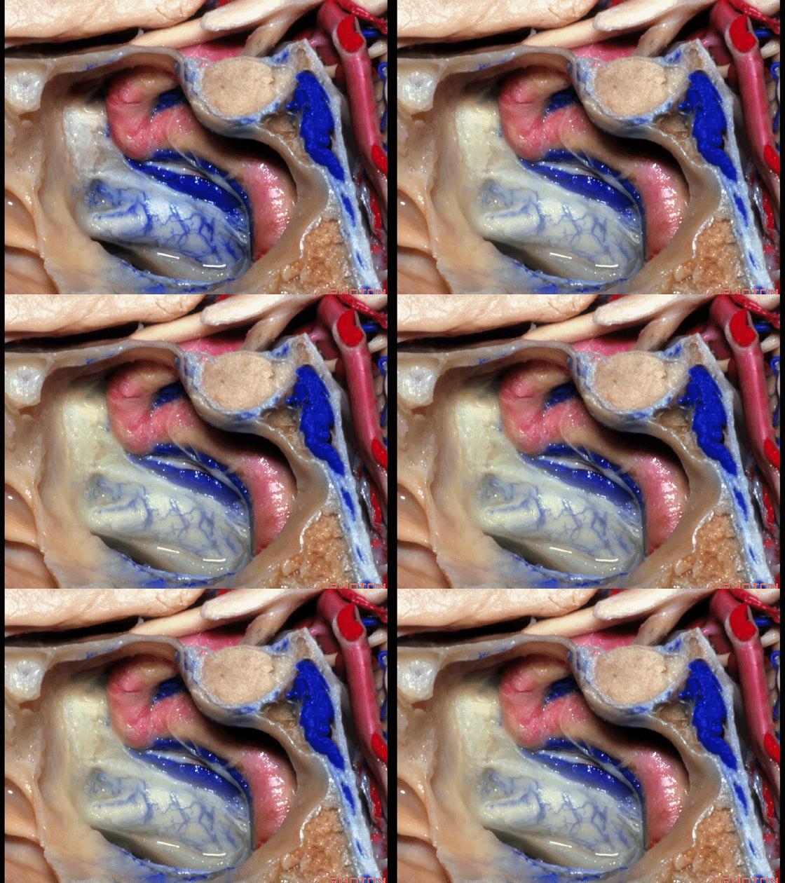
▼如果将黏膜切除,病人将丧失正常的黏膜纤毛运动,那么在6周后,腔内将满是分泌物、碎片,病人将抱怨常常闻到臭味。假如保留黏膜,仅仅切开蝶窦前壁和蝶鞍前壁的黏膜,那么6周后,窦腔内恢复正常。而如果剥除黏膜,则将满是肉芽组织。
If you pull out the mucosa, and look in 6 weeks later, you don't have that normal ciliary action in the mucosa that sweeps mucus and other product out of the sphenoid ostia. And you look in 6 weeks later, if there's no mucosa in the sinus, and there's granulation tissue, pars, mucus, often the patients complaint of it, feel smelling odor. While, if you leave the mucosa intact, just open it on the face of the sphenoid, and on the anterior sella wall, and look in 6 weeks later, the sinus looks perfectly normal. But if you take it out, it's just...it looks like granulation tissue.
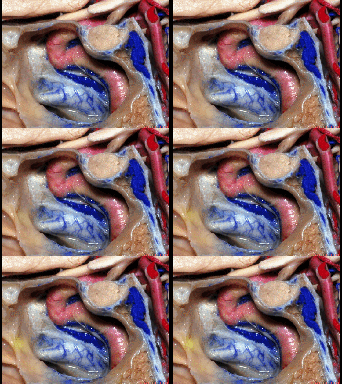
▼我在做经鼻入路时,填塞关闭蝶鞍后,我为何还要填塞鼻腔?我并非为了贴覆黏膜。填塞的目的在于防止术后出血。
And when I started doing this direct approach, we came to the face of the sphenoid here. And then we removed the tumor. We not strip up any mucosa here on the septum. And I was packing the nose initially. And I ask myself, what am I packing when I...after I close the sella why do I pack the nasal cavity? I'm not trying to put any mucosa back in place.But the reason I was packing to spanserve bleeding.
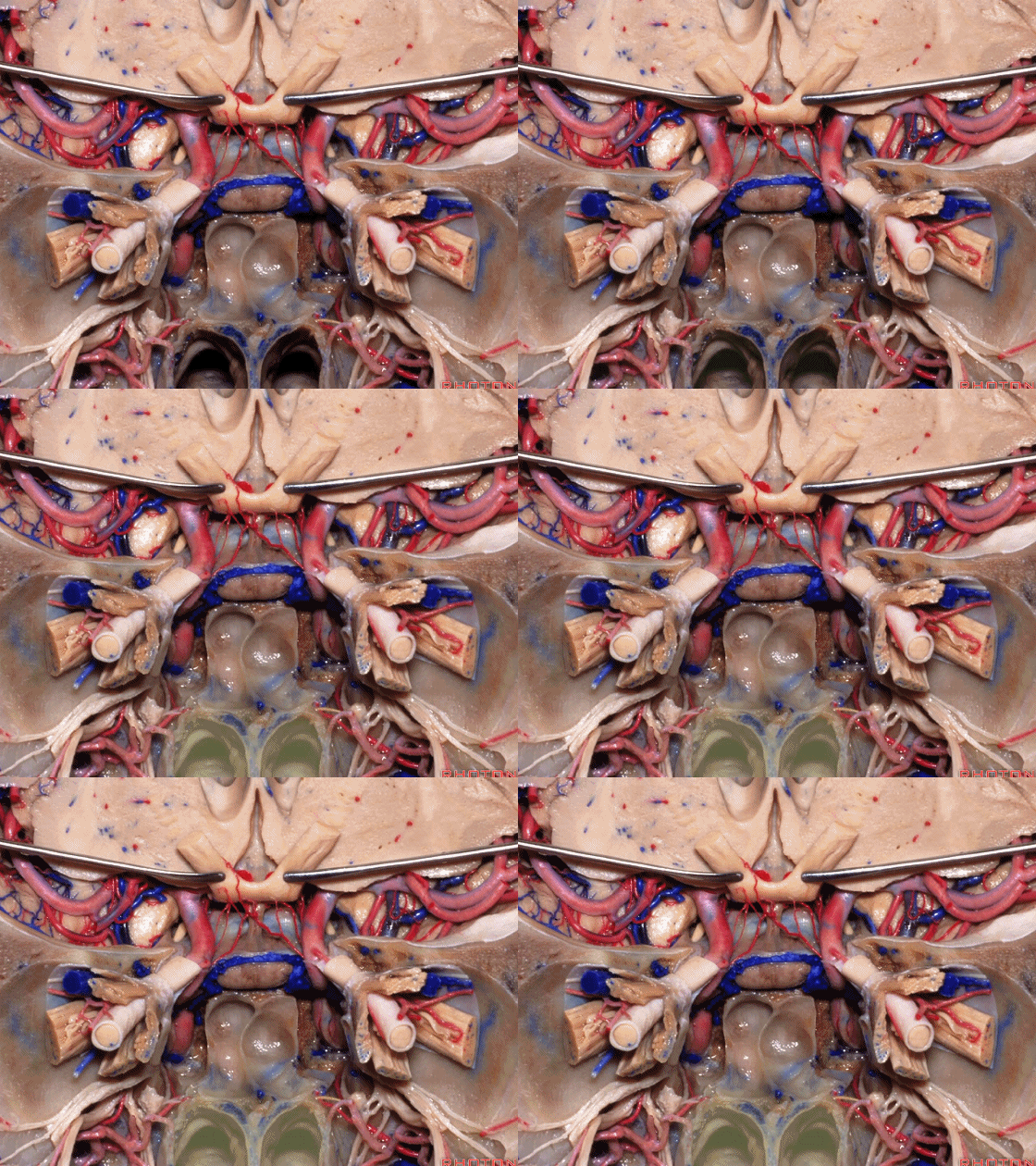
▼经蝶手术后最常见的出血来源是行经翼腭窝的上颌动脉分支,其供血鼻中隔。
And the most common side of postoperative bleeding after transsphenoidal surgery are these branches of the maxillary artery that come through the pterygopalatine fossa and supply the nasal septum.
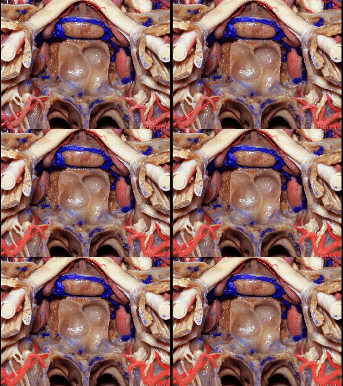
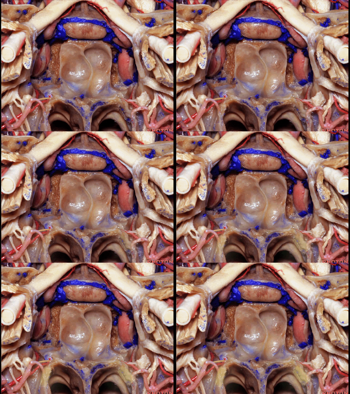
▼因此,如果术者止血彻底, 尤其针对这些侧方的沟槽内,一旦封闭了蝶鞍,就没有必要进行填塞。关闭蝶鞍,保留蝶窦黏膜,即可取出扩张器,结束手术。不用填塞的前提就是止血彻底,尤其在蝶腭孔处(下图)。
So that if you get good hemostasis in this area, in these later gutters, once you close the sella, there's no need for packing. You close the sella, you leave all the mucosa you can in the sphenoid sinus, and then you can pull out the speculum, you're done with the procedure. There's no need for packing if you have good hemostasis here at the sphenopalatine foramen.
![]()
▼下面复习一下鼻腔侧壁解剖,这里是下鼻甲、中鼻甲、上鼻甲、最上鼻甲,均为术野的外侧界。
Here we see the inferior, middle, superior turbinate, and a suspanme turbinate. in the lateral margin of the exposure.
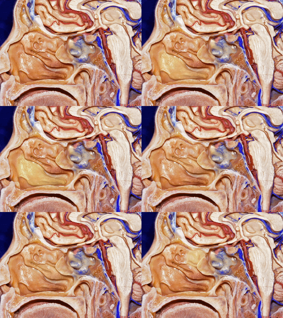
▼如果我们切除上述鼻甲,即可见上颌窦内侧壁
If we remove that, the upper turbinates, we see the medial wall of the maxillary sinus,
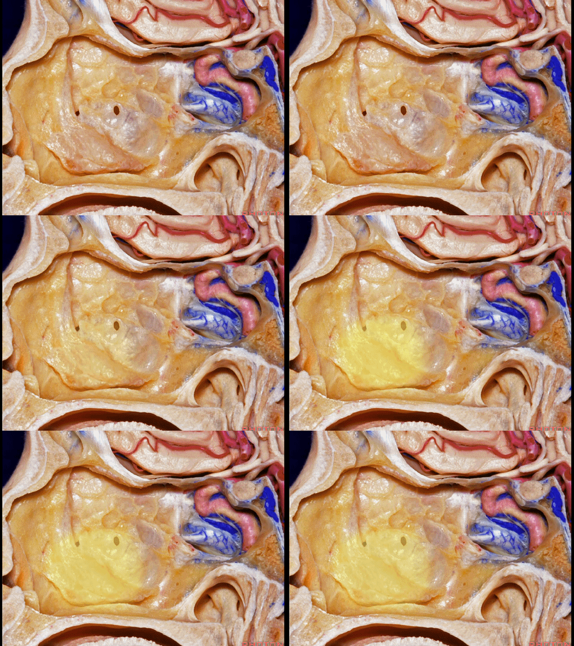
▼以及眶内侧壁。
and the orbit in this area.
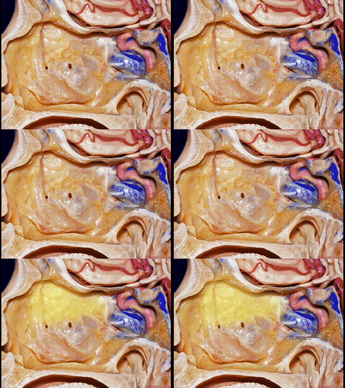
▼进一步向外侧暴露,即可进入上颌窦
If we move laterally, we're into maxillary sinus, orbit.
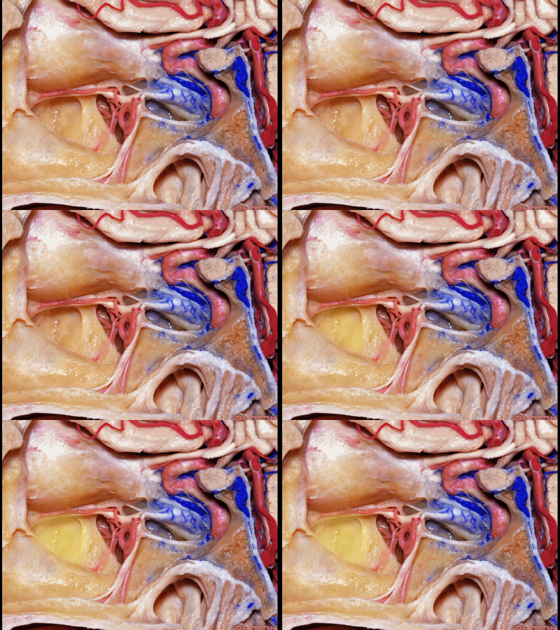
▼沿着蝶窦,沿着其底壁走行的是翼管神经。
Here, along the sphenoid sinus, along the floor, we see the vidian nerve.
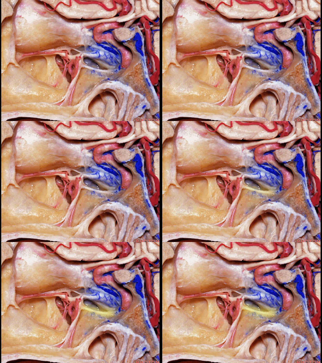
▼蝶窦侧壁有三叉神经上颌支
In the lateral wall we see V2
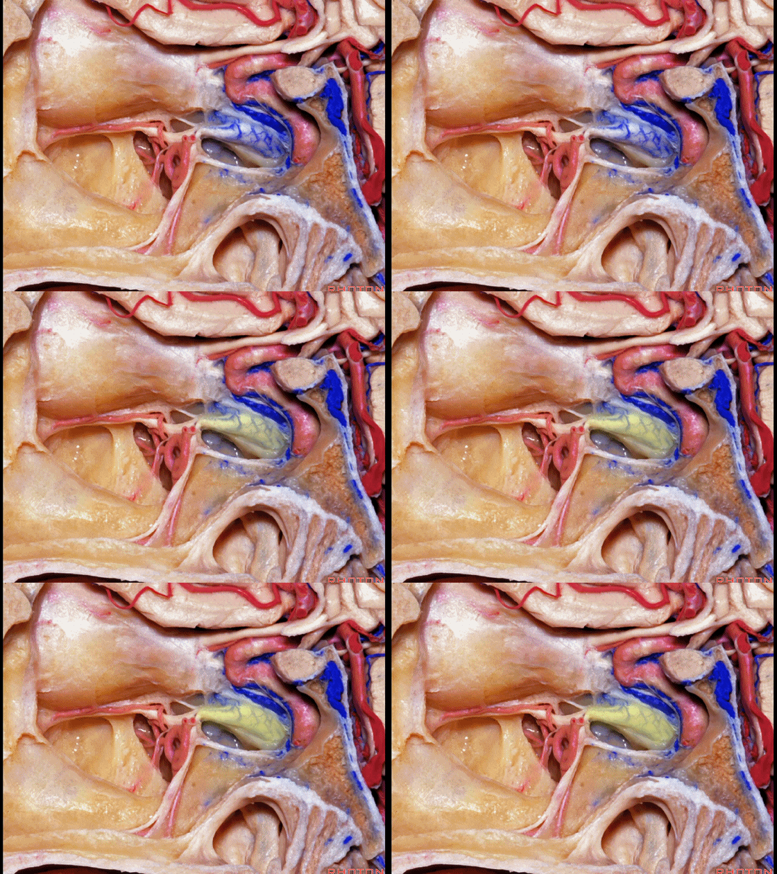
▼上颌支移行为眶下神经。
becoming the infraorbital nerve.
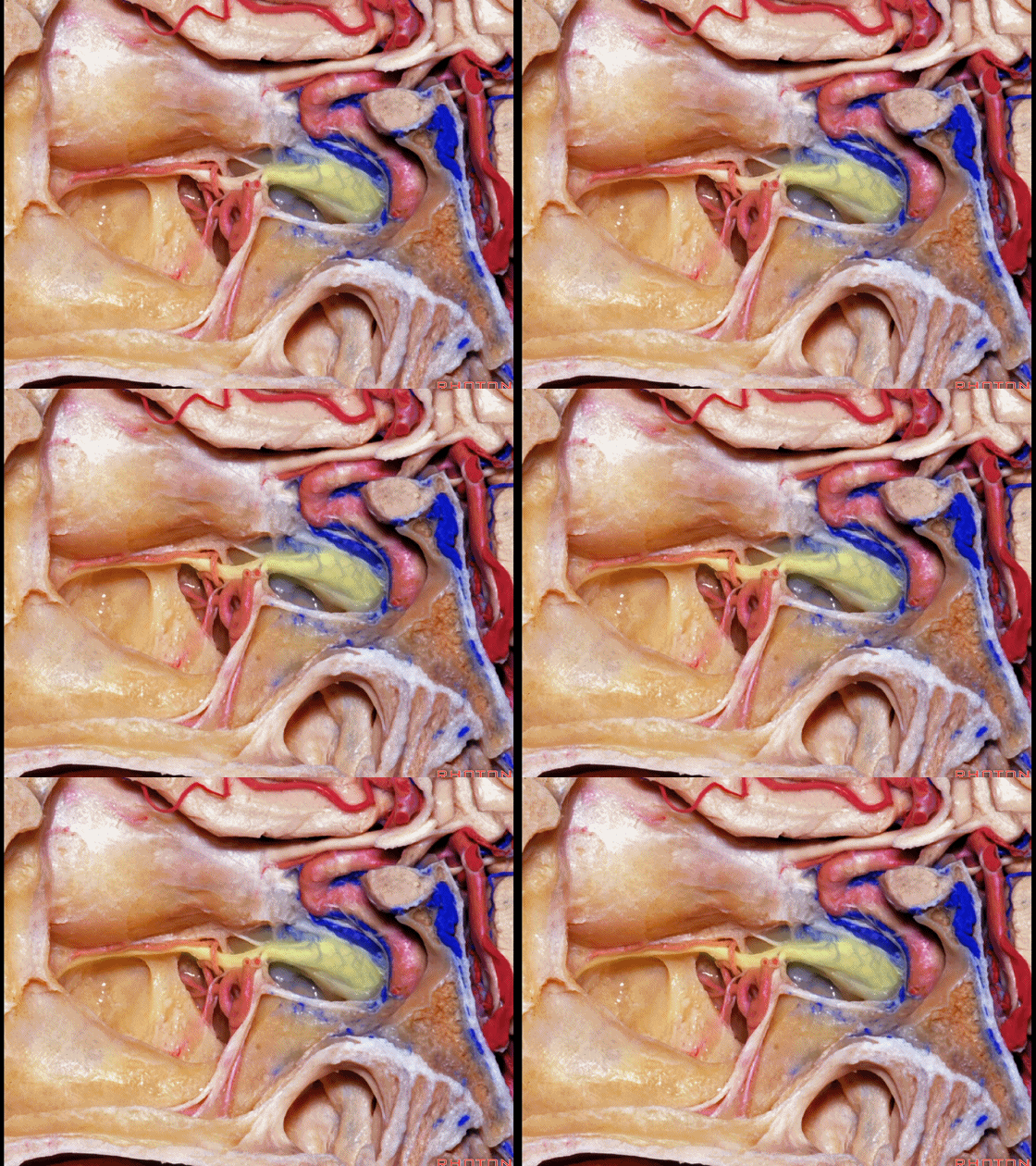
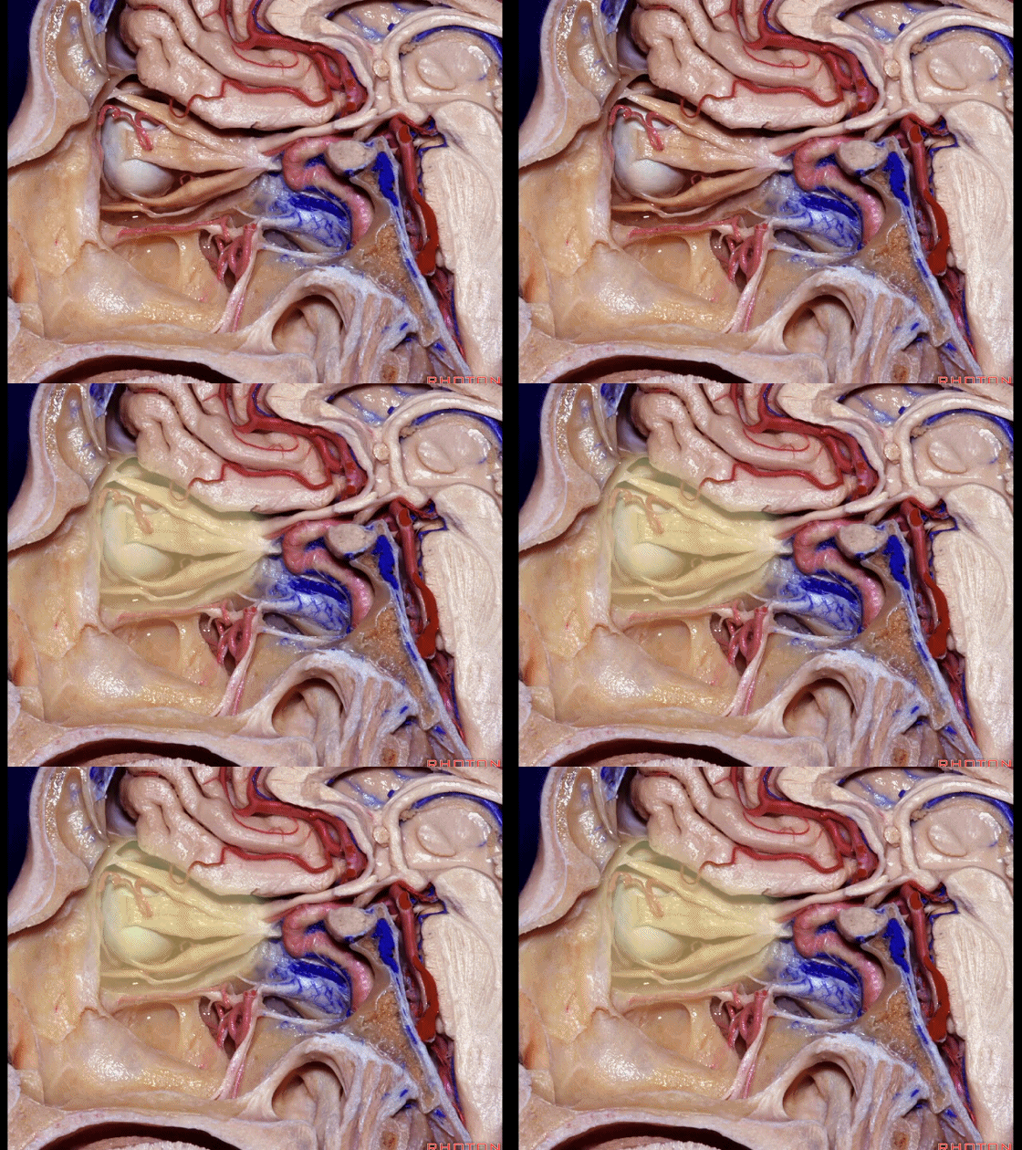
▼再向外侧即可进入眼眶。
And as we move laterally, we're into orbit.
▼再来看看左侧 蝶窦。
Here we're looking at the sphenoid sinus.
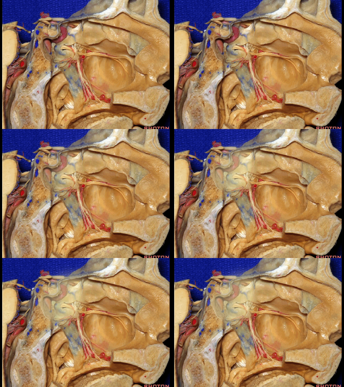
▼下方是翼管神经。
We see vidian nerve below.
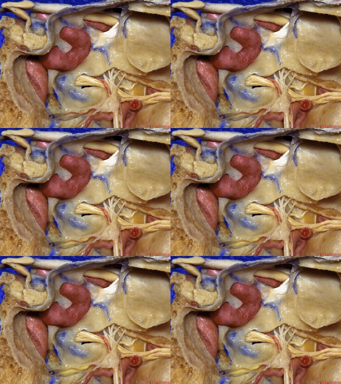
▼这是翼腭窝。
Here is pterygopalatine fossa.
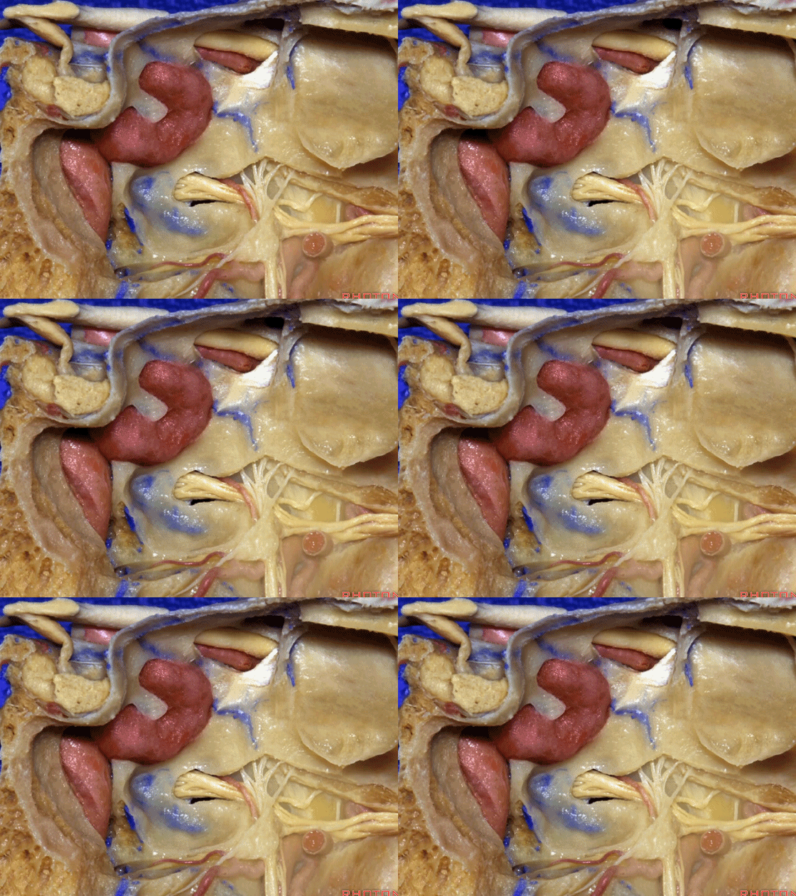
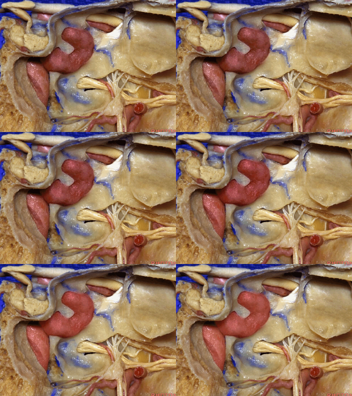
▼颈内动脉常常拱入蝶鞍前壁(下图)。如果术中偏离了中线,我们将遇到的第一个突起就是颈内动脉。因此,即使我们使用的是显微镜和扩张器,为确保前方不会误入颈内动脉,常可使用内镜辅助,以确保术中所直面的突起是中线处的蝶鞍。在内镜辅助下,我们可以轻易地识别出中线旁的突出物是否为颈内动脉。
And, always, the carotid arteries bulge forward of the anterior sella wall. And, if we come in off the midline, the first prominence we often see is the carotid artery. So, even if you're using the microscope and the speculum, in order to make sure that the opening directly ahead of you is not carotid artery, it's a good thing to have the endoscope available and to make sure that the prominence that you're looking at is the sella directly in the midline. With the endoscope, you can always identify the carotid prominences that are off the midline.
![]()
▼下面介绍经翼突入路。想要经过翼突至前方,可经中鼻道(下图)进入。
And if you're gonna go through a pterygoid process by, here we are again, middle meatus
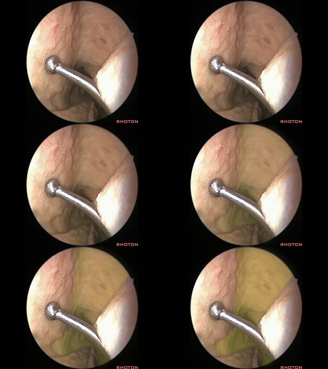
▼就在这个区域。剥除这里的黏膜。
right in this area.And then you start to elevate that mucosa,
▼剥除这里的黏膜。沿黏膜下分离
right in this area.And then you start to elevate that mucosa,
▼可见蝶腭孔内的蝶腭动脉(下图)。
and here we see the sphenpalatine artery at the foramen.
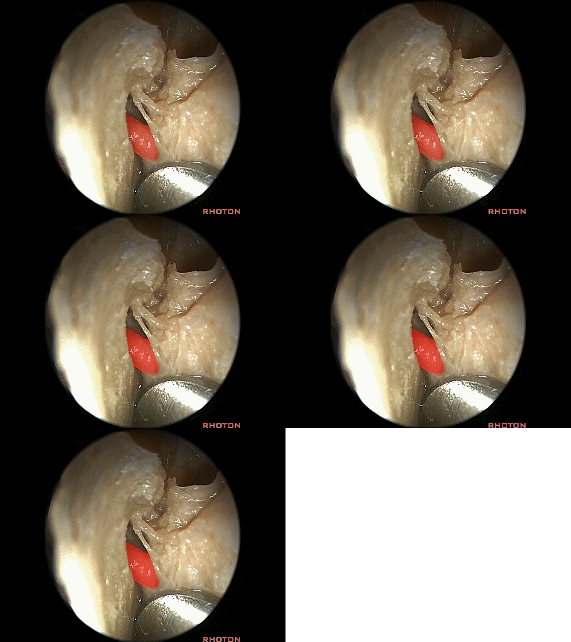
▼扩大该开口,即可暴露蝶骨的翼突(下图)
And you enlarge that opening, and here we have pterygoid process of sphenoid,
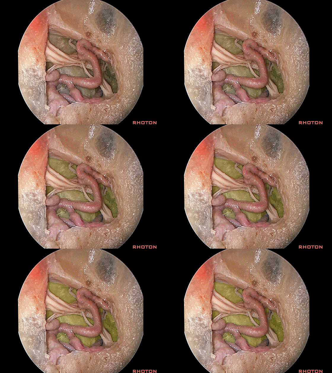
▼这是上颌动脉
maxillary artery
▼这是蝶腭动脉
sphenpalatine artery
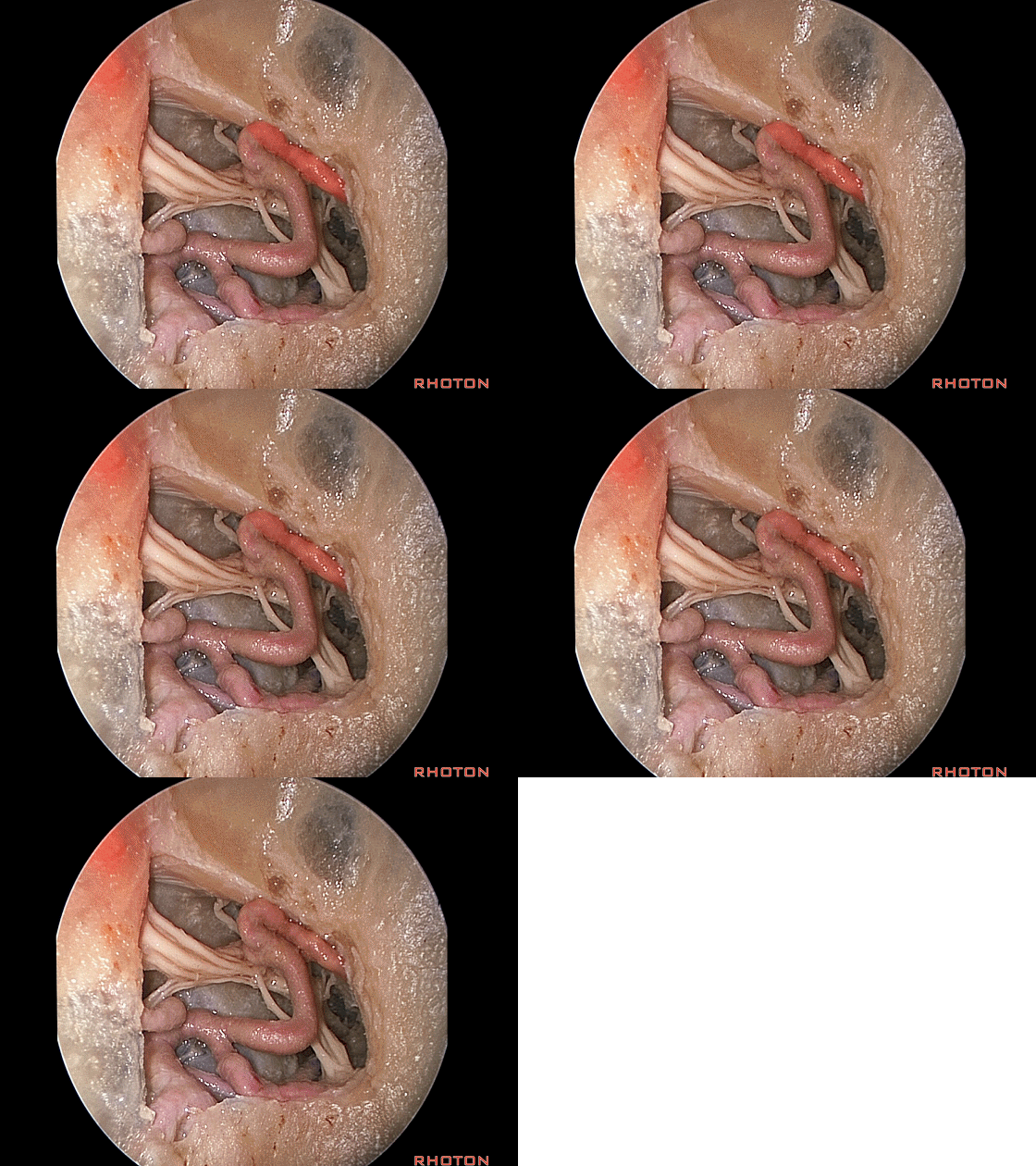
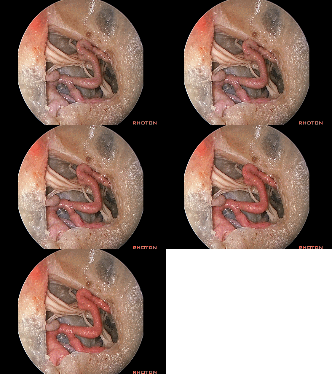
▼这是翼管神经
vidian nerve
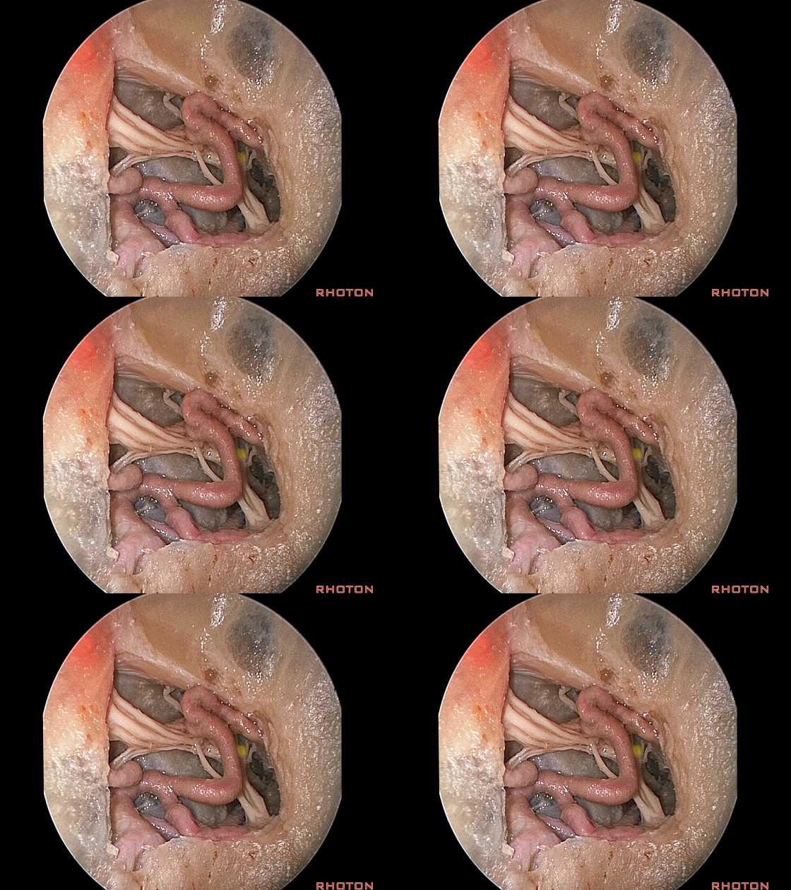
▼这是三叉神经上颌支出自圆孔。
V2 at rotundum.
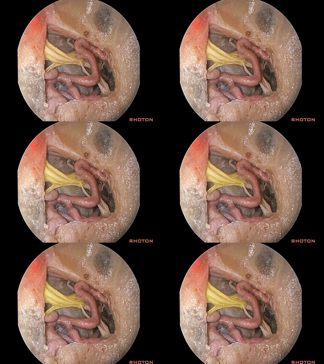
▼随后可进一步扩大开口,这里的操作最好都在黏膜下进行
And you can enlarge that opening, then, it's better if you can do this all submucosally,
▼即可显露翼突根部(下图)
to get into the base of the pterygoid process
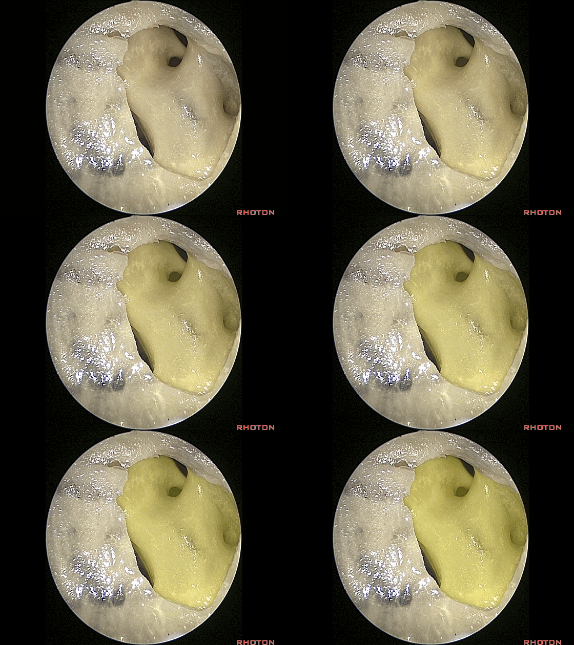
▼将翼突根部磨除即可获得通向斜坡的宽阔径路。
that can be drilled away to widen the exposure to give you access to the clivus.
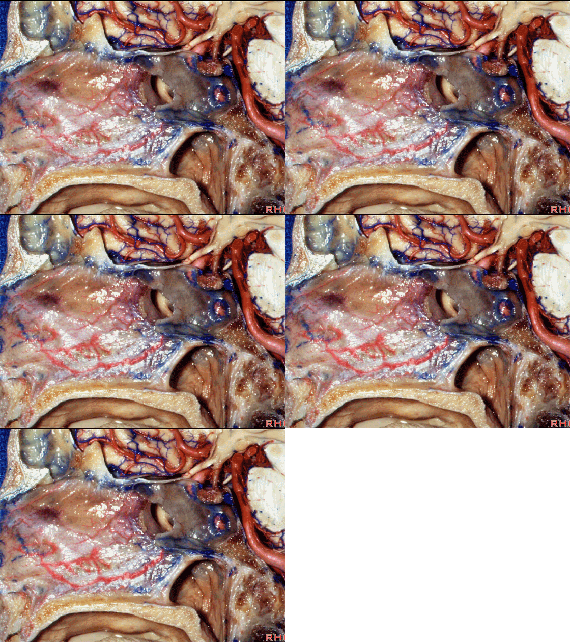
▼对于经鼻手术颅底重建,我们来看鼻中隔,上部供血动脉为筛前动脉,筛后动脉。
But for closure, here we're looking at the nasal septum, upper part supplied by ethmoidal arteries,
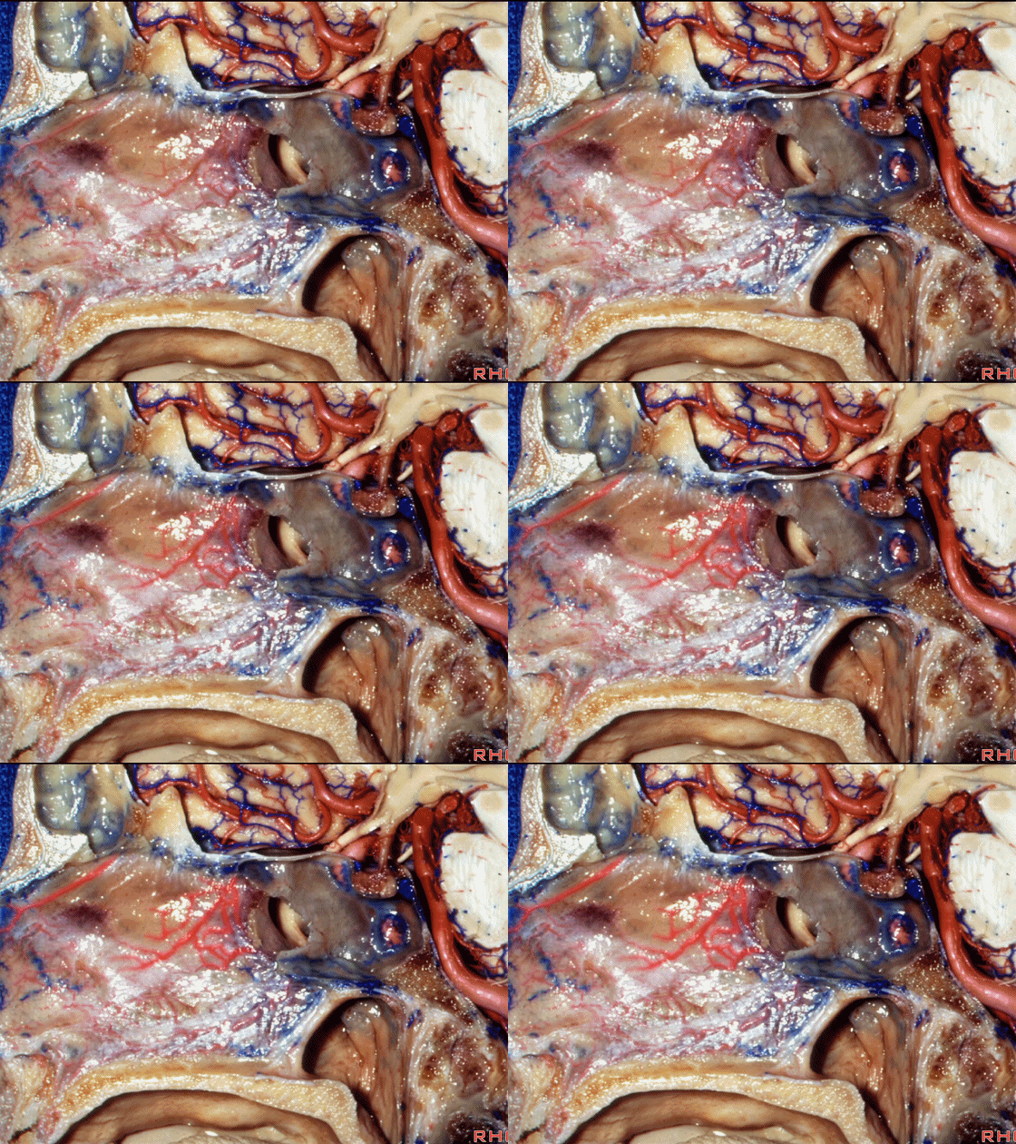
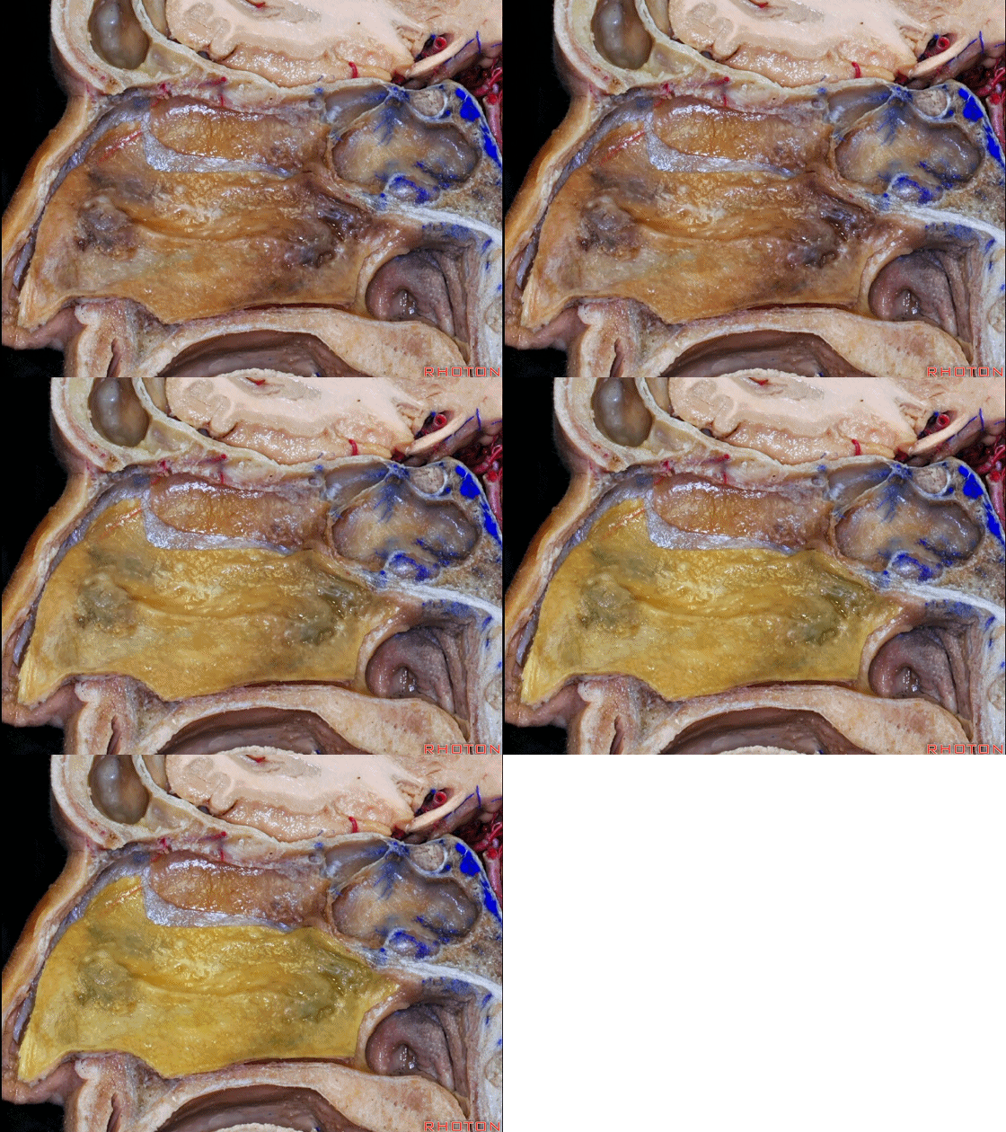
▼更大的下部则由蝶腭动脉来供血。
lower part, larger part, the greatest part by the sphenpalatine arteries.
▼黏膜辦的面积可很大,包括一侧鼻腔的大部分黏膜(下图)。
And these flaps can be very large, and include most of the nasal mucosa on one side.
▼我们需保留这些供血动脉(下图箭头)。我们也希望保留嗅黏膜。
You have to spanserve that arterial supply. You wanna spanserve the area of the olfactory mucosa.
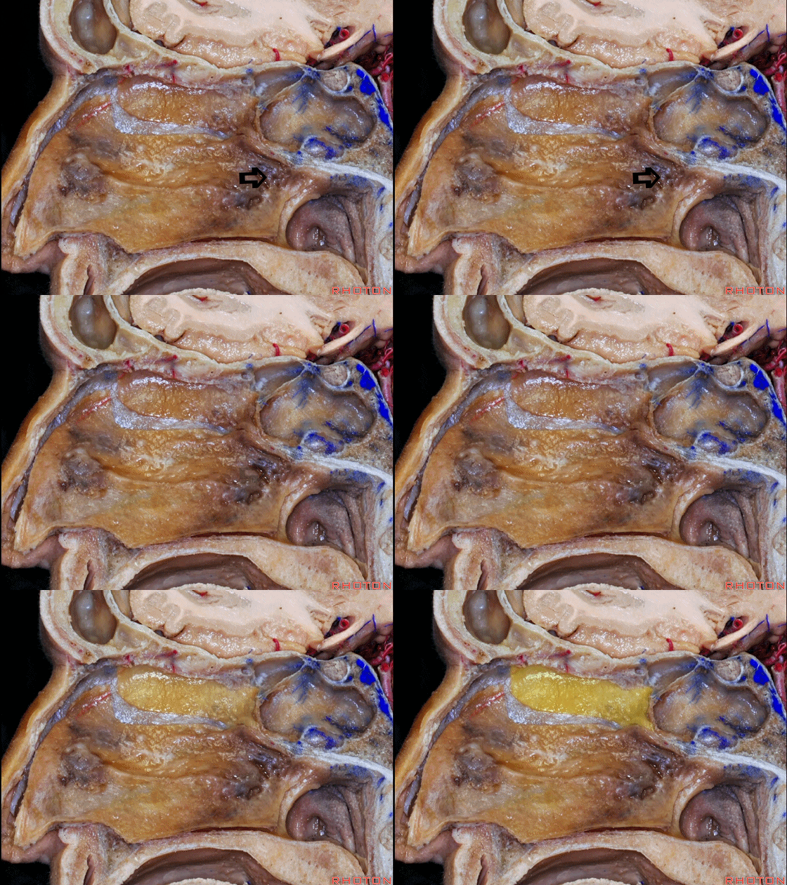
▼这片黏膜辦可用于修补前颅底的缺口
And, these flaps can be used to close large defects in anterior fossa
▼也可向下修补斜坡。
or clivus anterior fossa, or downward in clivus.
▼黏膜辦的取材来源有 鼻中隔(下图)以外,我们也可将黏膜辦向下延伸至硬腭。
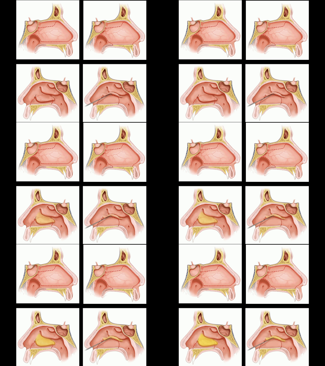
these flaps also in addition to including the nasal septum,they can extend down across the hard palate,
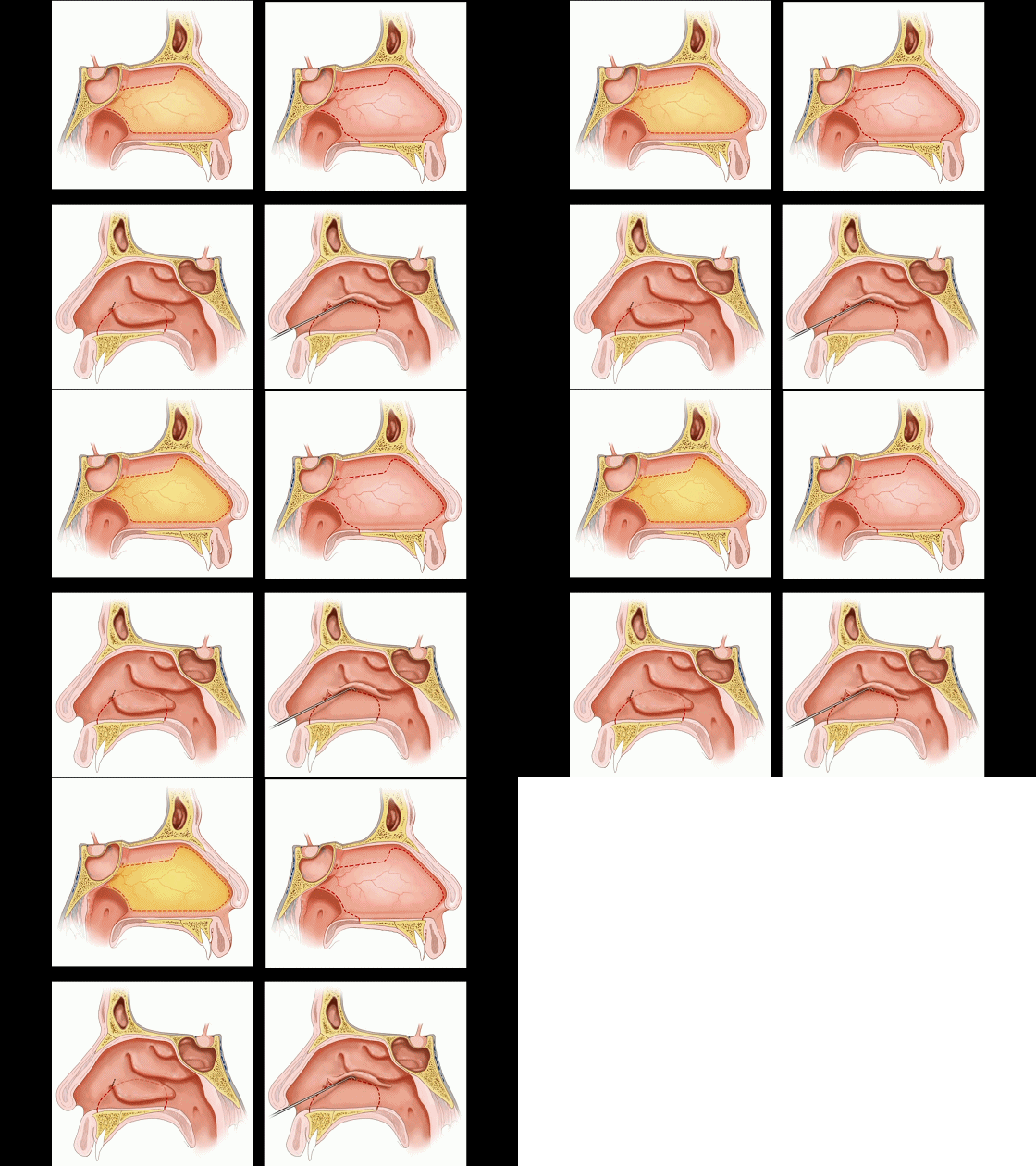
▼下图示硬腭
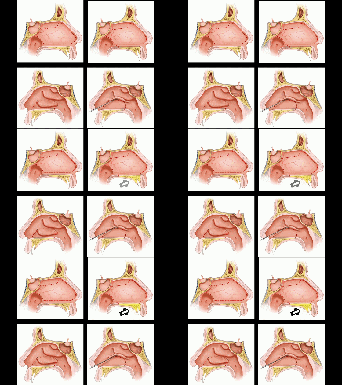
▼或继续延伸至鼻腔侧壁(下图左侧),或进一步向上延伸至下鼻甲下方(下图右侧)。因此,这一广泛的黏膜辦可从硬腭延伸至下鼻甲下方,用于切口修补。
to the lateral wall of the nasal cavity, up under the inferior turbinate. So, this extended flap along the palate up laterally under the inferior turbinate, gives a huge flap for aiding in these closures.
![]()
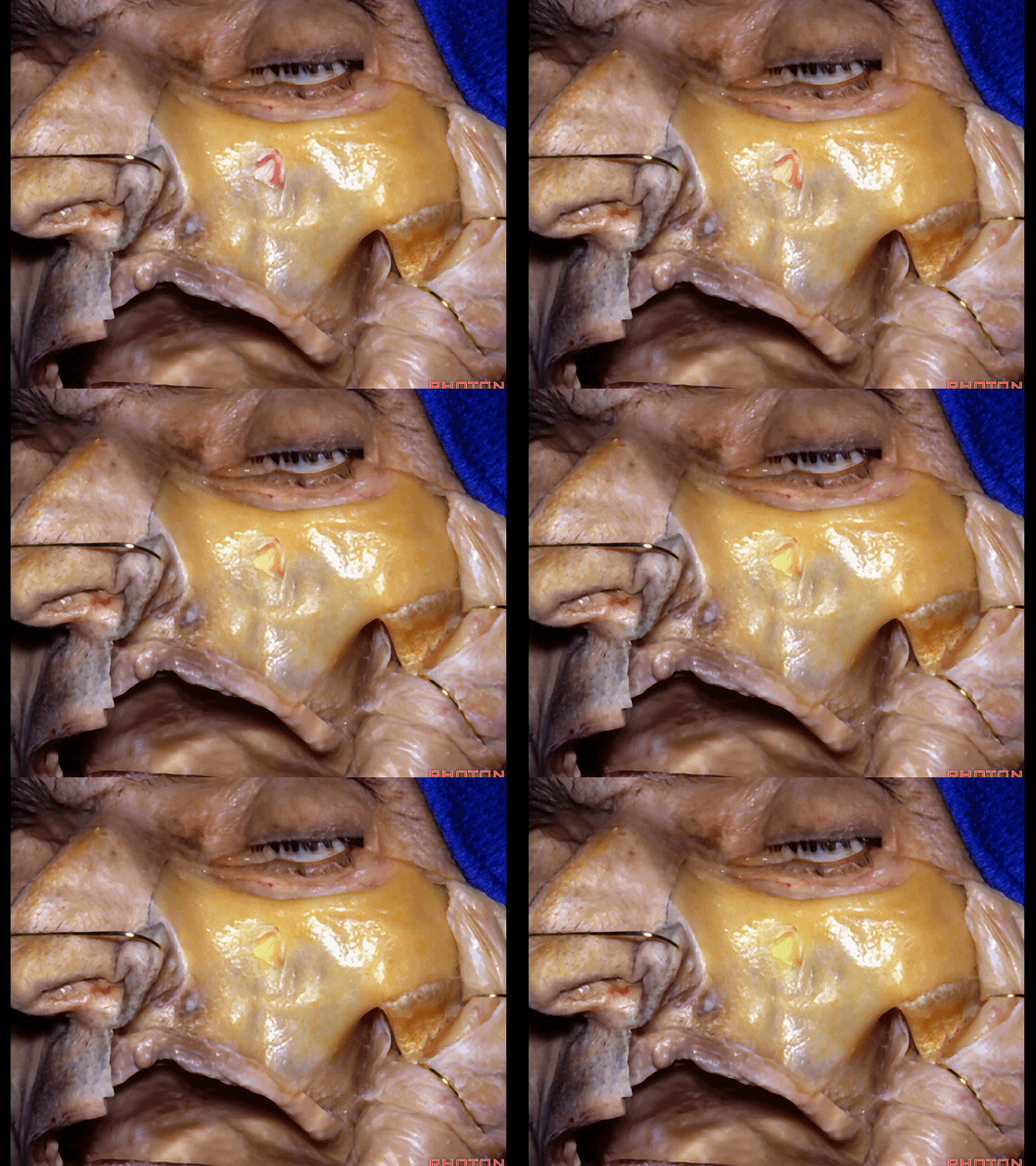
▼下面介绍经上颌骨入路。上颌骨可用作通往颅底的宽阔径路。
Maxilla can be used as a route to wide areas of skull base.
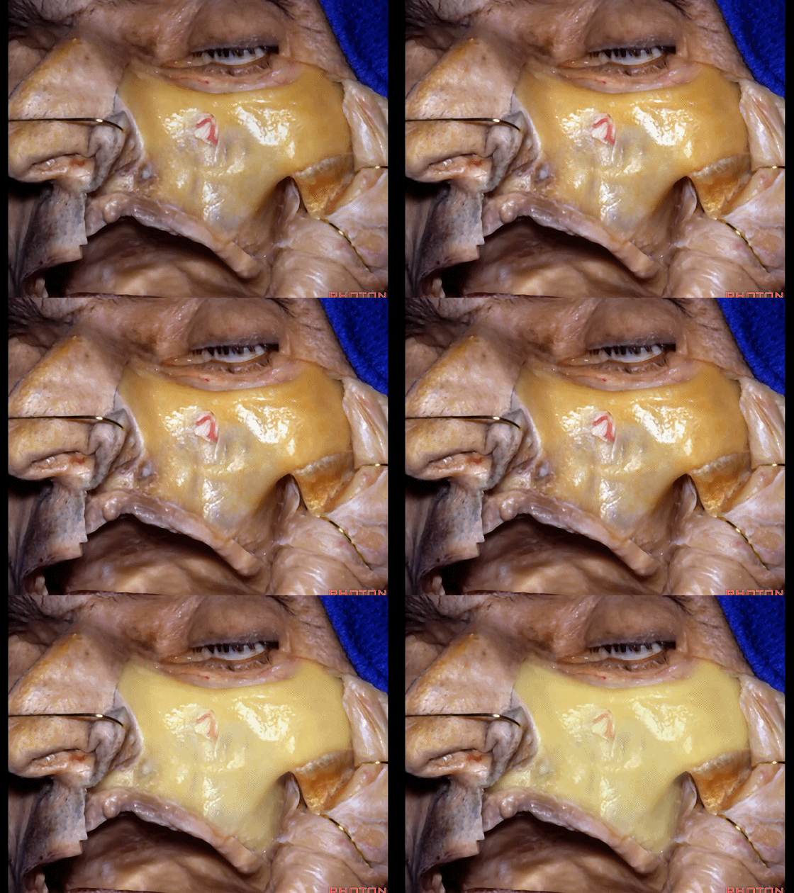
▼这里显露的是 眶下神经。
Here we're looking at the maxilla below the infraorbital nerve.
▼我们可进行下部上颌骨次全切除术。这一术式是由Robinson教授发扬光大的。它并不常用但确实是颅底外科中出色的入路。术者可行面部脱套术,可不留面部切口。随后行上颌切开术,位于眶下神经以下,经过腭骨,显露颞下窝(下图)
And you can do a lower subtotal maxillotomy. It's a procedure that was popularized by Prof. Robinson.I wouldn't say popularized but it's not a common procedure, but it's a great procedure for accessing skull base.You can just deglove the maxilla, no incision on the face. And then you do a maxillotomy below the infraorbital nerve, come through the palate and gives you infratemporal,
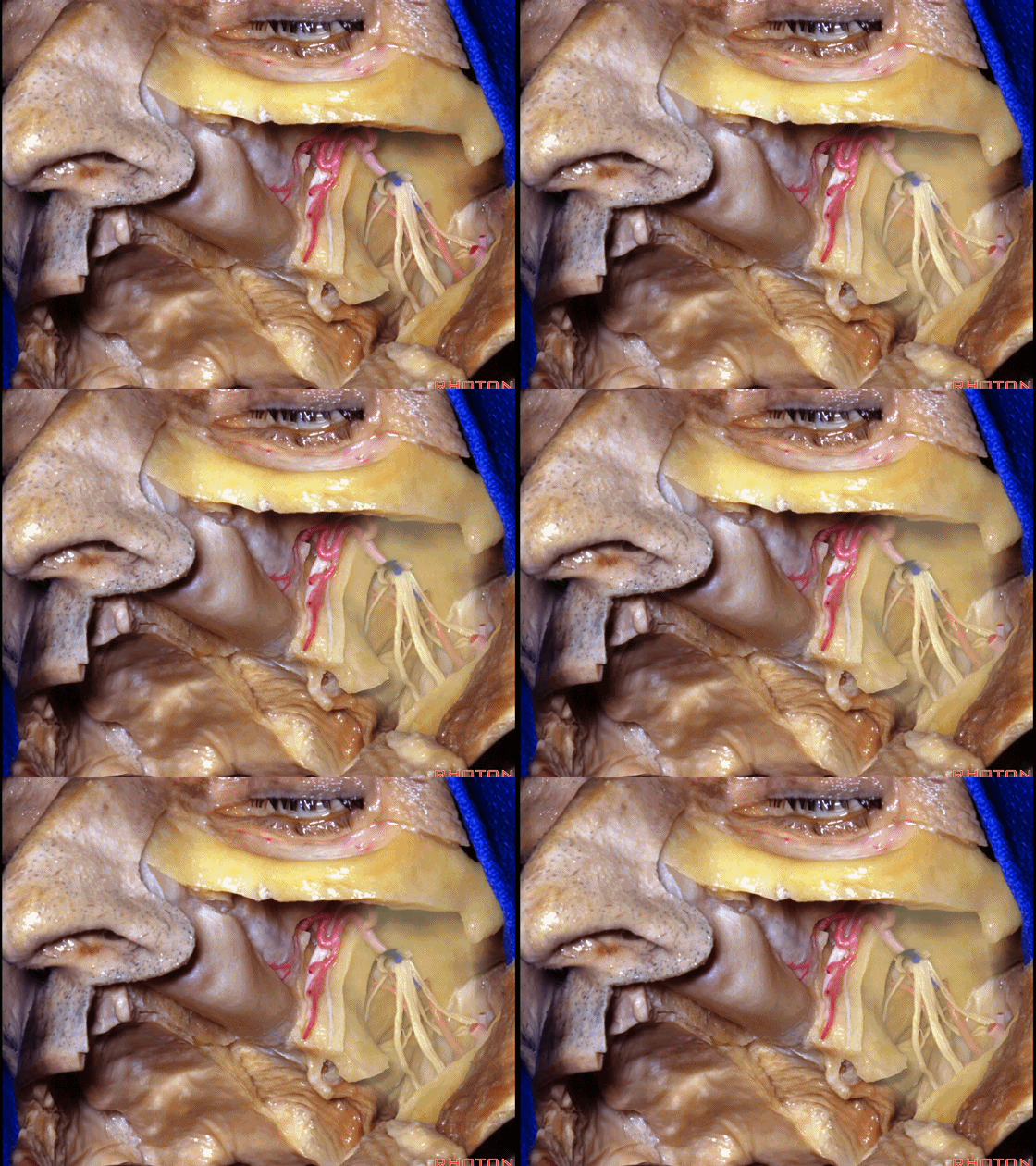
▼翼腭窝
pterygopalatine fossa
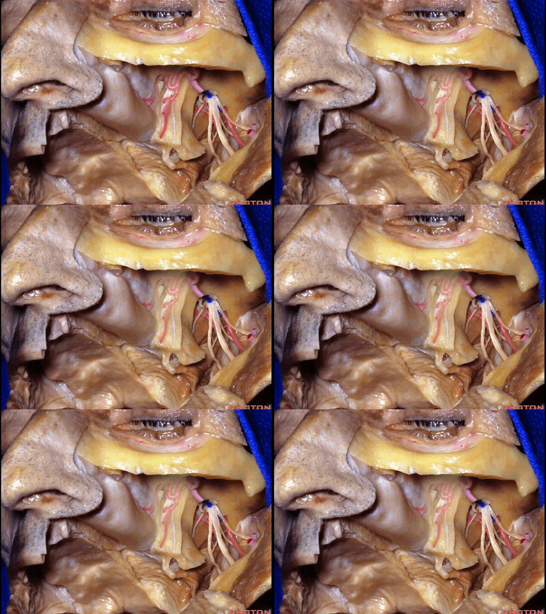
▼这是鼻腔黏膜
Here we see nasal mucosa.
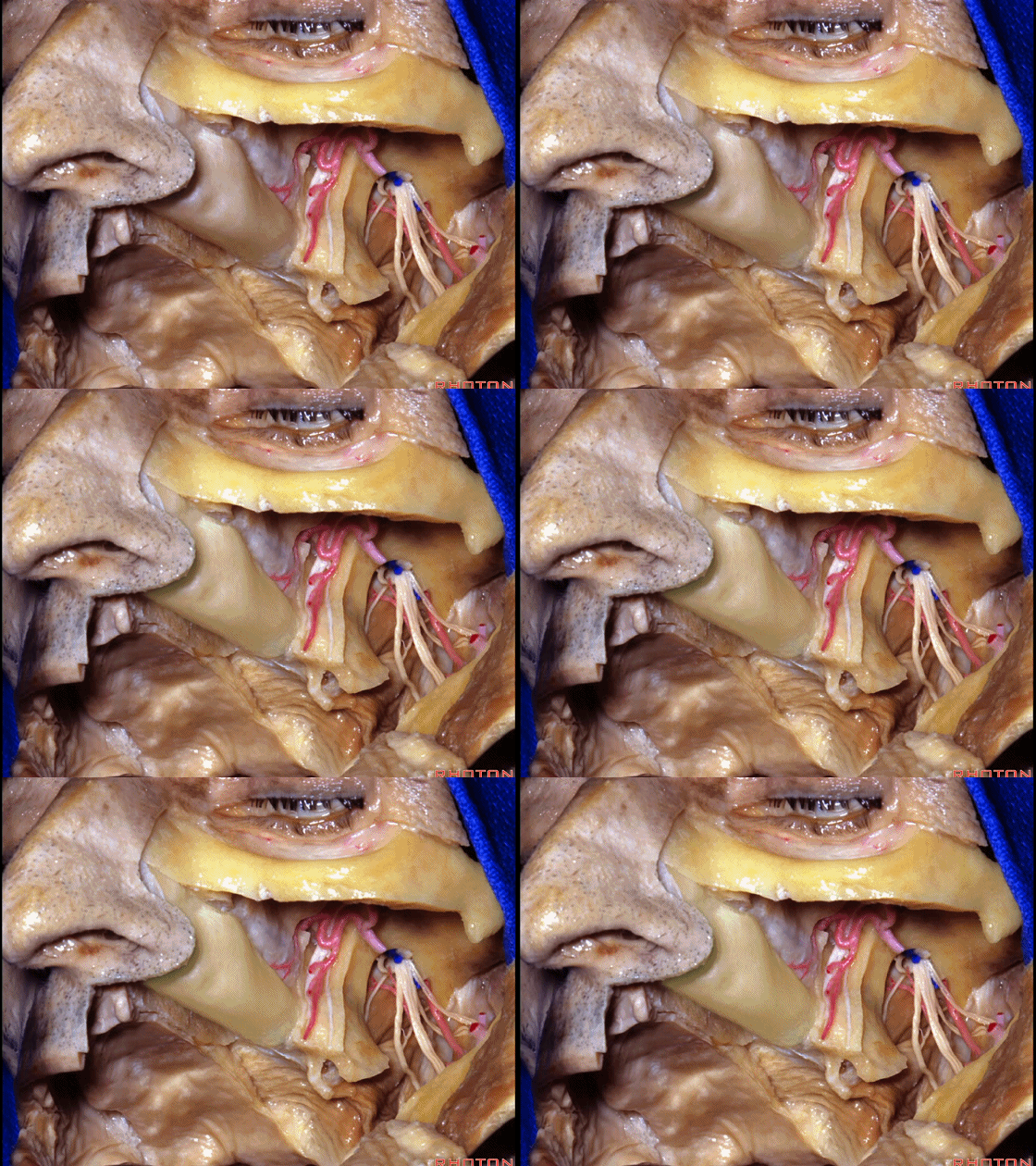
▼将鼻腔粘膜牵开,向内侧可暴露寰椎、枢椎、下斜坡。
You can retract that mucosa, work medially along C1, C2, lower clivus.
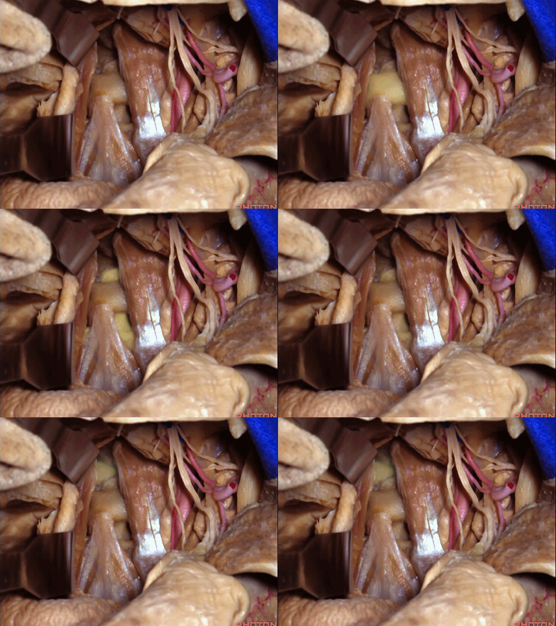
▼经此入路可暴露颞下窝
And it gives you access to infratemporal, pterygopalatine fossa,
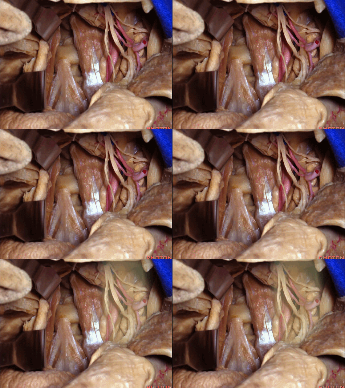
▼可显露翼腭窝、鼻腔、上颈椎、斜坡。
nasal cavity,upper cervical, clivus.
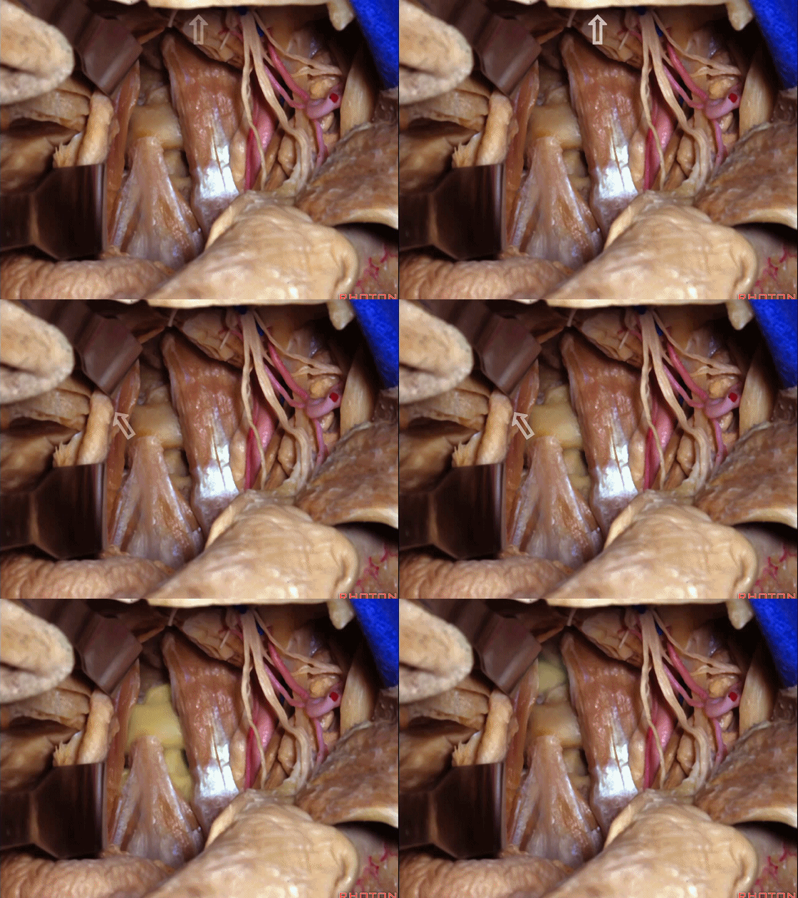
▼经斜坡(下图)可显露蝶鞍
You can get to the...through the clivus, you can get to the sella,

▼或通过这一下部上颌骨切开入路暴露前颅窝。下图示蝶鞍
or the anterior fossa using this lower maxillotomy approach.
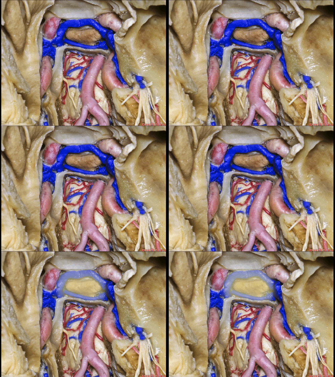
▼涉及上颌骨的另一入路是 上部上颌骨切开入路,可由此暴露中颅窝,于眶底下方(下图)切开上颌骨
Another approach involving maxillotomy that you can access also middle fossa is the upper maxillotomy where maxilla is opened below floor of orbit,
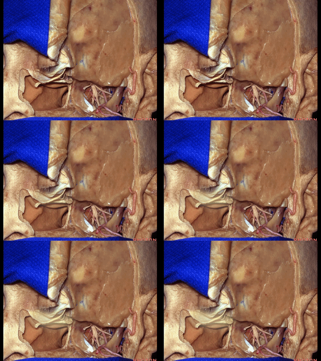
▼下方至牙槽嵴
above the alveolar ridge,
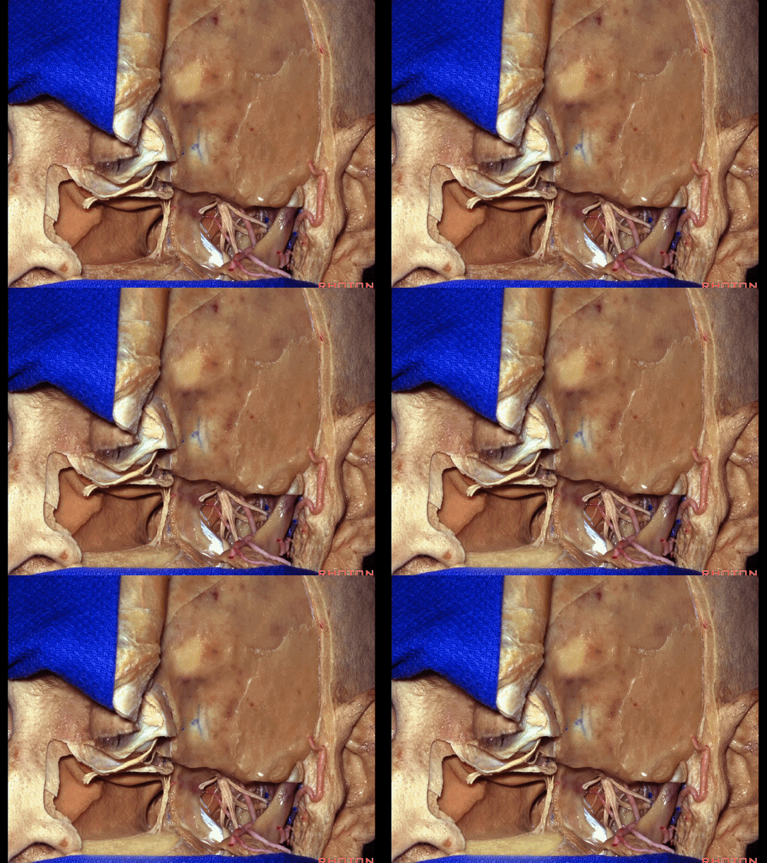
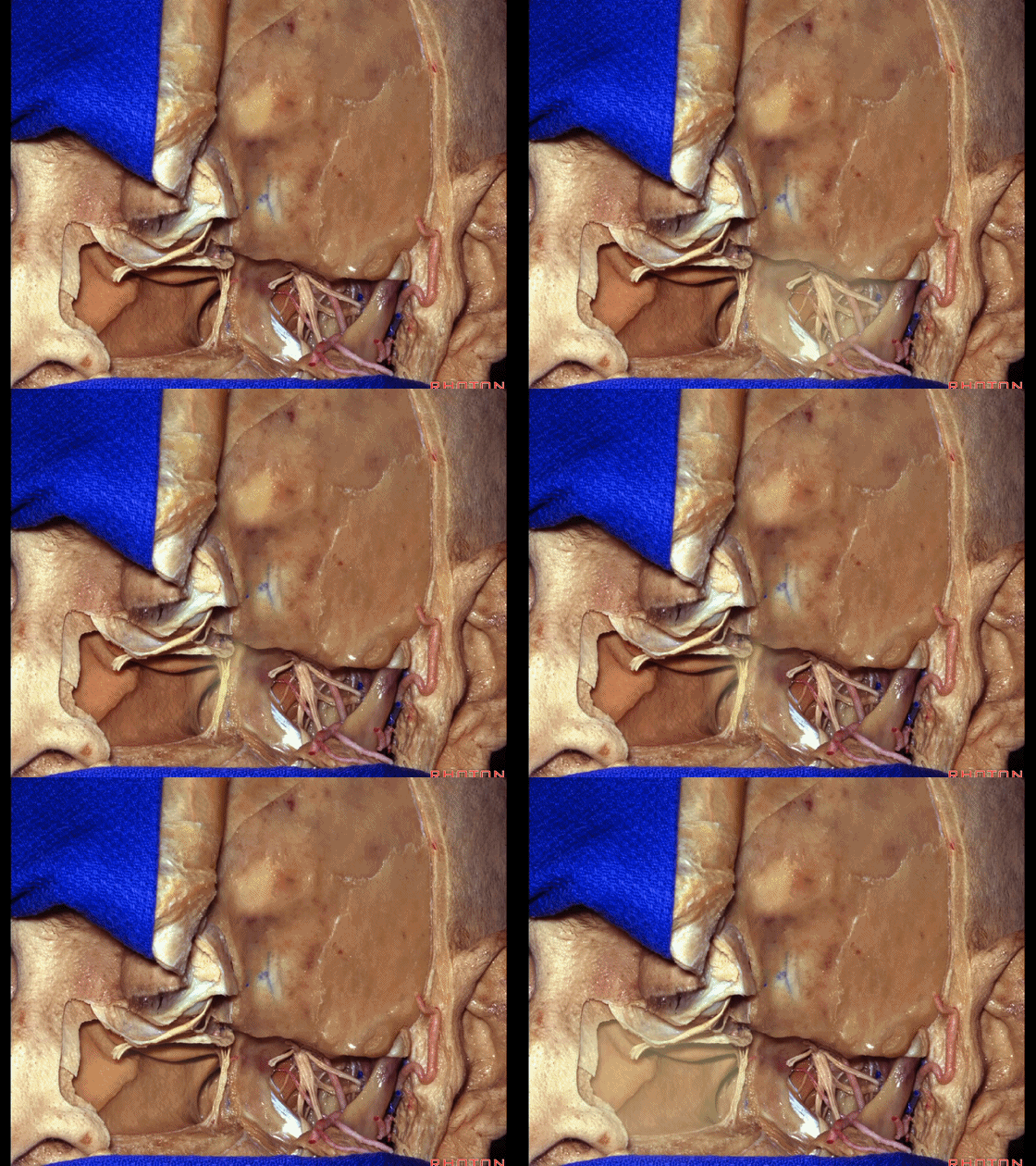
▼其暴露范围可至 颞下窝、翼腭窝、鼻腔。
and it gives you access to infratemporal, pterygopalatine, the nasal cavity.
点击继续阅读
前颅底(2)与鼻腔解剖---Rhoton解剖视频学习笔记系列(下)


