The Rhoton Collection视频合集中的《Anterior Skull Base, Part 2 前颅底第二部分》包含了《The Nose for Neurosurgeons 鼻腔解剖》的部分内容,本文将两部视频内容合二为一,并将其中的“经口入路部分”归入《Head and Neck Anatomy for Neurosurgeons》(将于近期推送)。
对于热爱神经内镜手术的医师来说,鼻腔解剖知识显然是非常重要的,本文所展示的图片不论是对初学者还是有一定手术经验的医师都具有极高的研究价值。

前颅底(2)与鼻腔解剖
Anterior Skull Base and Nose
▼这一集我们将讨论神经外科相关的鼻腔解剖。目前,使用内镜技术进行颅底手术已成为世界范围的趋势。我们可推动该领域的发展,但是,大家真正了解鼻腔的解剖吗?
现在让我们来看鼻腔的骨性结构。下方我们可见下鼻甲,中鼻甲。
Well, I wanna take the last few minutes and talk about the nose for neurosurgeons. Today I see all over the world increasingly endoscopic approaches being done to skull base. And we may stand there and help. But do we really understand the nose? Now we look into a nasal cavity. Here below we see, inferior turbinate,middle turbinate
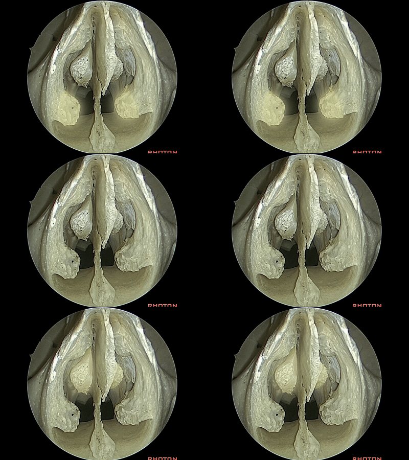
▼这是 筛骨垂直板
perpendicular plate
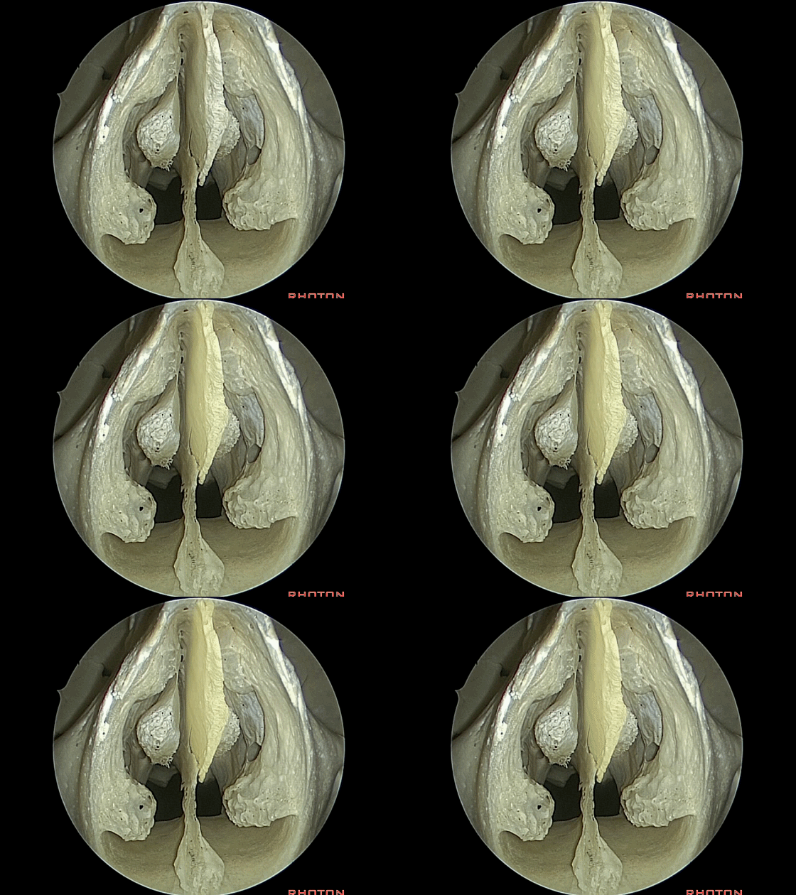
▼这是 犁骨
vomer
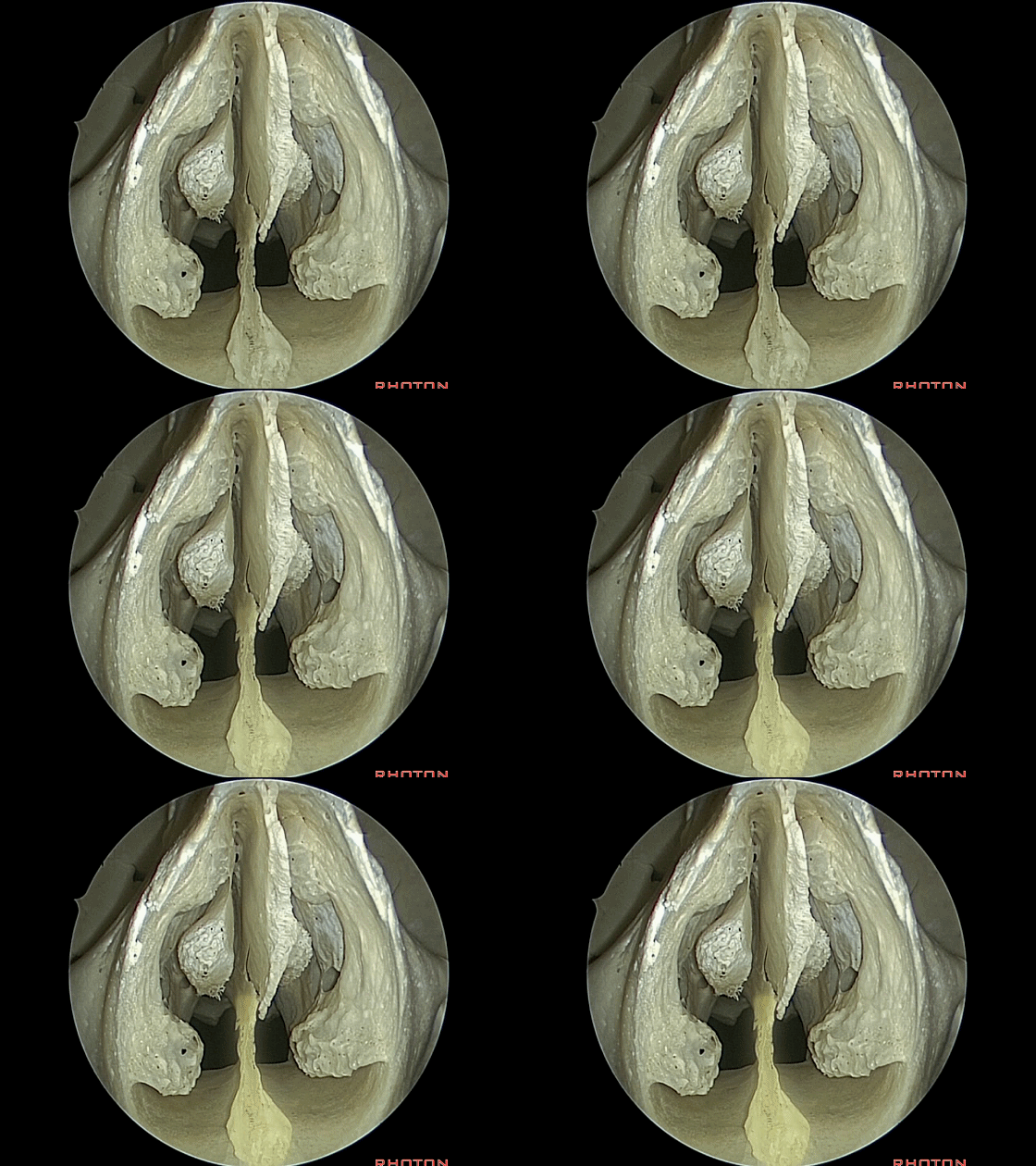
▼当我们从鼻前孔看向鼻腔内,这第一个突起是鼻泪管(nasolacrimal duct)。其开口位于下鼻甲下方。
But if you're looking at the anterior nasal aperture that's opening in the anterior opening into the nasal cavity, what's the firs prominence you see here?What is that?Anyone? Please help me. That's...it opens below the inferior turbinate, so that's nasolacrimal duct.
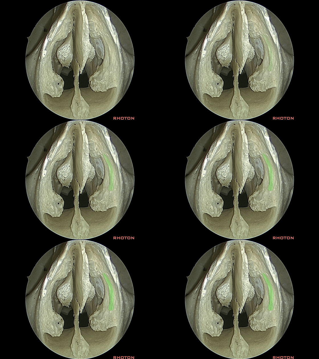
▼这是 筛板。因此通往筛板的径路确实很狭窄。
cribriform plate, It really is a very narrow route up to the cribiform plate.
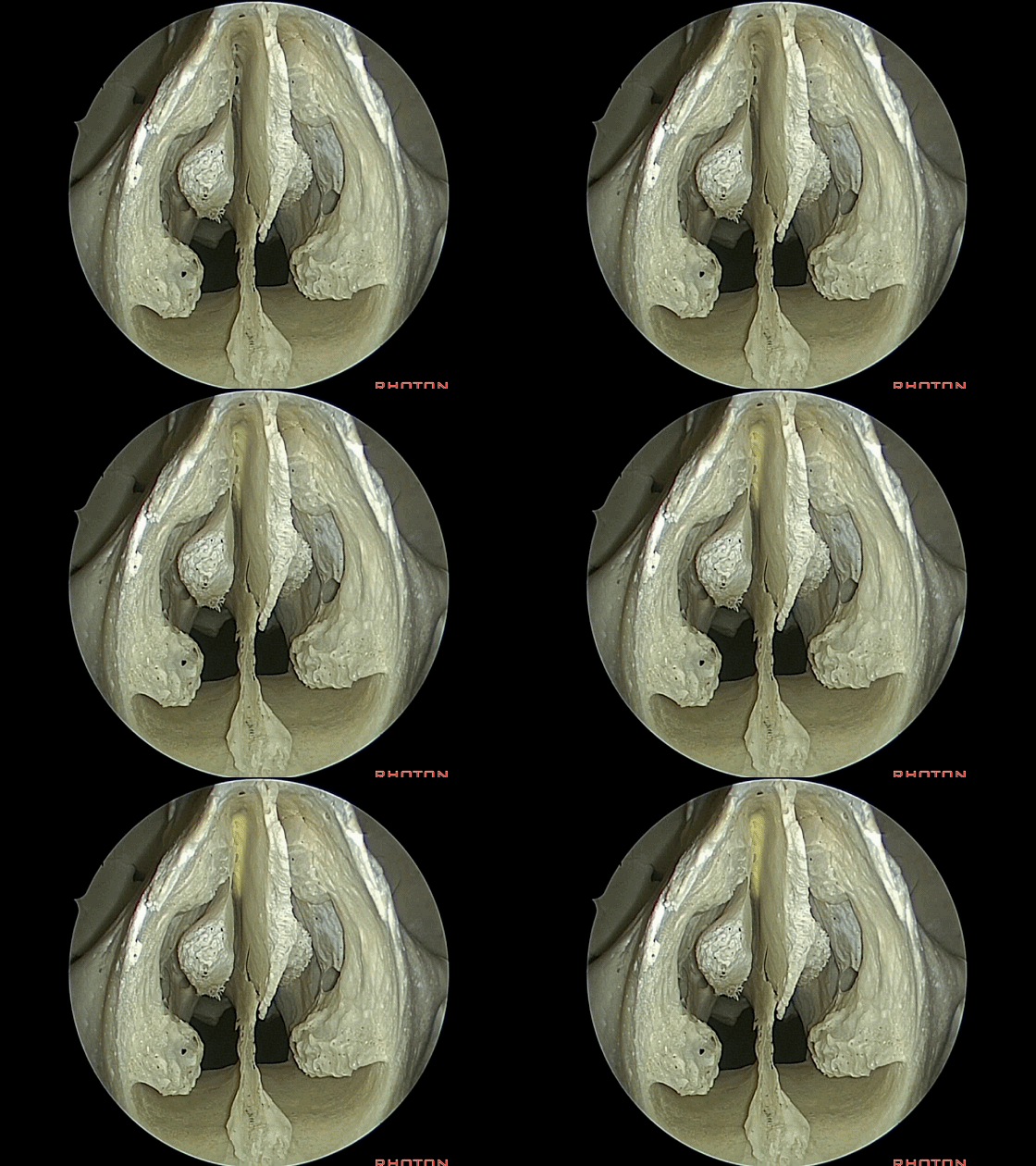
▼内镜进一步深入,这是中鼻甲。
So, here's middle turbinate.
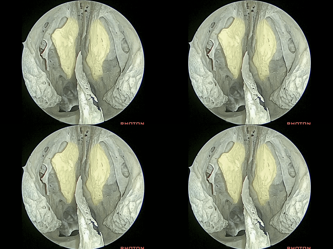
▼这是筛骨的钩突。
Here's the uncinate process of bone.
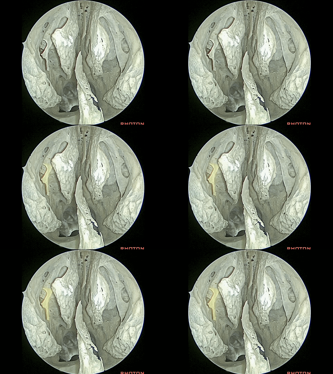
▼钩突前方的开口称为前囟,前囟由黏膜覆盖。这让人联想到小儿神经外科的囟门解剖
And the uncinate process...if you talk to ENT, anteriorly there's an opening, that's the anterior fontanelle,anterior fontanelle is covered by mucosa, So this area is like pediatric neurosurgery with fontanelle,

▼钩突后方的开口称为后囟。后囟也由黏膜覆盖。
and then along the posterior edge of the uncinate process in the bone it's open, that's the posterior fontanelle. the posterior fontanelle covered by mucosa.
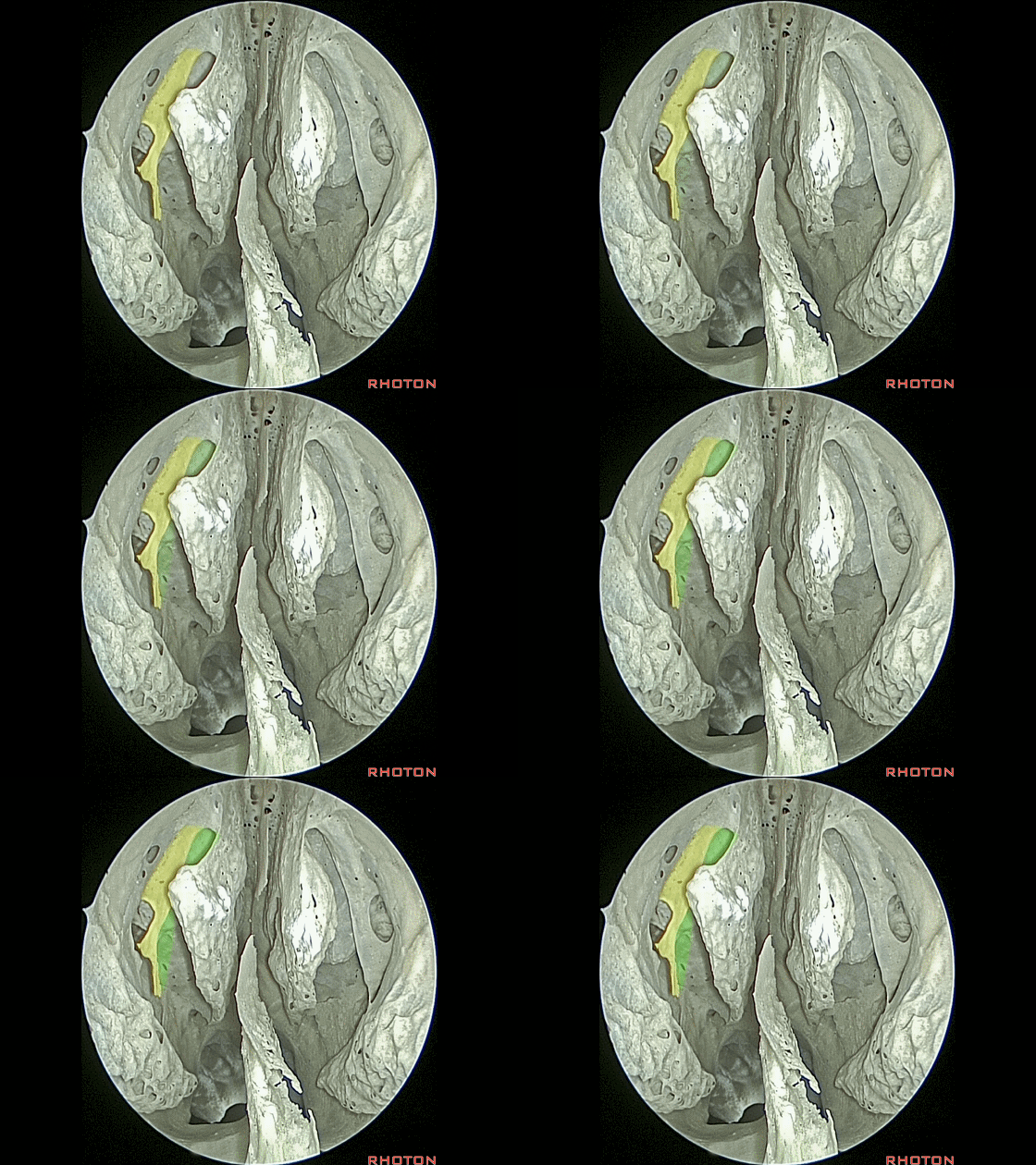
▼在其后方,可见筛泡。
And then, back of it, you begin to see the bulla.
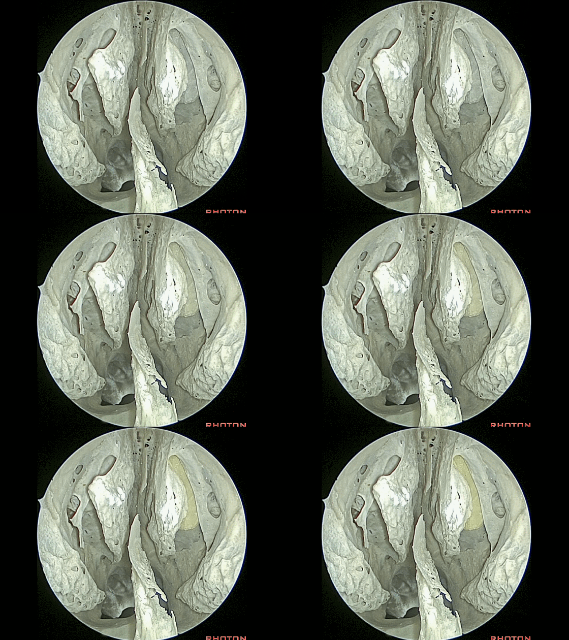
▼在双侧中鼻甲之间向上可见筛板(cribiform plate)。
So, now, if you look upward between the middle turbinates you see the cribriform plate, really is a tiny narrow channel.
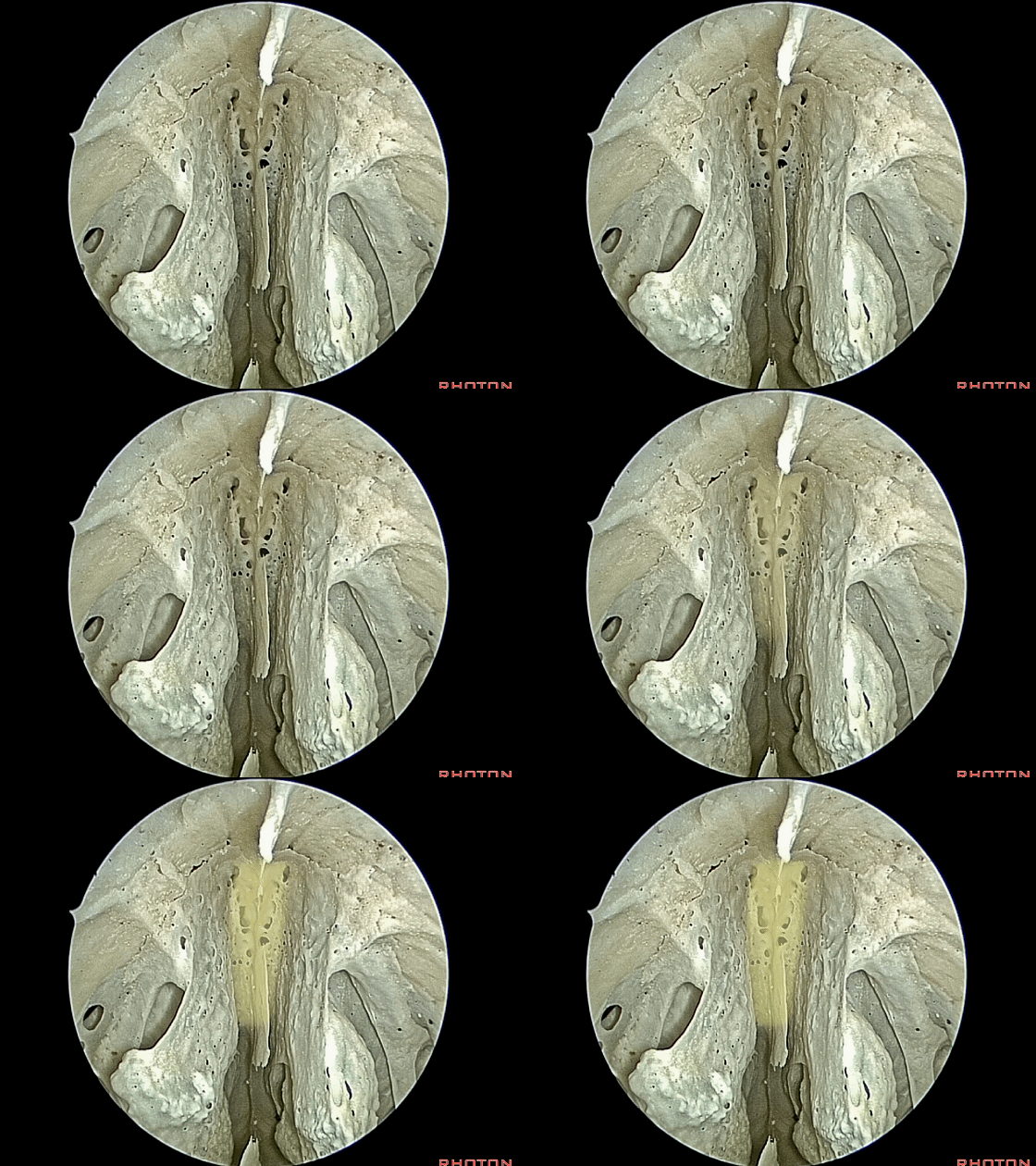
▼该区域十分狭窄。因此,如何从此处向上完成双侧眶内侧壁之间的广泛暴露?
and this really is a very narrow area. The question is how do you go from here looking up there to here, to an opening that extends over to the medial edge of the orbits?
▼这是嗅球间距。筛板的宽度并不大于两侧嗅球间的距离。
So this is bulb width. How to...the cribriform plate is no wider than the olfactory bulb.
▼这里我们打开筛泡(下图),从而获得了宽敞的路径。
Now the bulla are open here lateral to the cribriform plate to give you that wider access.
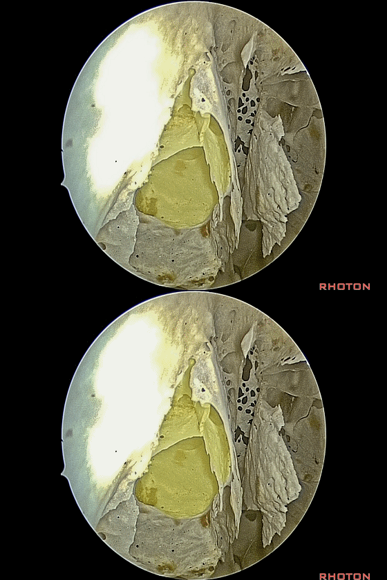
▼这是筛板。位于筛泡的内侧
And, now we see cribiform plate.
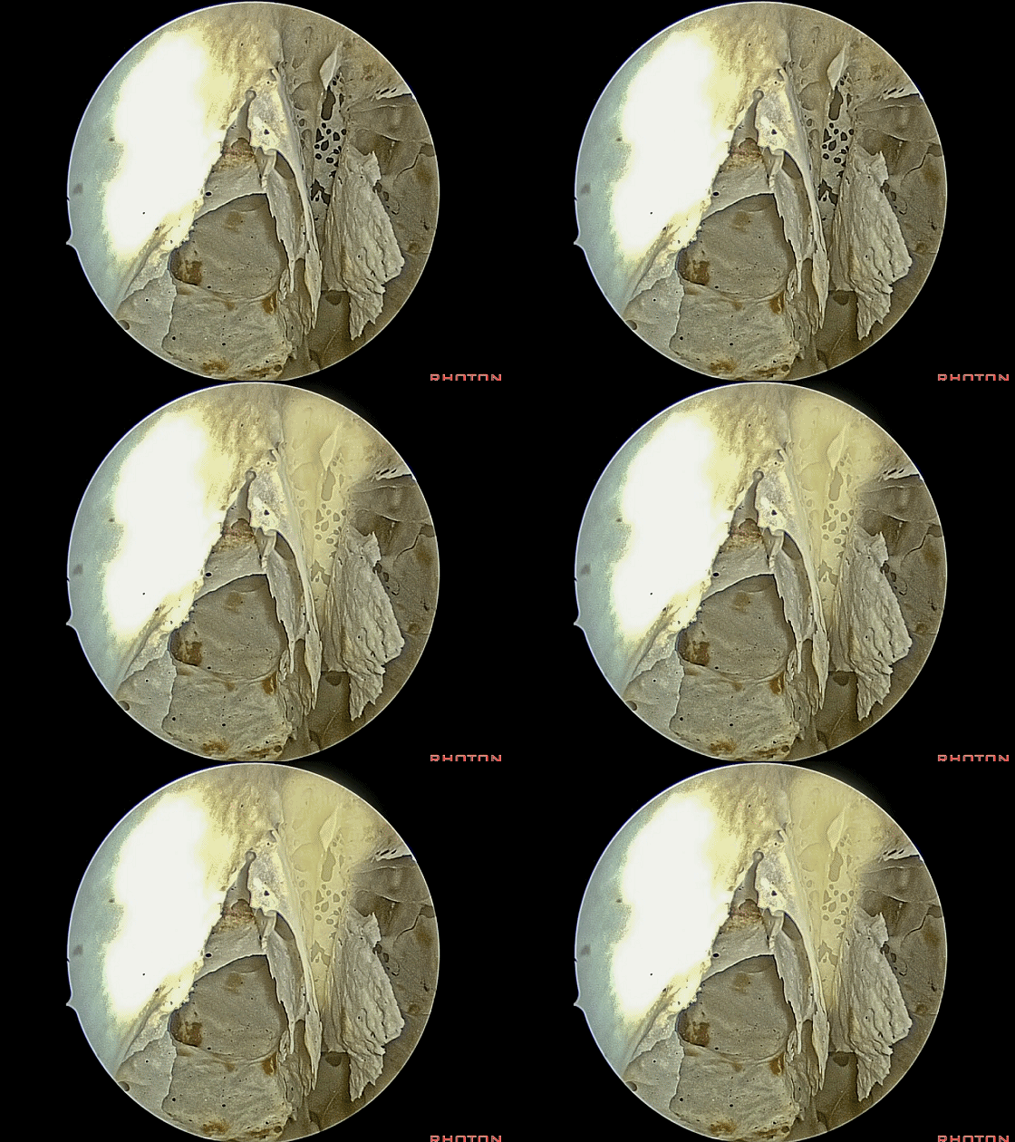
▼我们可打通这复杂如迷宫的筛房。在筛板外侧进入前颅窝底。即可获得宽敞的术野。
And you can work through this complicated maze of ethmoid air cells.So by going through that, you see how much, on just one side,by opening the ethmoids, how wide an opening you get.

▼这是构成眼眶内侧壁的筛骨纸板(下图)
And the bone here laterally on the ethmoids forms the medial wall of the...if you open this you're into the...anyone? Orbit.
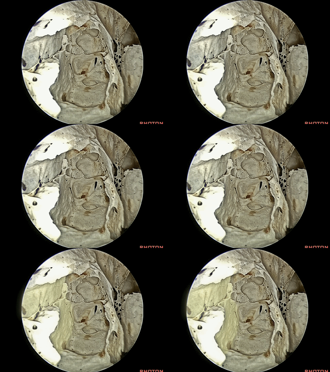
▼我们可进一步向前方,通过解剖中鼻甲,磨除筛窦迷路,即可实现前颅底的暴露。
And you can work forward here,going through that middle meatus, removing ethmoid labyrinth,you get this exposure of anterior fossa,
▼这里,我们已打开了双侧的筛房。
So, here we've opened the ethmoid air cells on both sides.
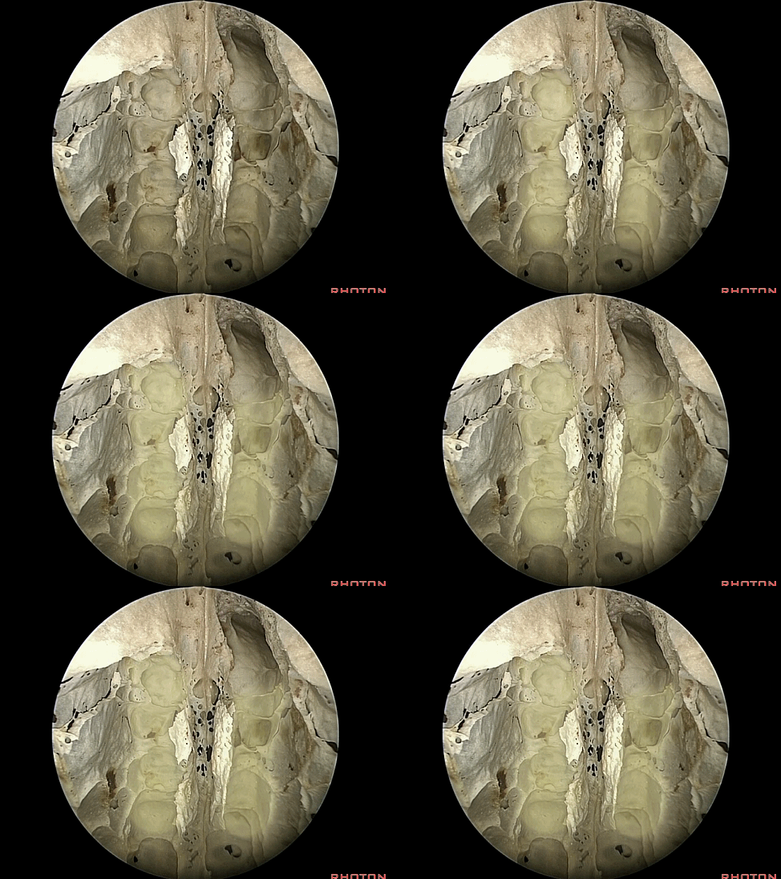
▼如今可利用 成角磨钻 打开 额窦底(下图)。
There're angle drills today that allow you to open the floor of frontal sinus.
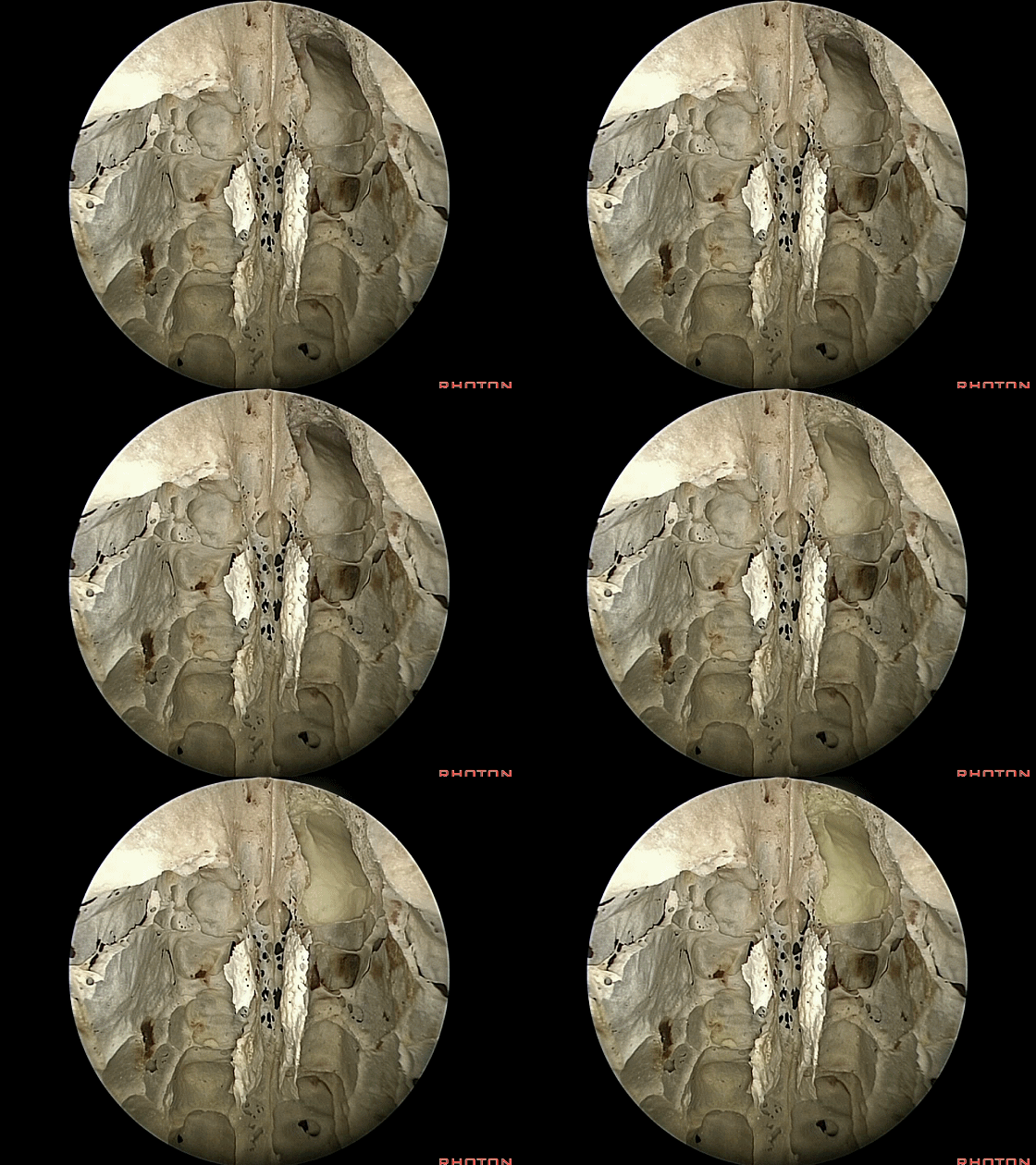
▼通过成角内镜观察 额窦。这是筛板的前界(下图)。
This is just looking up into the frontal sinus from below with an angled endoscope. Here's the anterior edge of the cribiform plate.
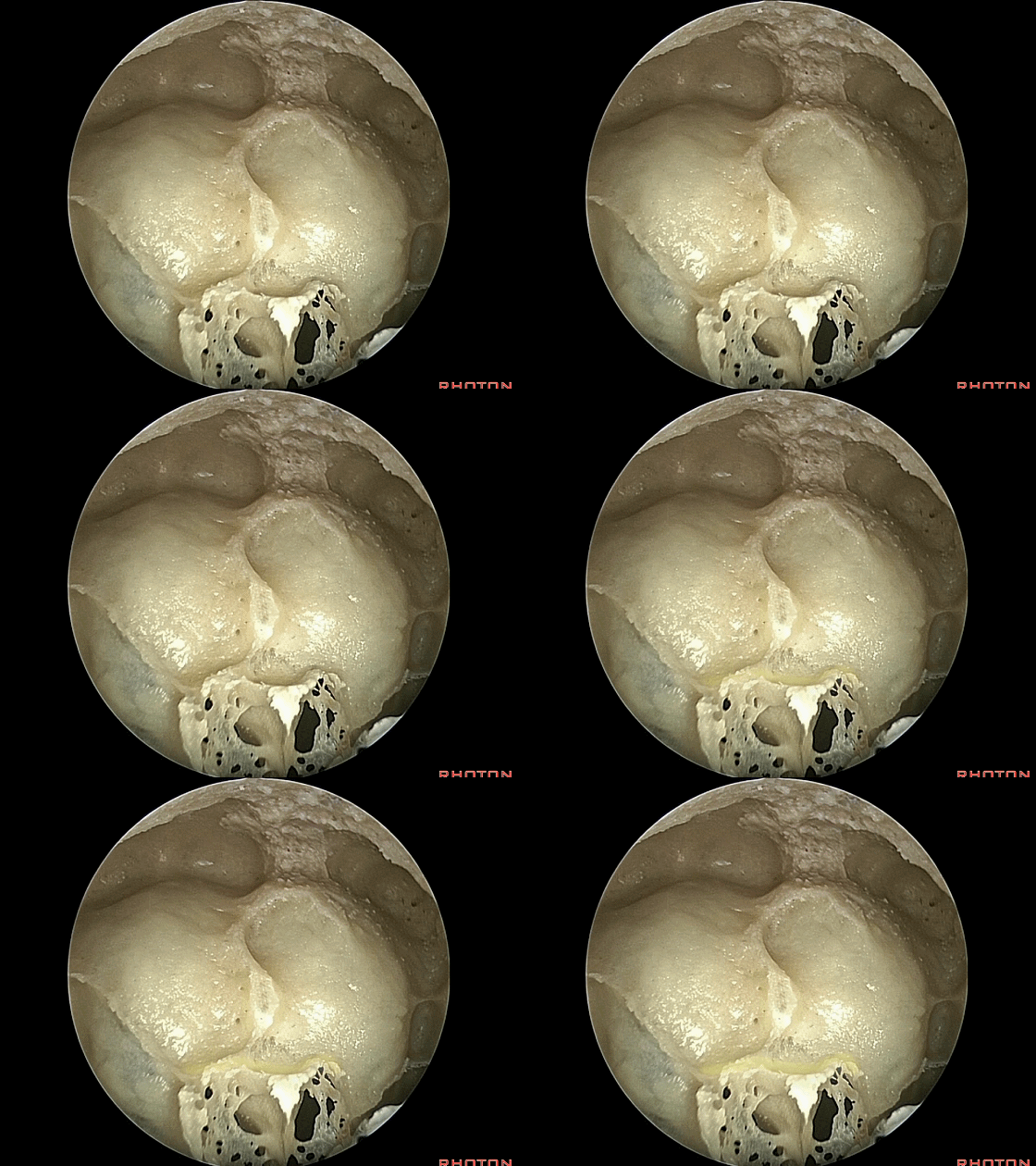
▼穿行于筛窦内的血管是筛前动脉。
coming through the ethmoids is the anterior ethmoidal arteries.
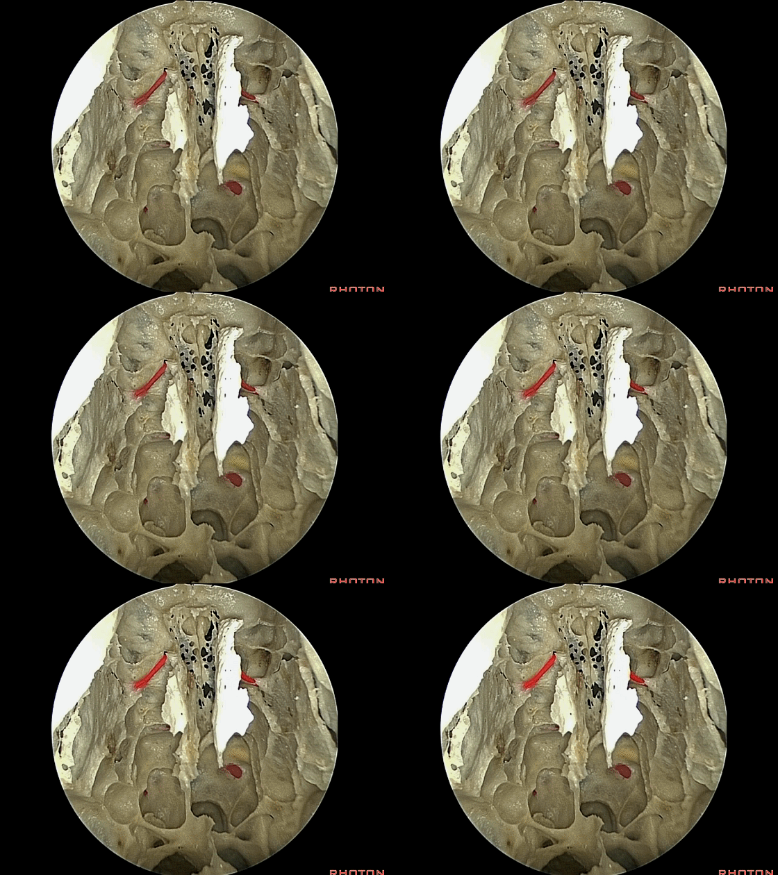
▼后方的是筛后动脉。
Back here are posterior ethmoidal arteries.
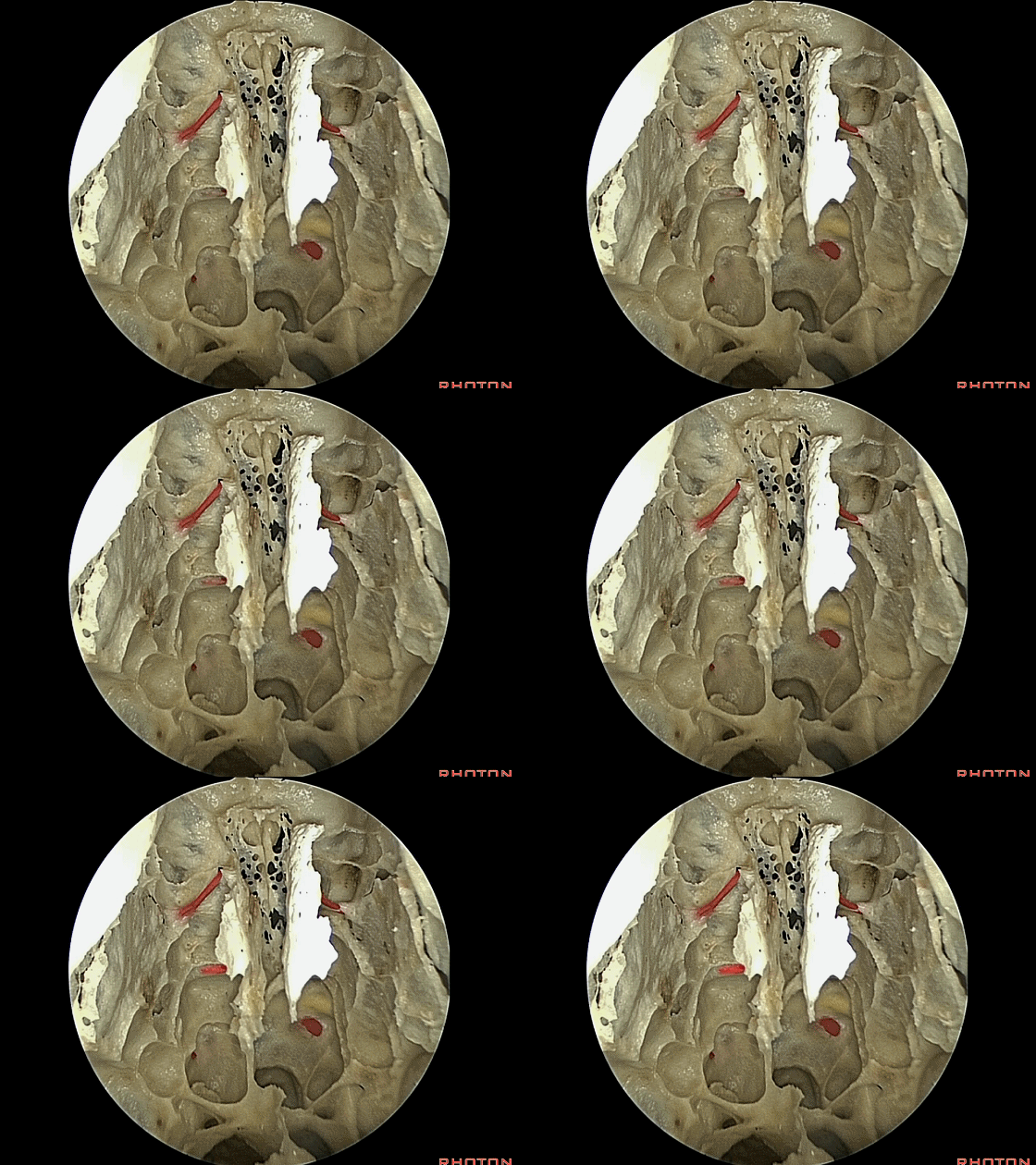
▼向后通过蝶窦,可见视神经管(下图)
You look back into the sphenoid, this is optic canal

▼这是颈内动脉表面的一处自发性骨质缺损。
and a spontaneous dehiscence over the carotid artery.
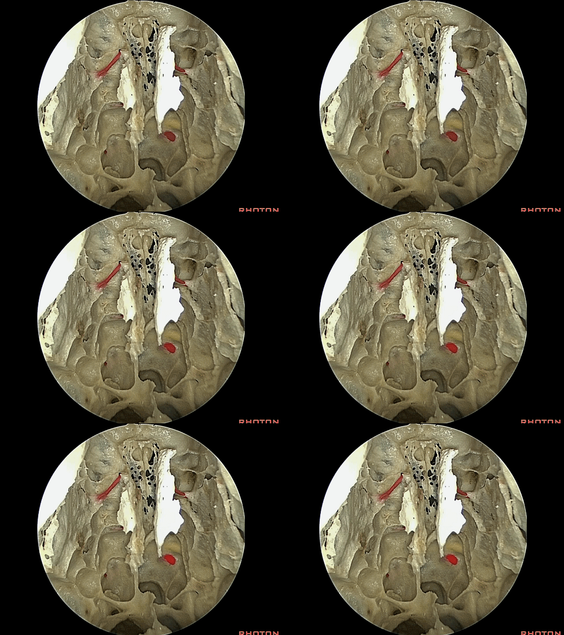
▼这是腭骨的蝶突。
And here is sphenoid process of palatine.
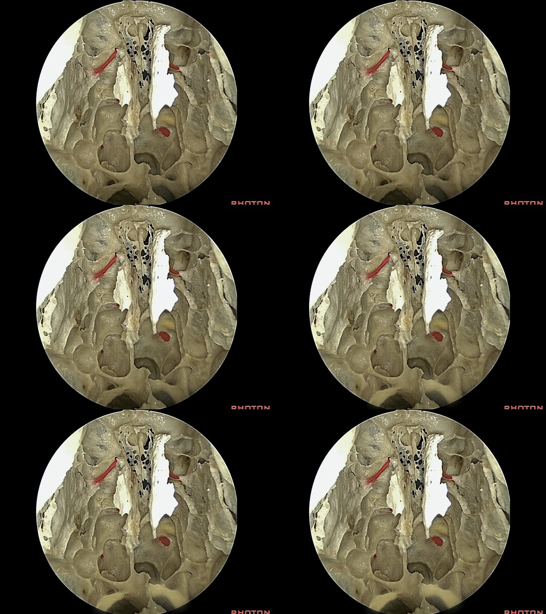
▼这是翼管。
So this is vidian canals.
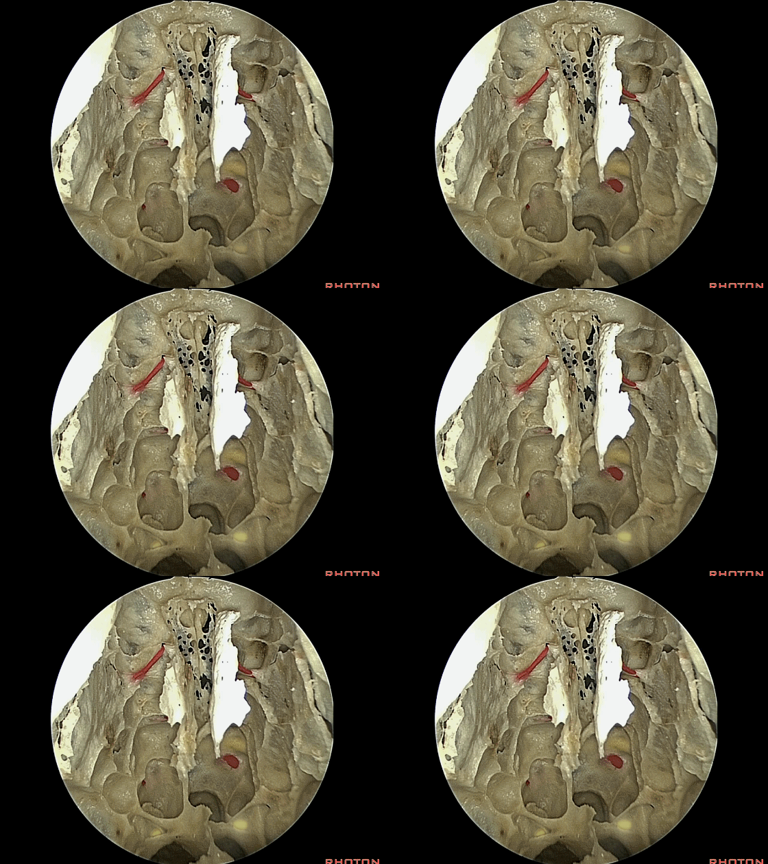
▼来看看这片区域的全景图,这是蝶窦。
But just a view...a panoramic view of all of this area, you can get into by opening sphenoid,ethmoid,frontal sinus from below.
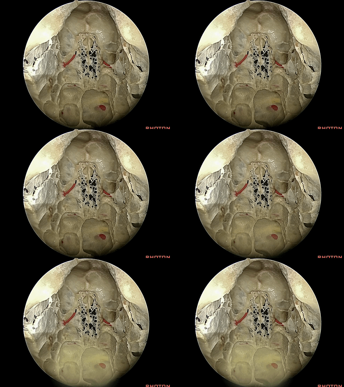
▼这是筛窦
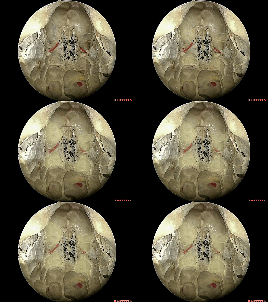
▼这是额窦
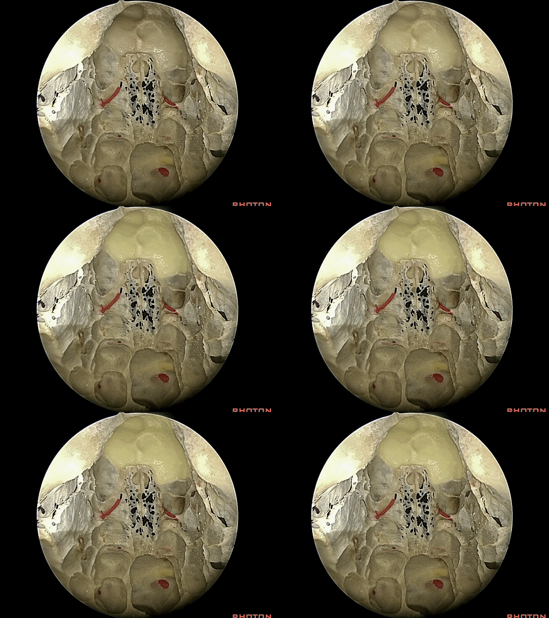
▼如果在术闭时需要大片骨膜瓣修补,只需在此处的额骨钻一小口,在不开颅的情况下取下骨膜瓣,随后向后翻转之,根据瓣膜的大小,可一直翻转至蝶窦。
If you need a large pericranial flap after you worked in these areas,well you can just drill a little opening here in the frontal bone, take a big pericranial flap without doing a craniotomy, and you can fold that flap, all the way back, depending on how big a flap you take, all the way back to the sphenoid sinus...
▼这是暴露后的全部术野。该入路还可向下暴露至寰椎、枢椎
But you have access then to all of this area. You can work down medially, even to C1, C2.
▼再看看这个结构。在右侧鼻腔侧壁,这是 腭骨
So you get a chance to look at this. As you look at the lateral wall of the nasal cavity,
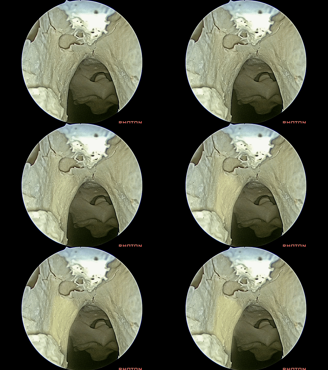
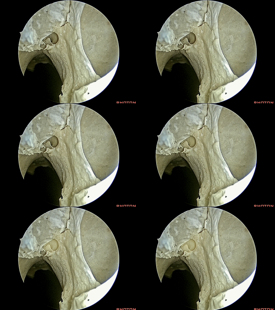
▼腭骨上方的这个区域,这是 蝶腭孔(sphenopalatine foramen)
in this area, above the palatine bone, we see the sphenopalatine foramen,
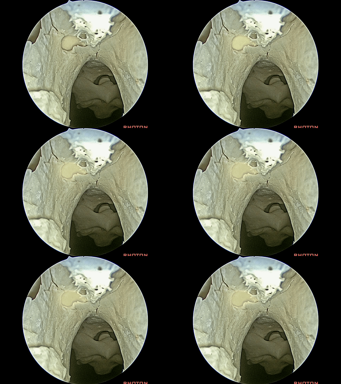
▼这是翼管的前方开口
and the anterior opening of the vidian canal.
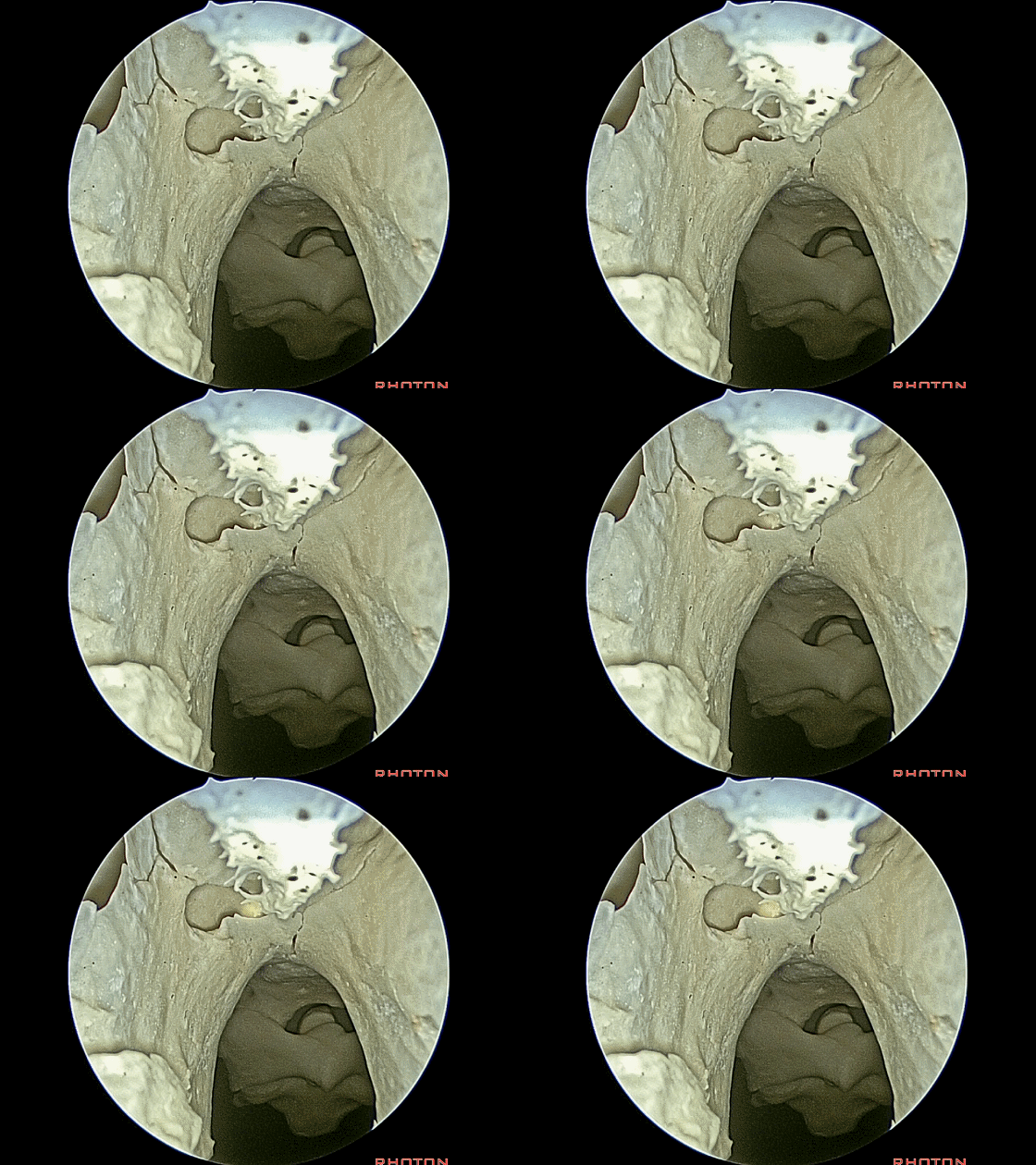
▼这是左侧 上颌窦
Here's the other side. Here, maxillary sinus, left side,
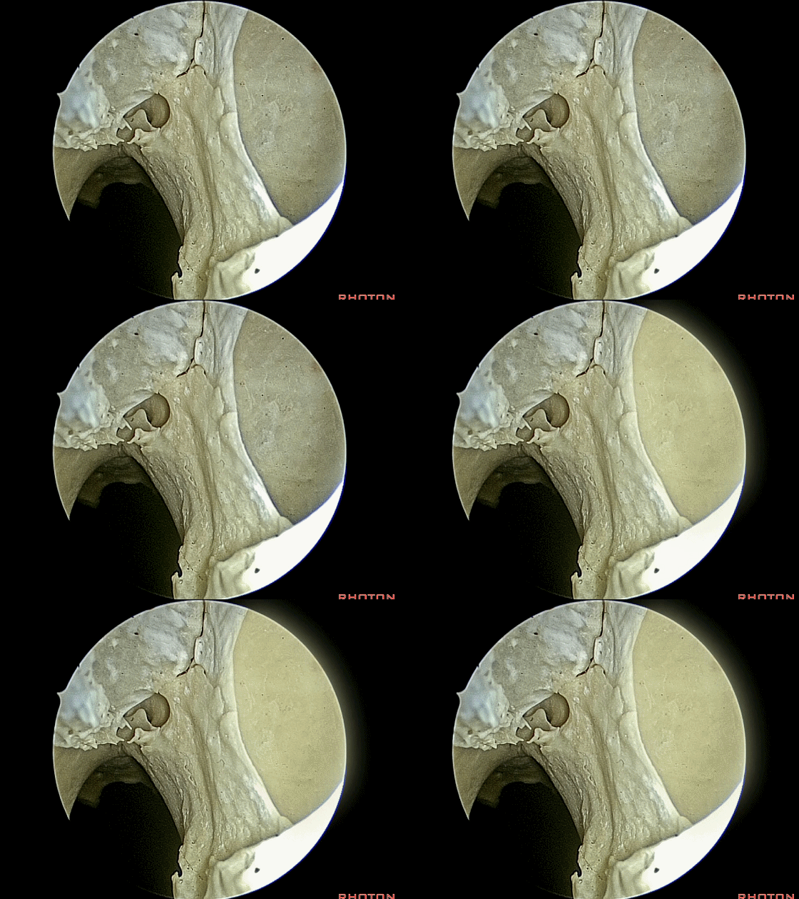
▼这是左侧 蝶腭孔。
sphenopalatine foramen.
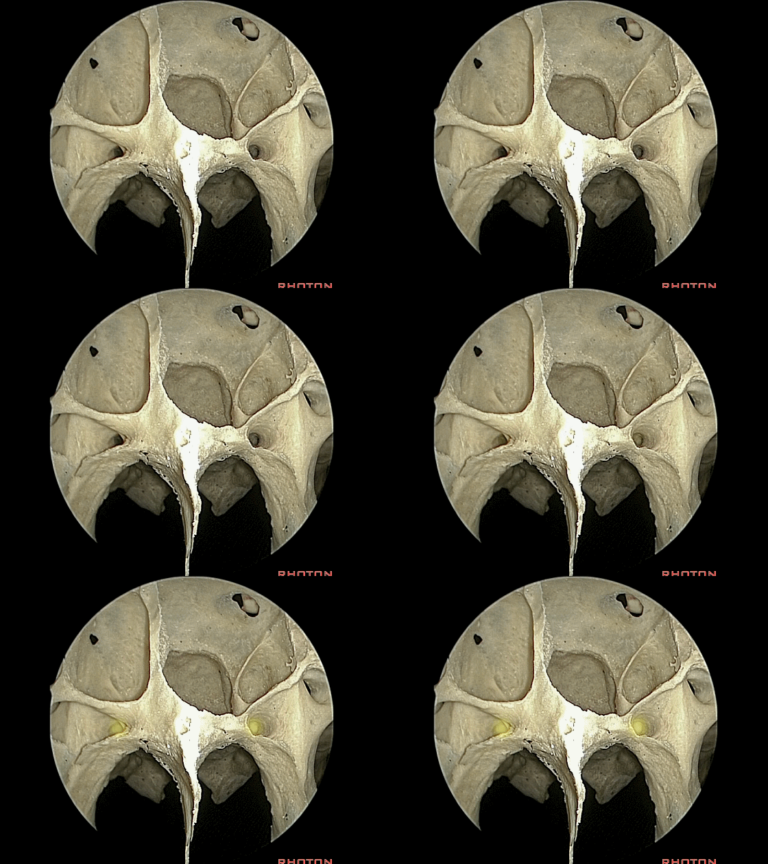
▼再看一下蝶窦,这是翼管
Just a view of the sphenoid, the vidian canals,
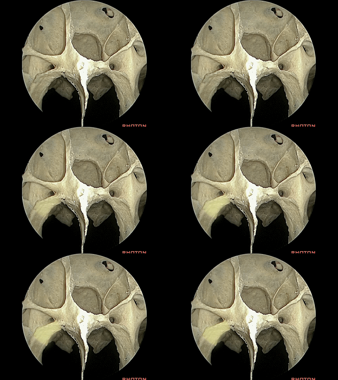
▼这是腭骨蝶突
sphenoid process
▼这是圆孔(rotundum [rəʊ'tʌndəm]),其内有三叉神经上颌支
V2 here
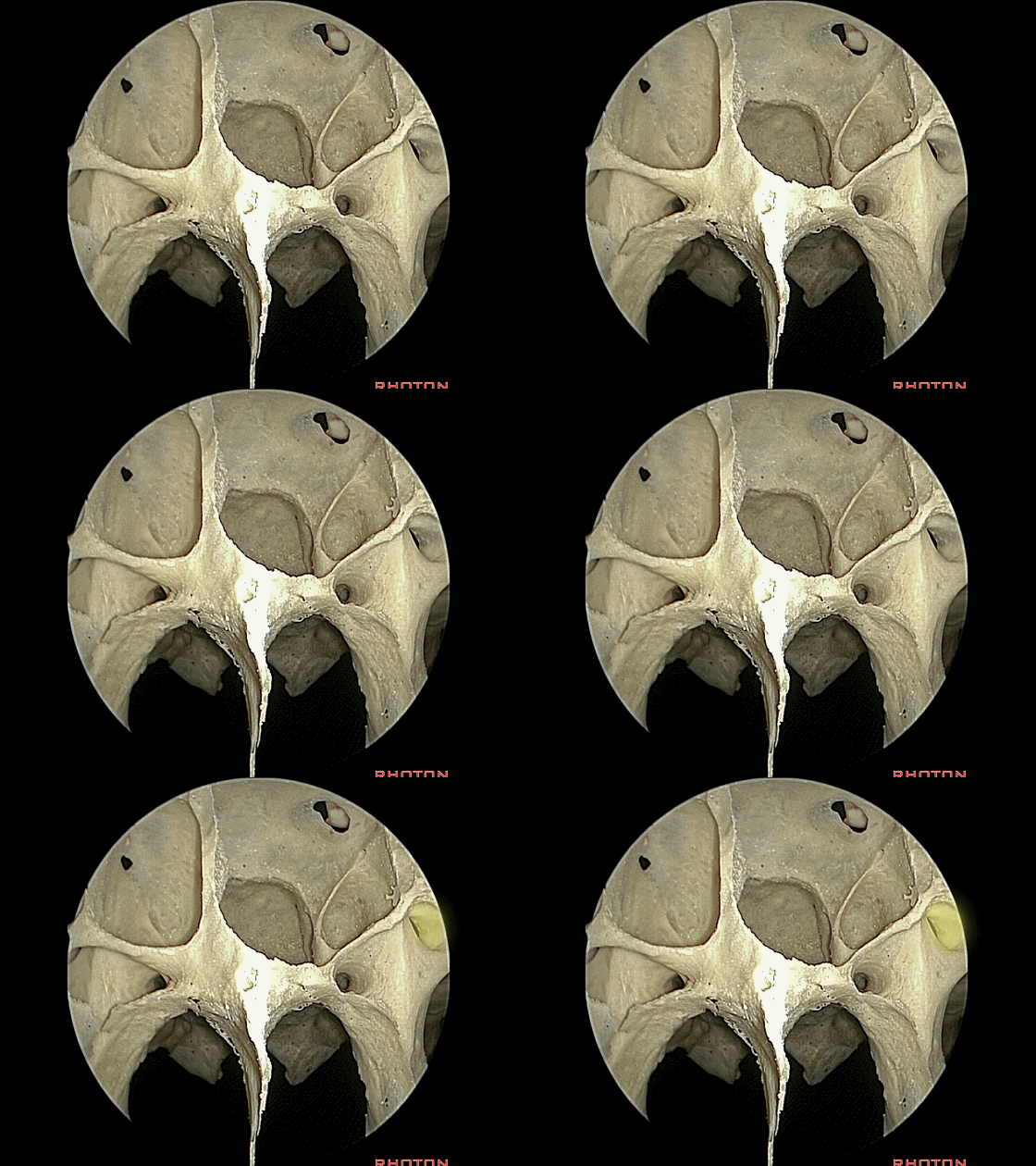
▼后方是三叉神经下颌支 通过 卵圆孔(ovale [oʊ'veɪl])进入颞下窝。
V3 back in infratemporal fossa.
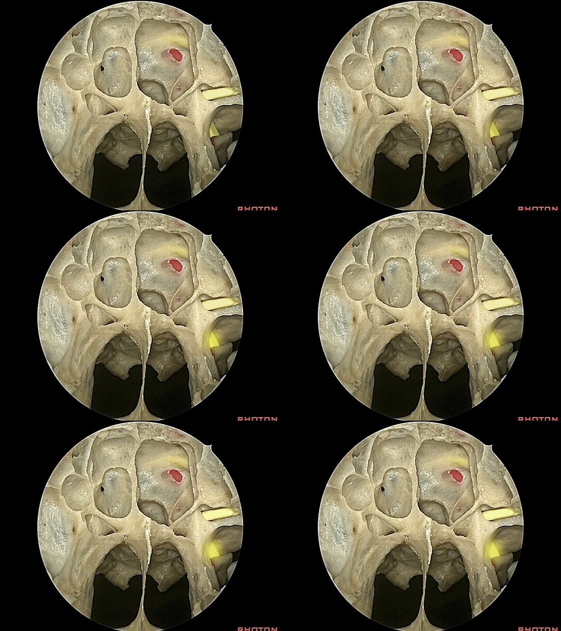
▼这就是蝶窦开口,位于鼻甲后界。
Now, and here we just see this sphenoid ostia in the back edge of the turbinate here.
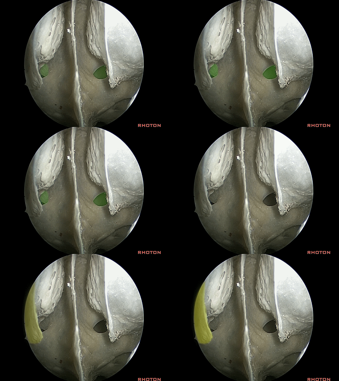
▼这是菲薄的蝶窦前壁,上有蝶窦开口,我们称其为 蝶甲,是蝶窦的前壁。
And this area, the thin anterior wall of the sinus through which the ostia open, is the sphenoidal concha, just this thin anterior wall.
![]()
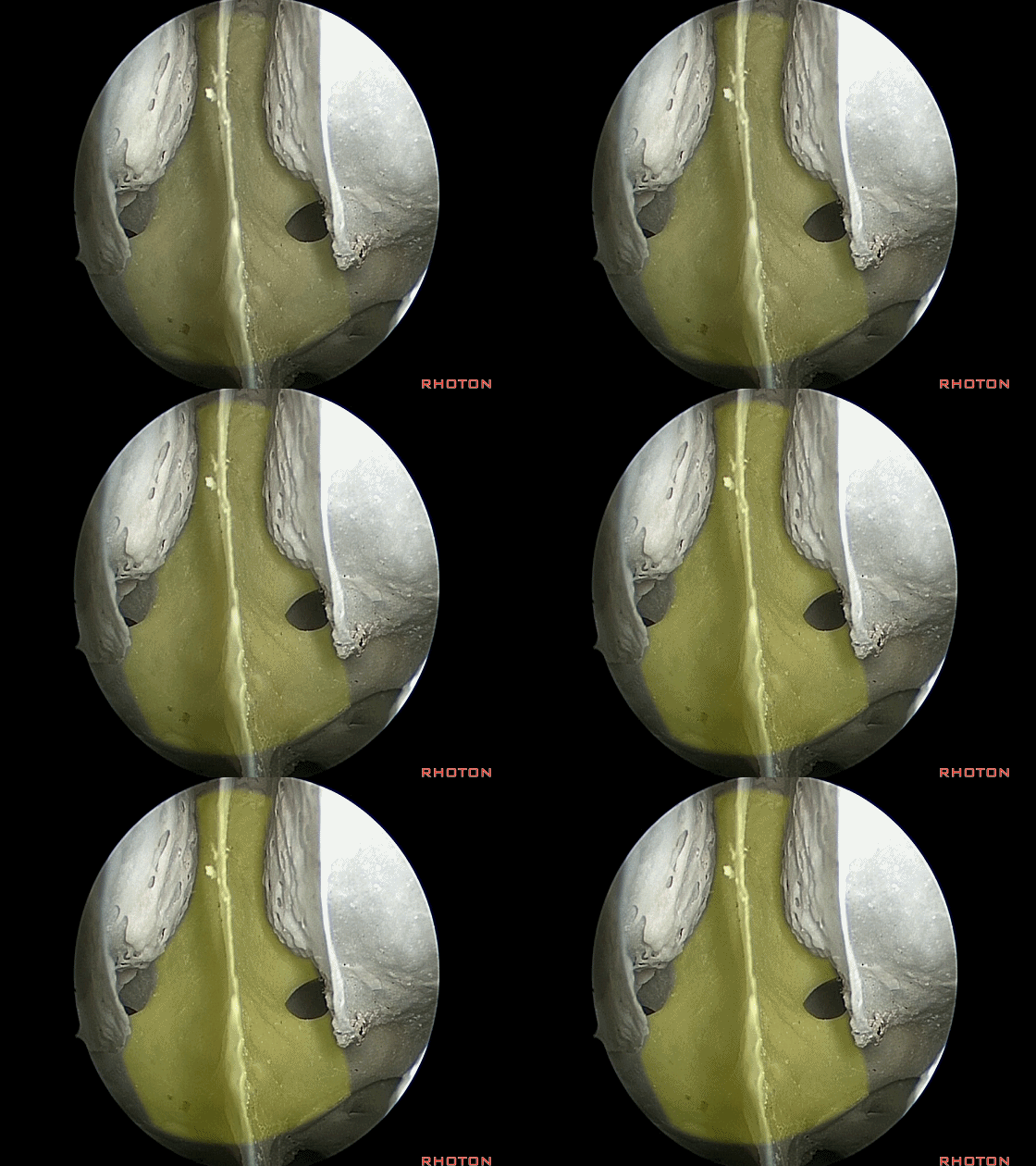
▼现在从内镜下进入右侧鼻腔,这是鼻中隔
So, now we look in and we're on the right side,septum,

▼这是内镜下所见的下鼻甲
and with the endoscope, an inferior turbinate
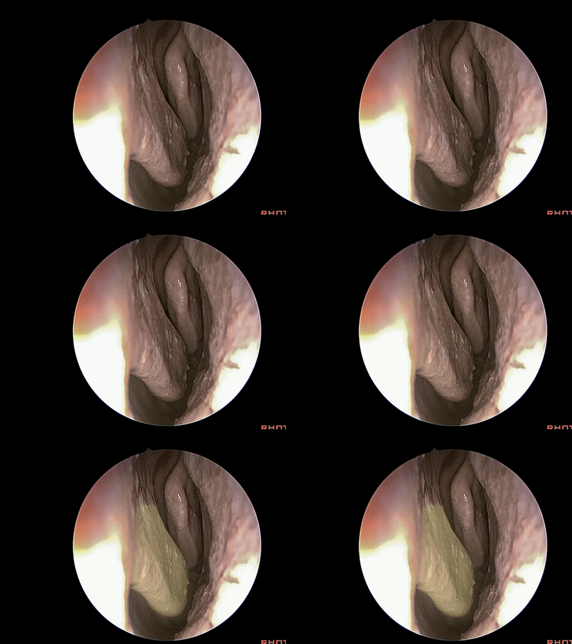
▼这是中鼻甲
and a middle turbinate here.
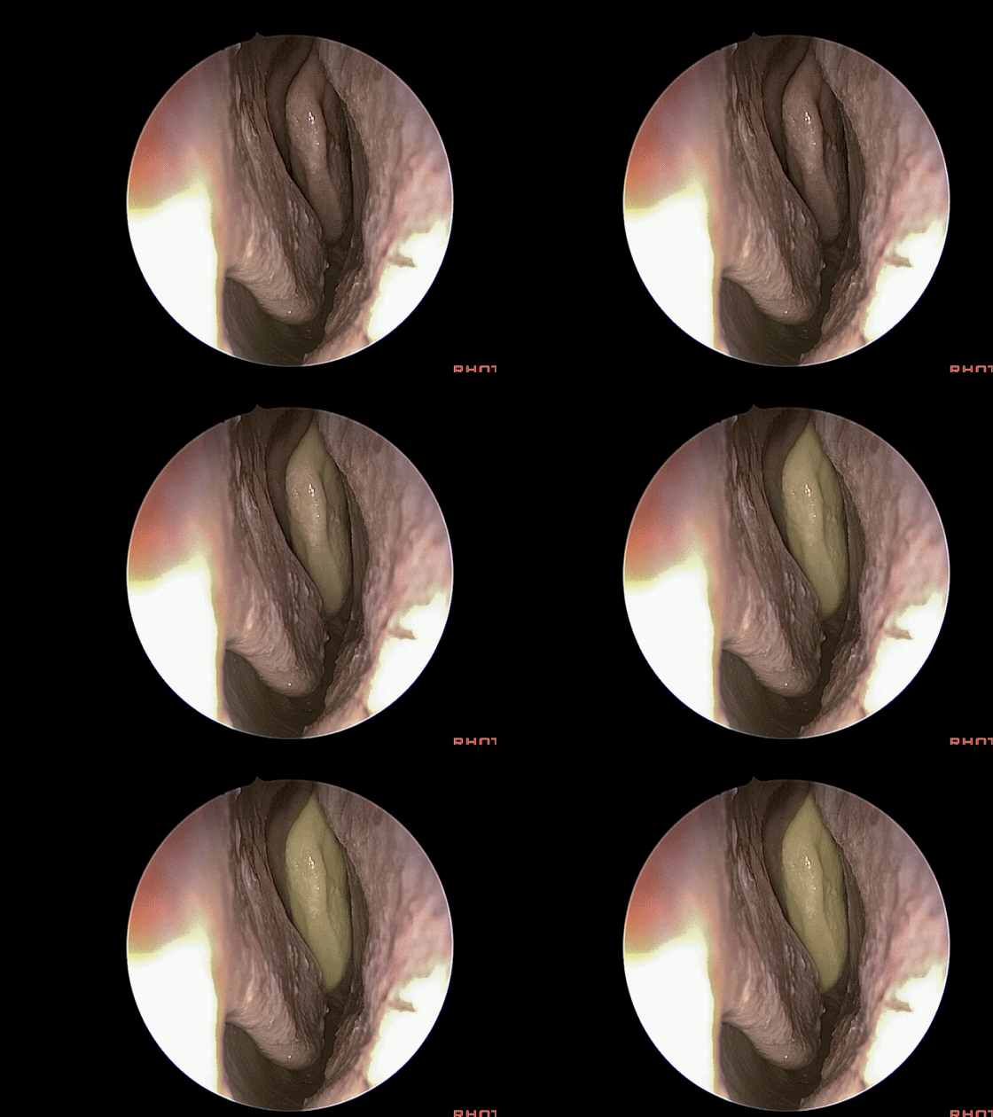
▼在中鼻甲内侧继续向上探查可见上鼻甲。
The septum is here. And then we look above and we see a superior turbinate.
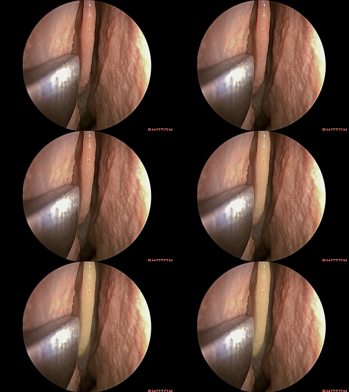
▼有时可见其上方为 最上鼻甲。
And then above that,sometimes a suspanme turbinate.
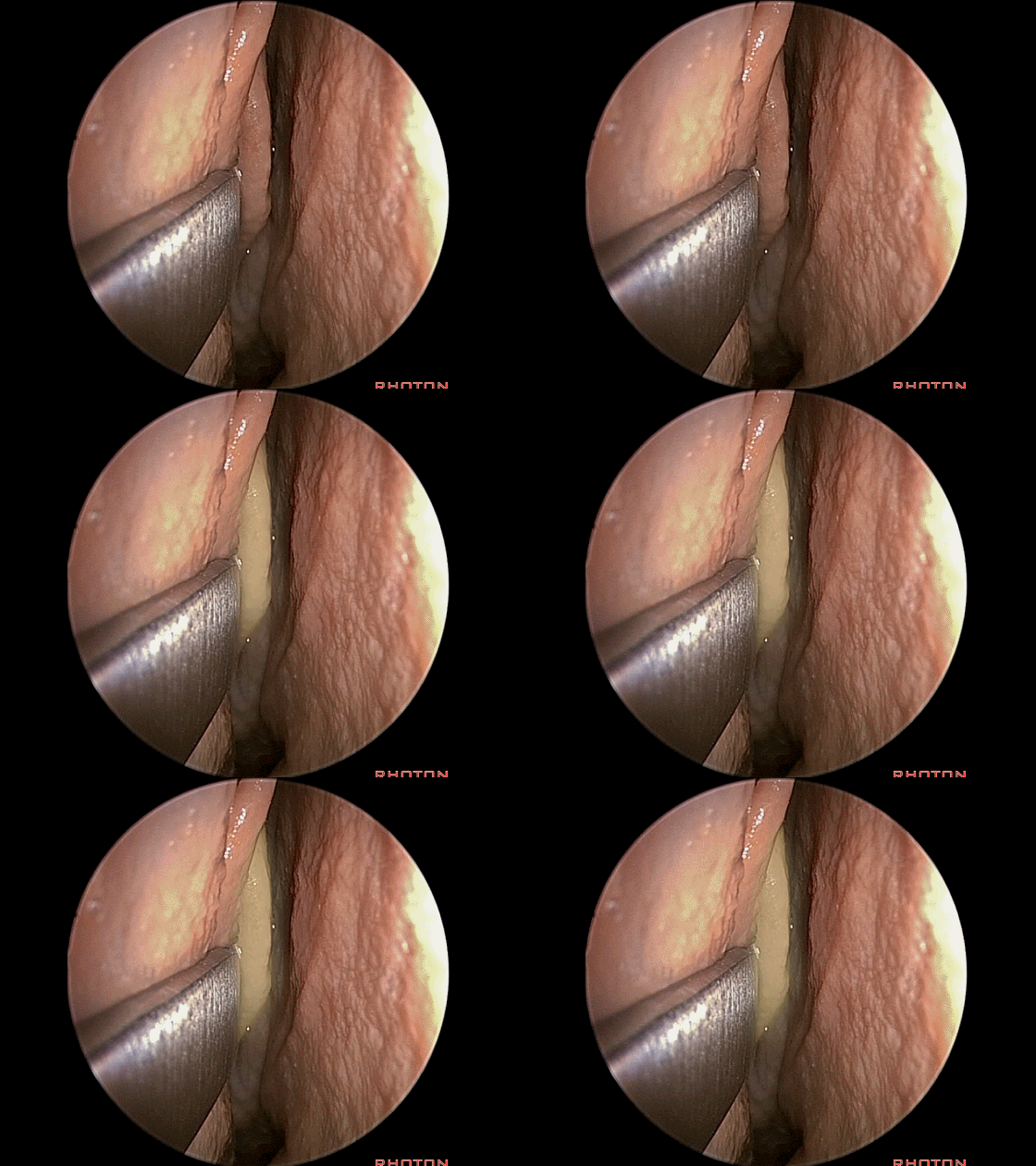
▼无论哪一个是最上方的鼻甲,蝶窦开口(下图)均位于其后界稍上方数毫米处。
but the sphenoid ostia is gonna be at the back edge of the turbinates. Usually, just a few millimeters above the back edge of, whatever the highest turbinate is, right here.
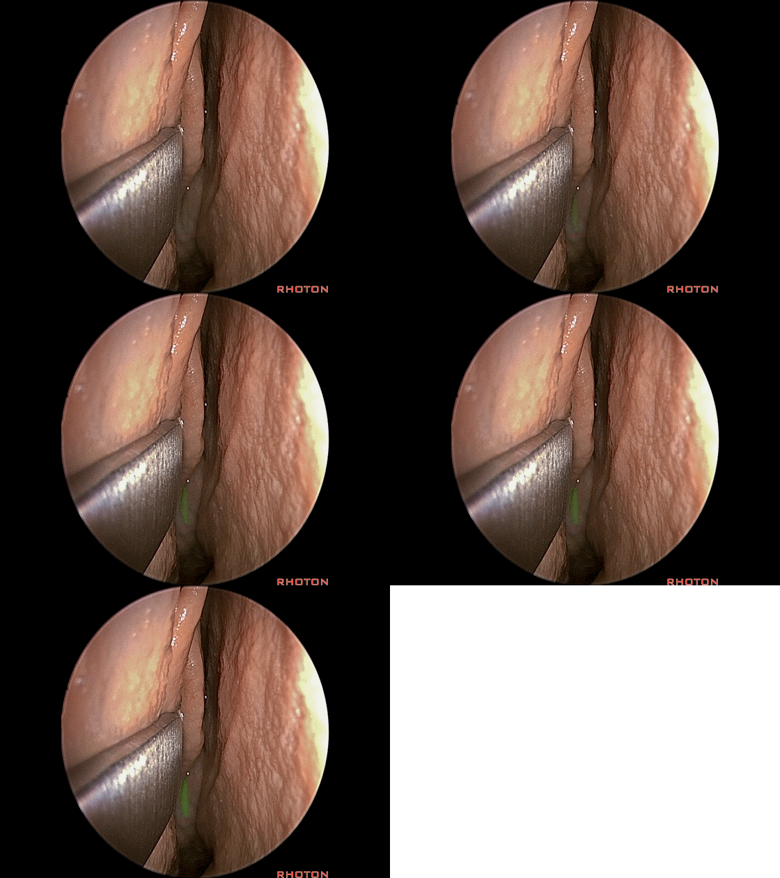
▼这是鼻泪管。
Now, we see the nasolacrimal duct.
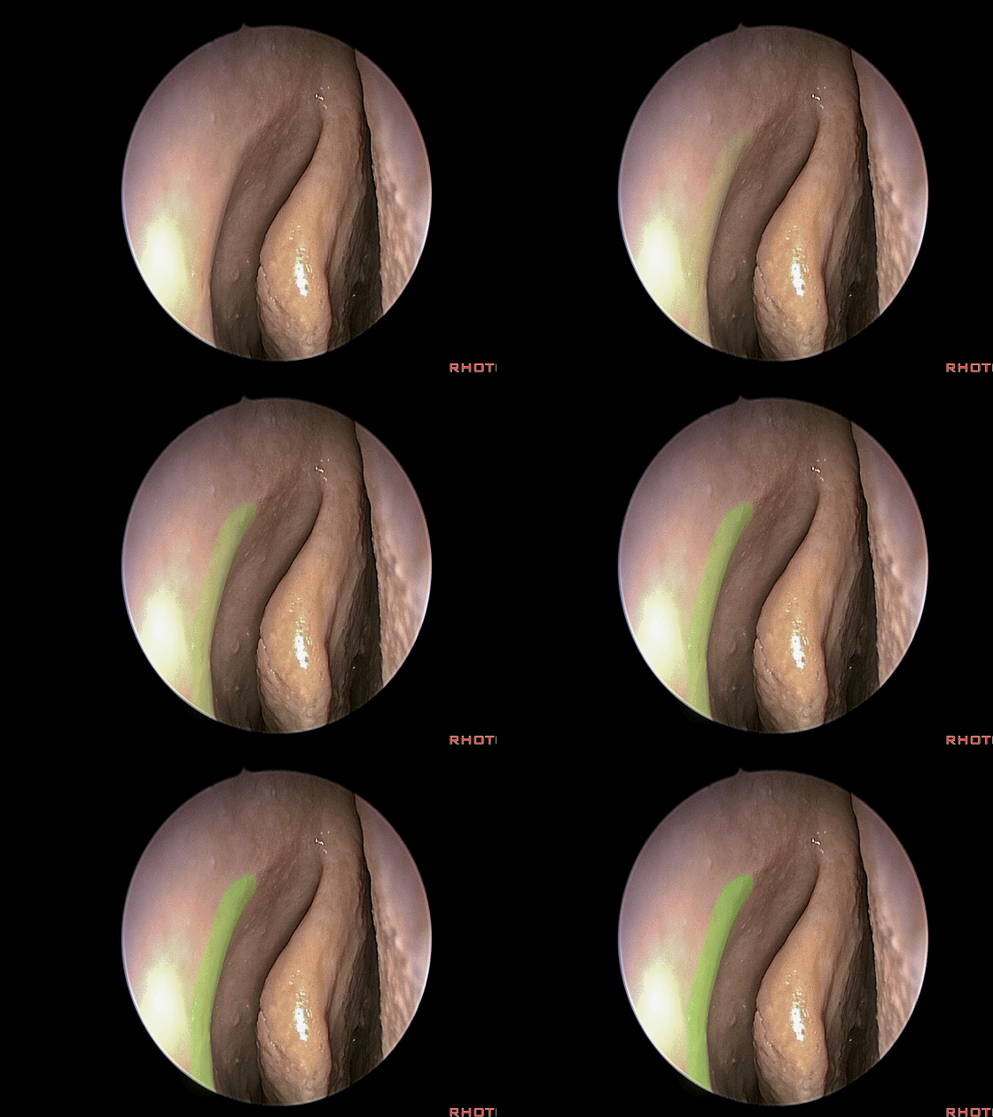
▼中鼻甲(下图)外侧连于筛骨。
We see the middle turbinate attached laterally to the ethmoid.
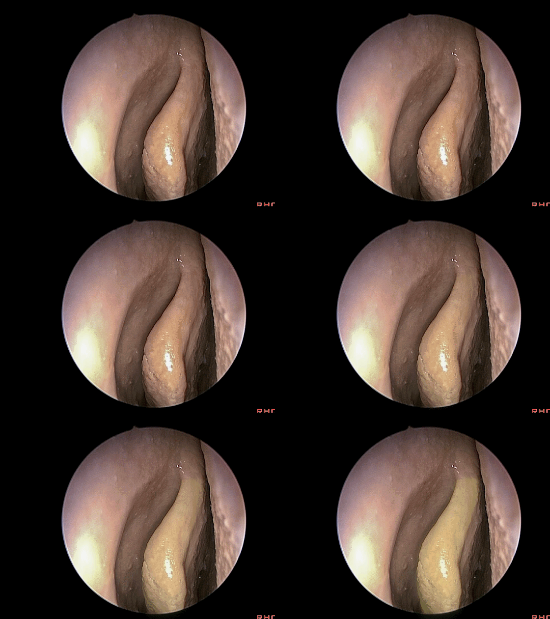
▼将中鼻甲牵向鼻中隔,这是钩突(uncinate process)。
And then we pull the middle turbinate toward the septum, and here's the uncinate process.

▼钩突后方,这个初露端倪的结构是 前组筛房 (筛泡 anterior ethmoid air cells; ethmoid bulla )
And behind the uncinate process, Here's...we begin to see that. What is that structure? That's anterior ethmoid air cells.
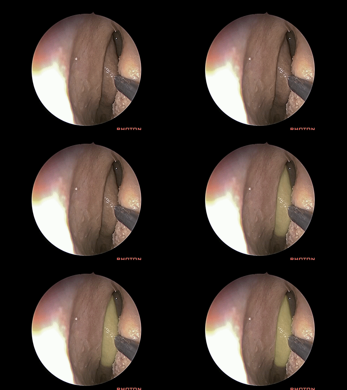
▼牵开中鼻甲,抵近观察 前组筛房(筛泡),打开筛泡就是进行前颅底扩大暴露的关键步骤。
And here, when we retract that turbinate, these are anterior ethmoid air cells,Now opening the bulla is the key to the floor of the anterior fossa of widening it.
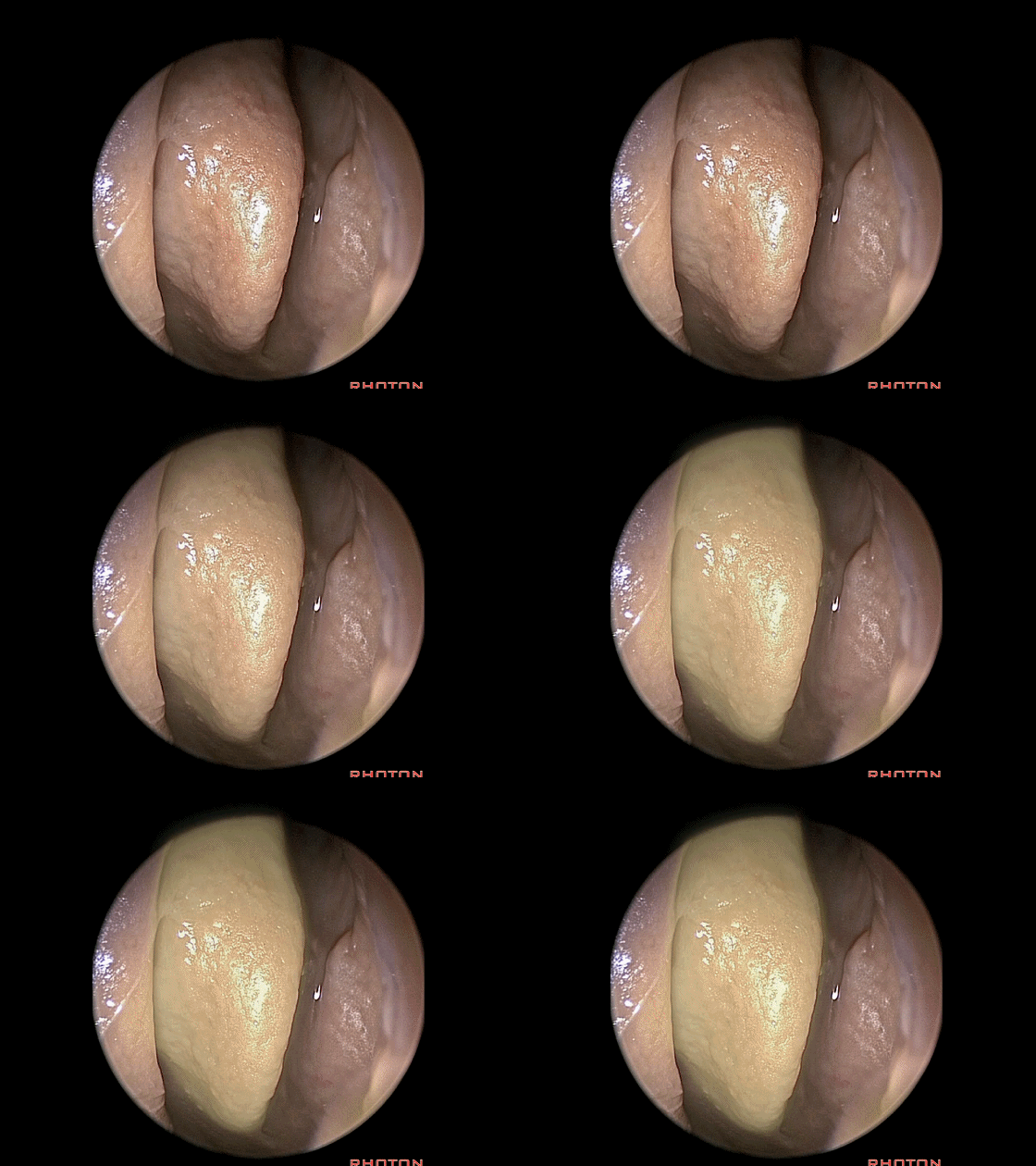
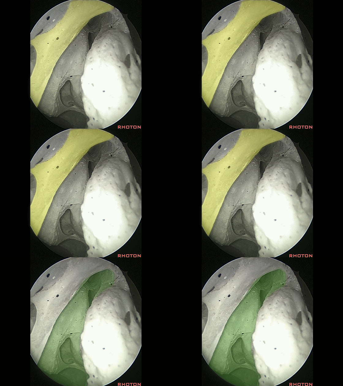
▼在钩突后方的结构是 半月裂孔(下图),其位于钩突与筛泡之间。
And behind the uncinate process that overlies...well, that bony process is the semilunar hiatus between the uncinate process and the bulla.
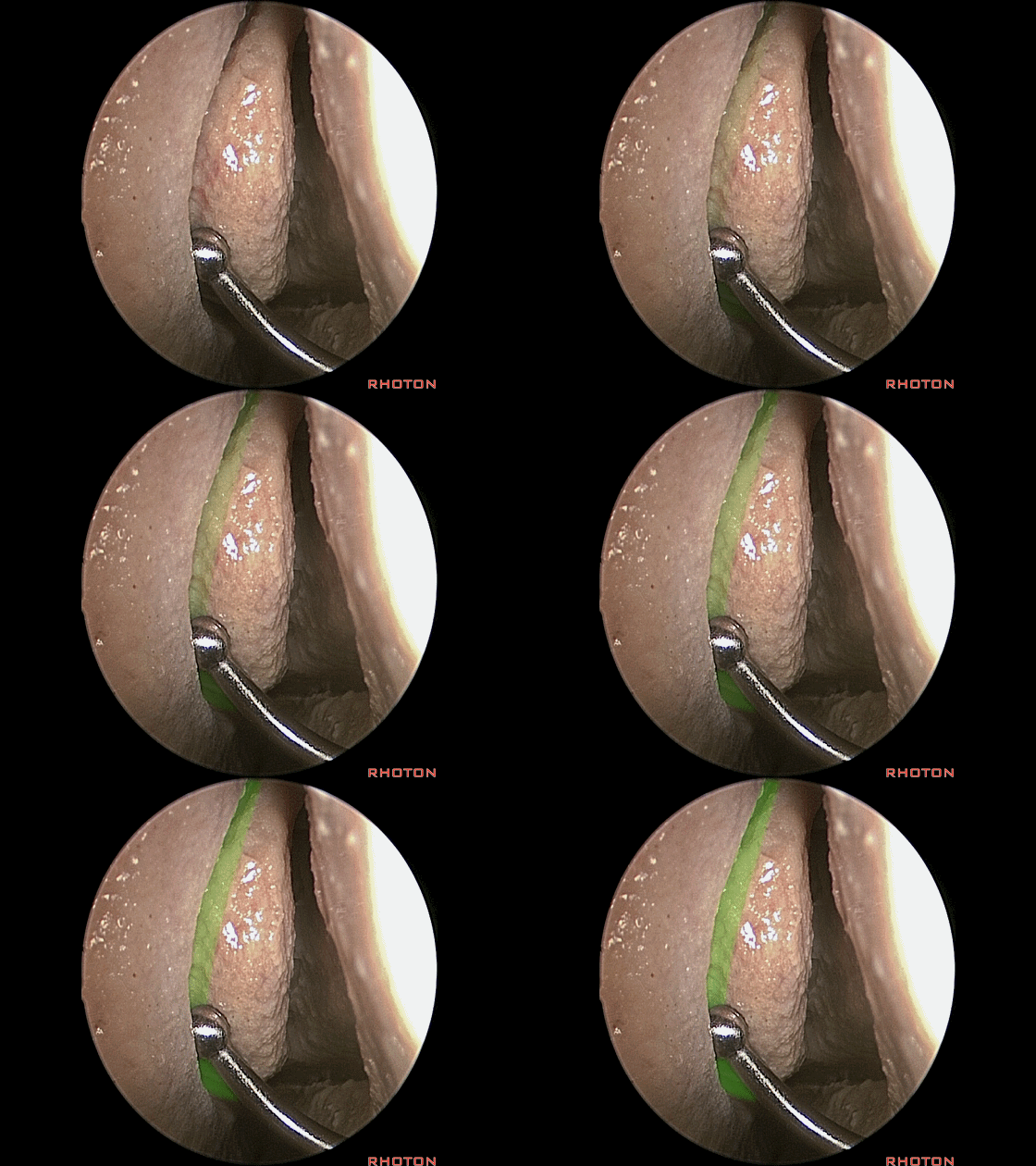
▼半月裂孔(绿色)位于钩突(黄色)后方
And this hiatus, in back of the uncinate process,
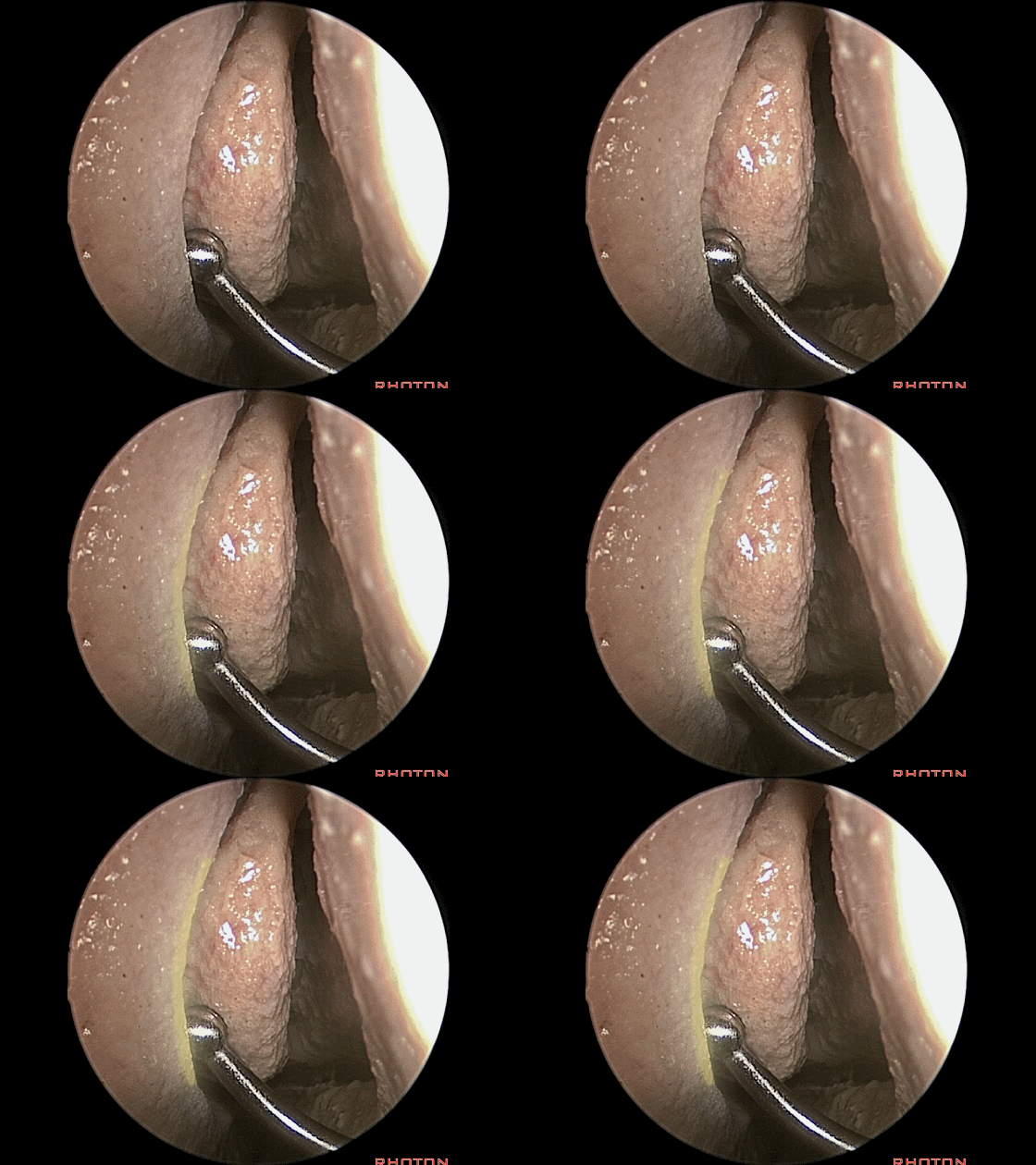
▼在内镜下我们进入钩突(下图)后方
So that, and, if you're looking in back of this with endoscope,
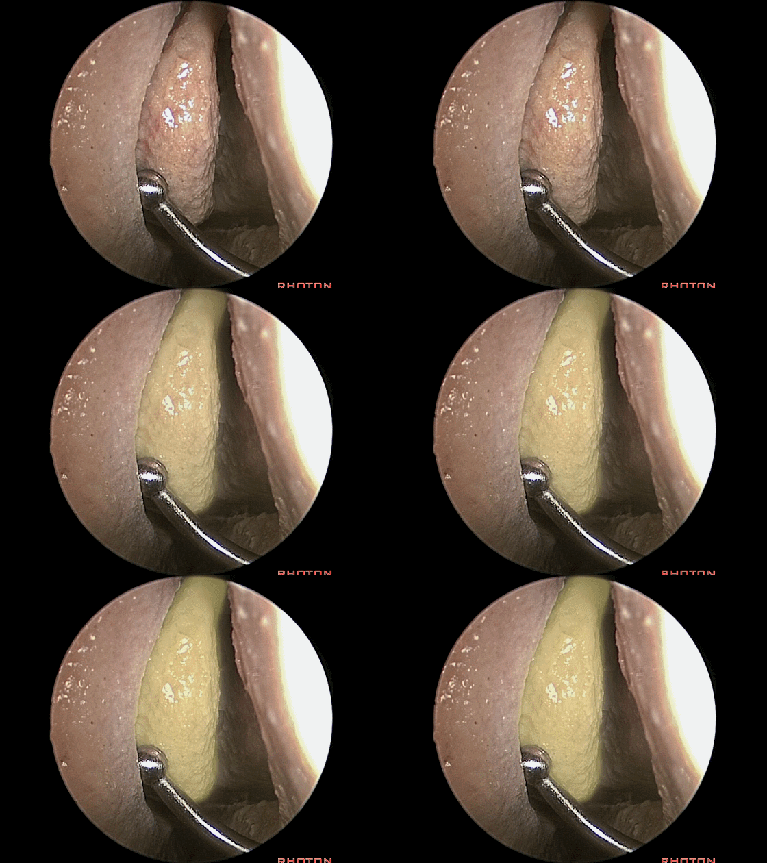
▼这是前组筛房(筛泡)。
the air cell.
▼半月裂孔,正是额窦开口(下图)以及部分筛窦开口的位置。
the semilunar hiatus, is where you see the drainage of the frontal ducts, some of the ethmoids.
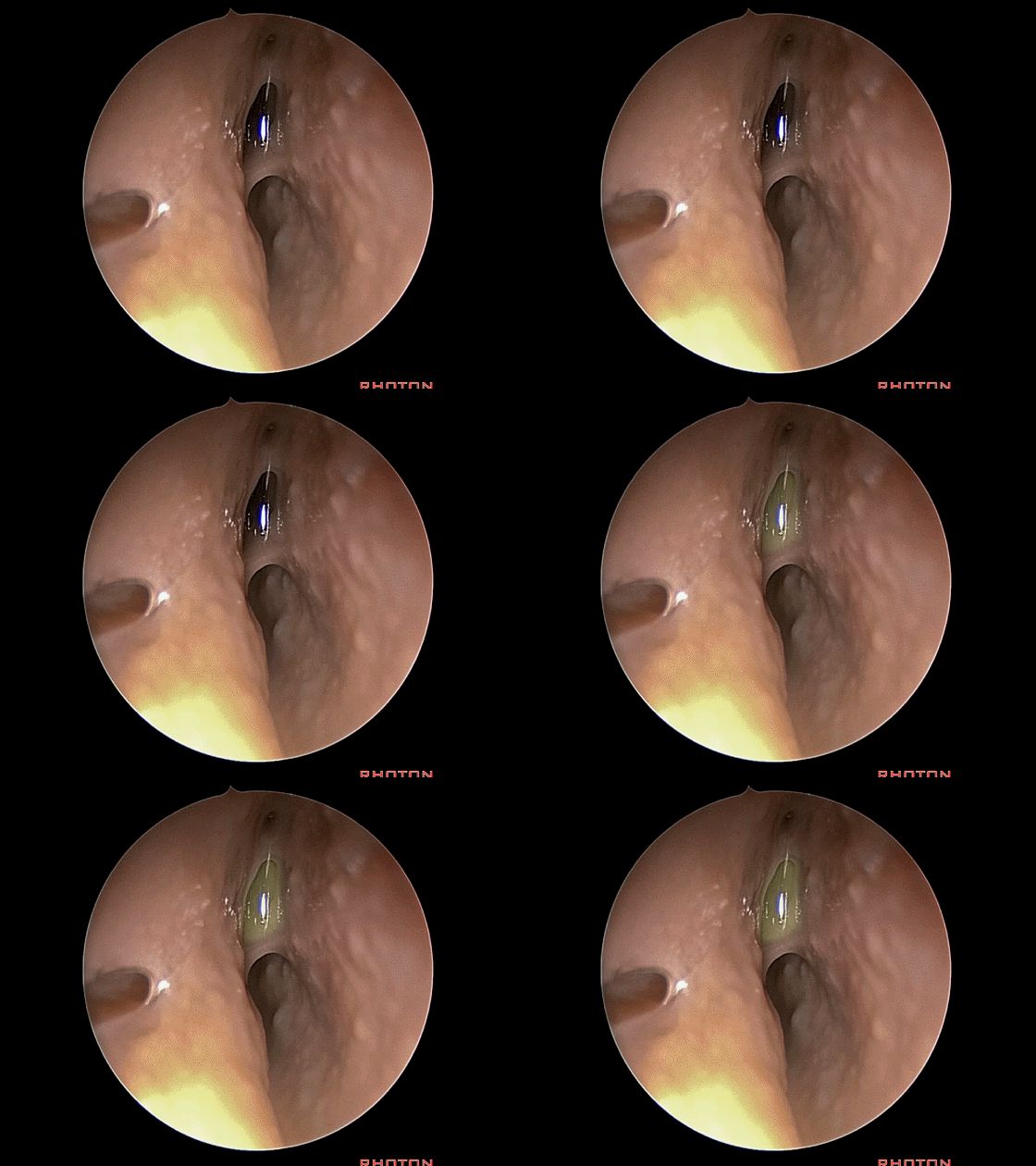
▼这是筛窦开口。
and the ethmoids ducts
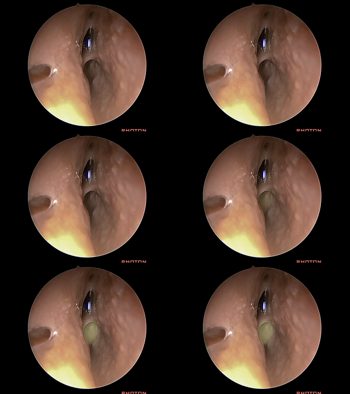
▼沿着该裂孔向下,即可见上颌窦开口(下图箭头方向)。
And if you follow the hiatus down, you see the drainage of the maxillary sinus.
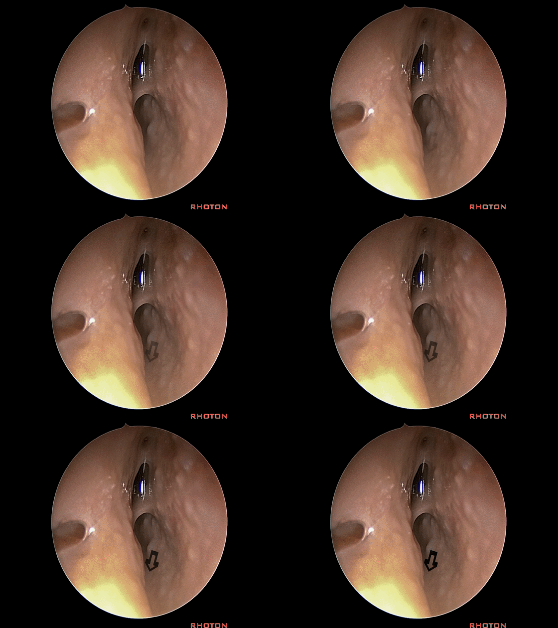
▼因此,该入路中重要的一个步骤就是分离钩突的上端(下图)和下端。
So, a step along the way to widening this approach is you divide the uncinate process above and below.
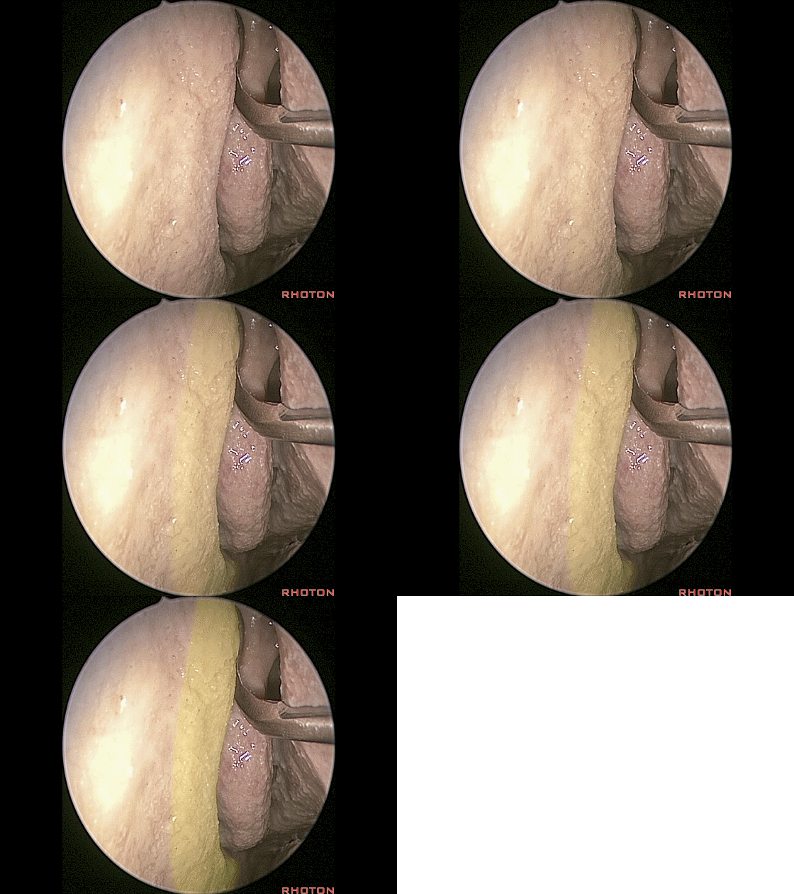
▼这是分离钩突的下端
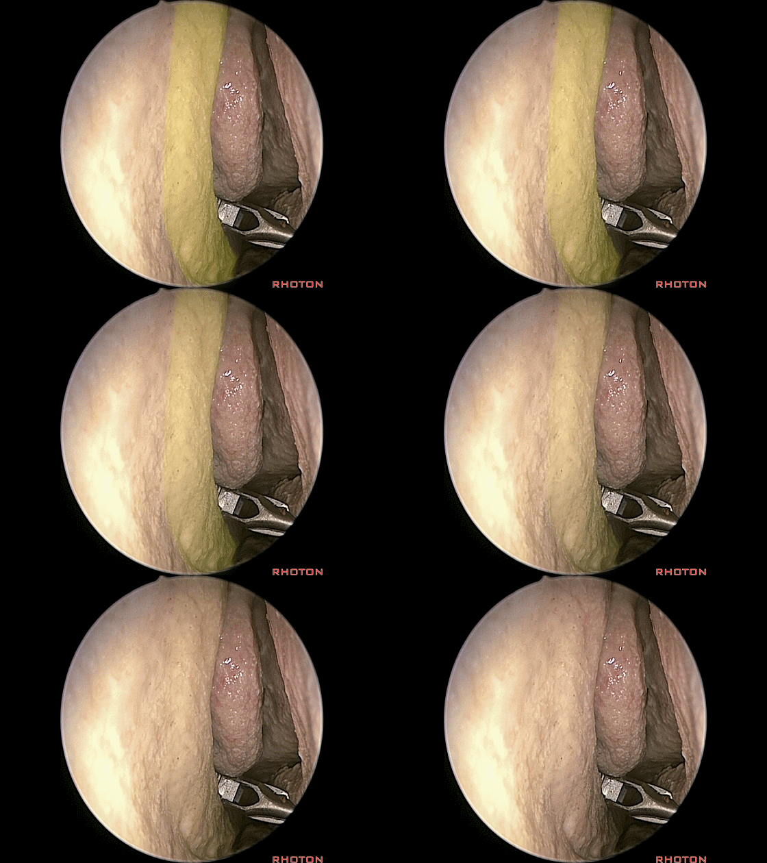
▼随后可翻转或咬除钩突,并有足够的空间取出。
And then you can fold it or bite it off, usually it's big enough for a shaver takes it off.
▼现在即可暴露体积巨大的筛泡,即前组筛房的一部分,位于中鼻道内。
and we see how big these ethmoid air cells are.And then you're looking at the bulla, the air cells of the ethmoidsin the middle meatus.
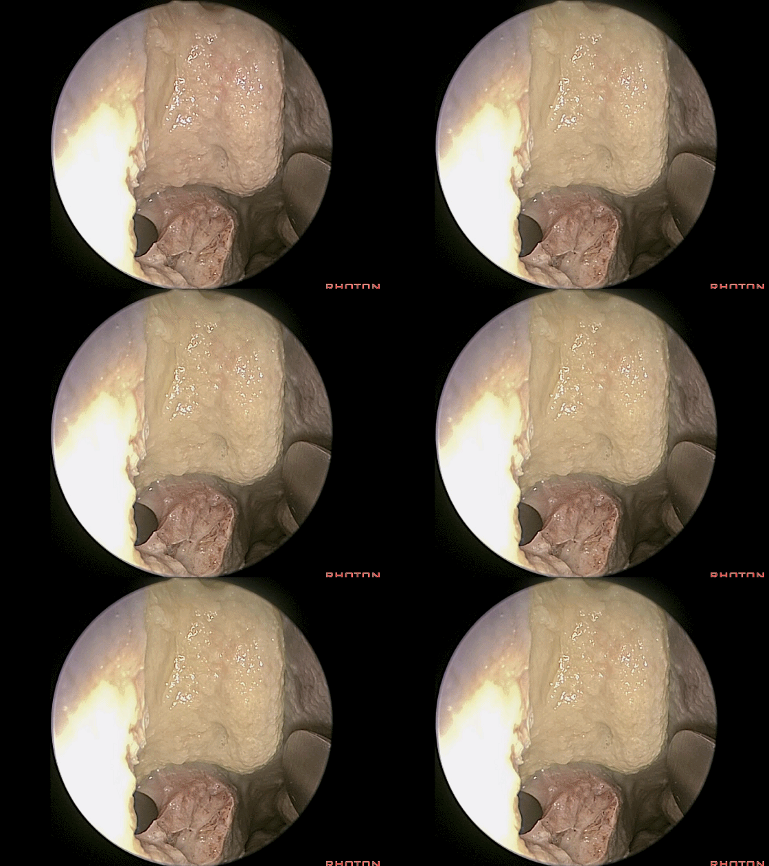
▼我们也可以在中鼻道内扩大上颌窦开口。
You can get enlarge the maxillary duct opening out of the middle meatus.
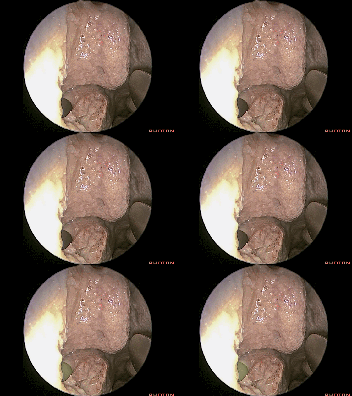
▼这里显露的即为上颌窦的巨大开口。
And here's a large opening into the maxillary sinus.
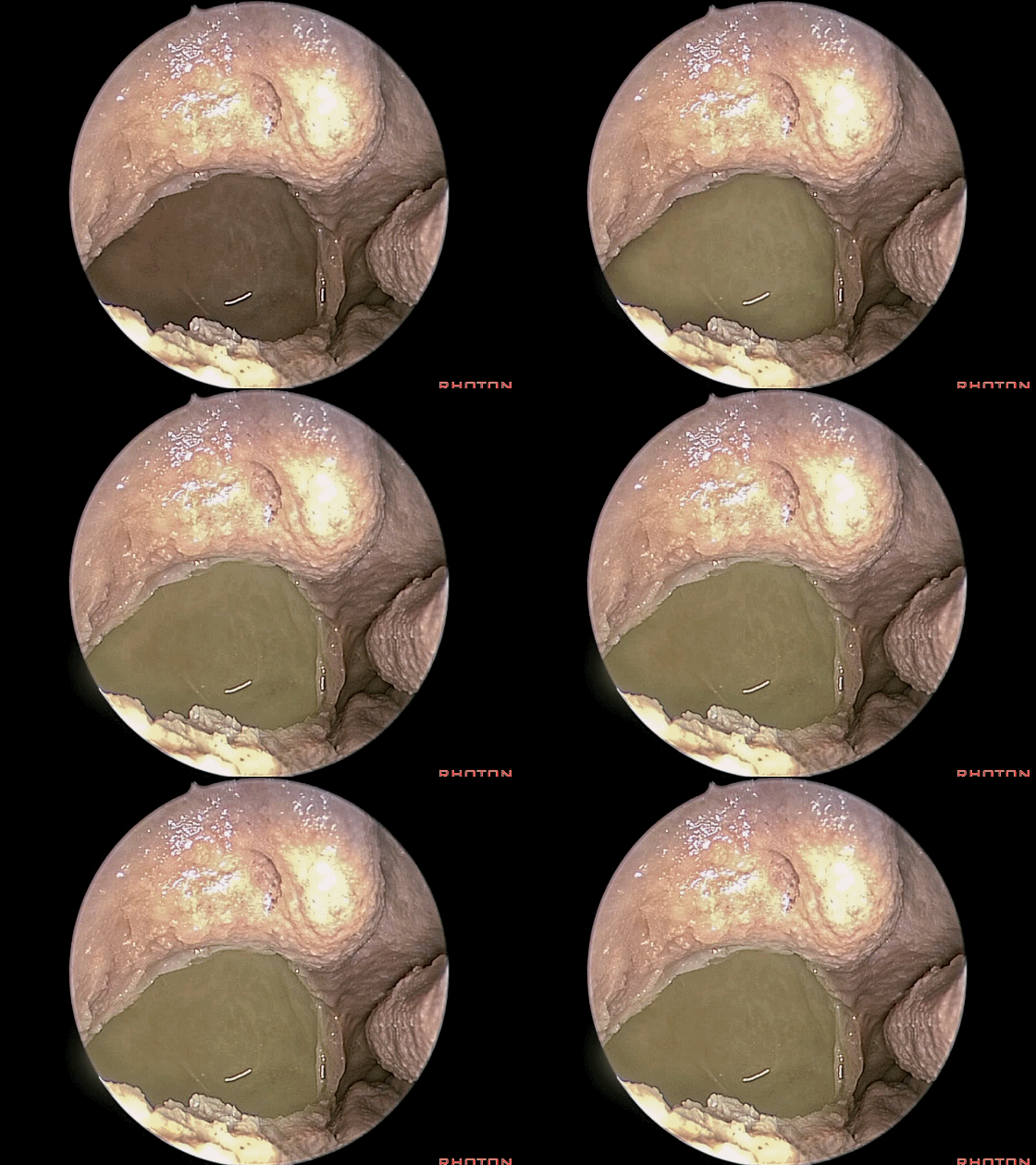
▼这是前组筛房(筛泡)。
This is the anterior ethmoid air cells.
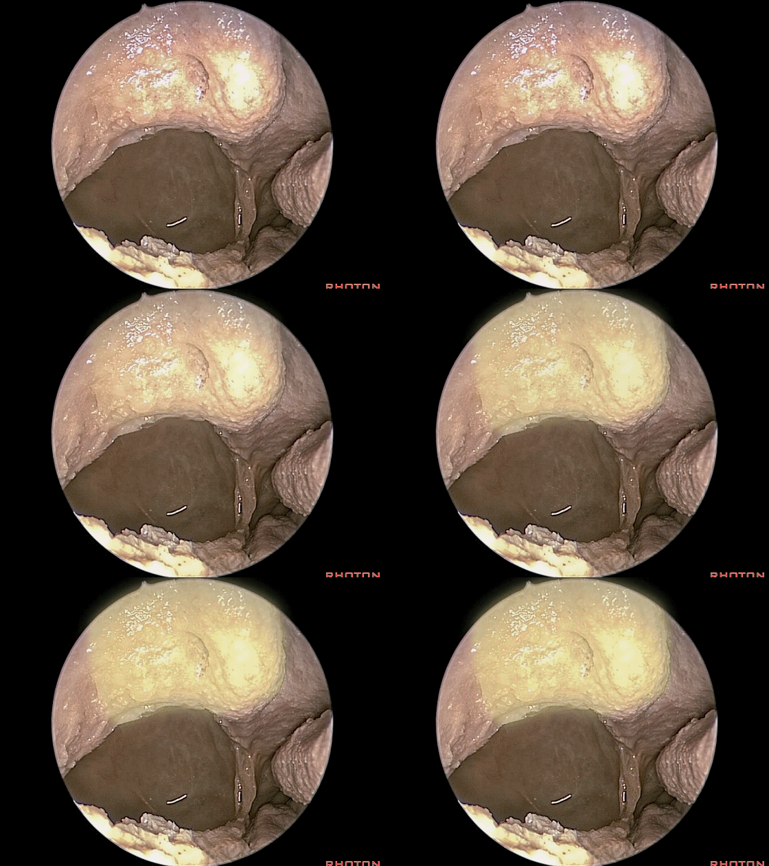
▼接下来,我们打开右侧的筛泡(下图)
and work your way back. So, here we made an opening in the ethmoid air cells on the right side,
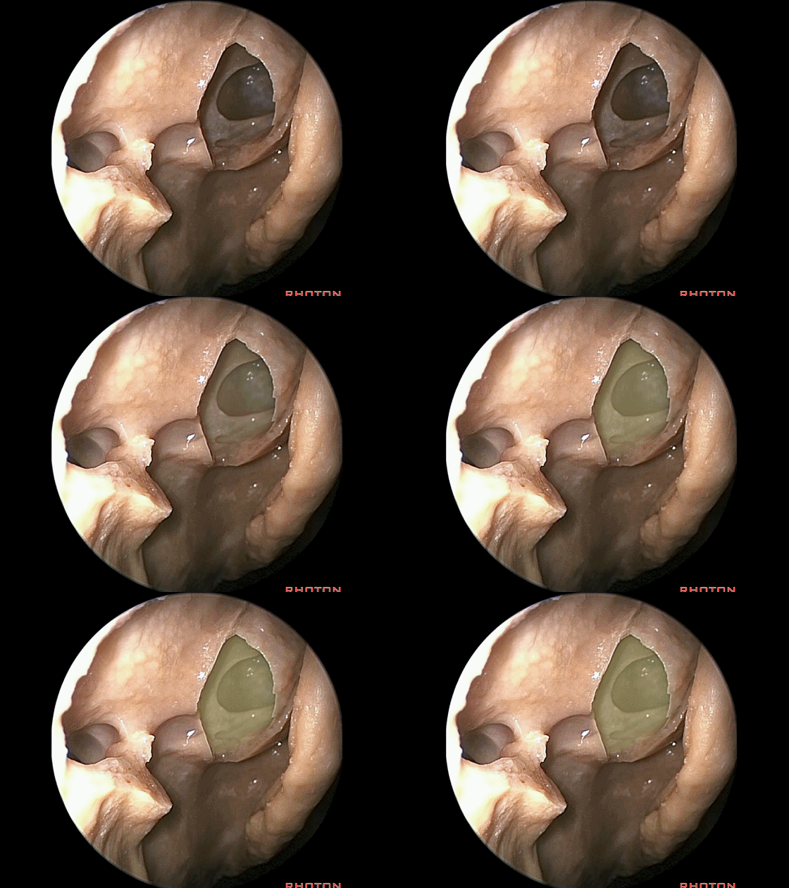
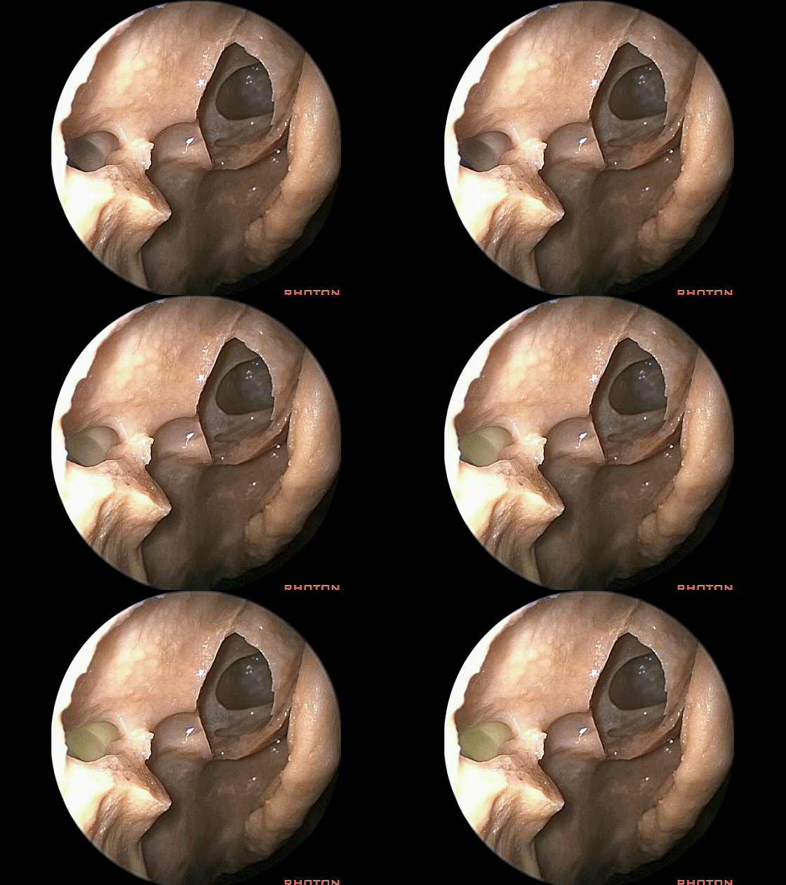
▼这是上颌窦开口
the opening into the maxillary sinus,
点击继续阅读
前颅底(2)与鼻腔解剖---Rhoton解剖视频学习笔记系列(下)


