Developmental Venous Anomaly in the Frontal Lobe
基本信息 Basic Information
男,48岁
48-year-old male
症状:头痛
Symptoms: headache
影像资料
Imaging Data
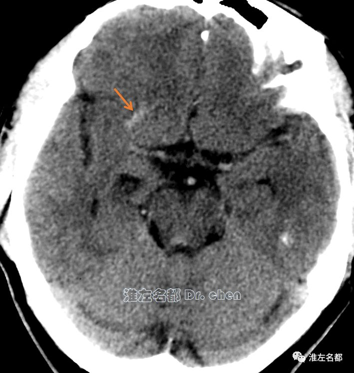
△头颅CT平扫:右侧额叶条状高密度影(橙箭)
△Cranial CT showed a strip-like hyperdensity in the right frontal lobe(orange arrow)
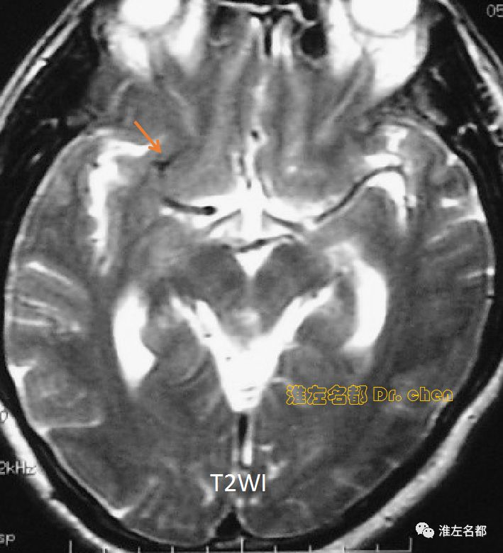
△T2WI:右侧额叶条状低信号(橙箭)
△T2WI showed a strip-like hyposignal in the right frontal lobe (orange arrow)
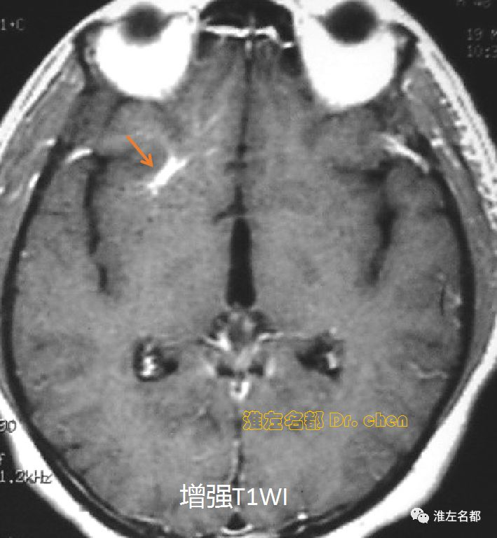
△增强T1WI:右侧额叶条状增强信号(橙箭)
△Enhanced T1WI showed a strip-like enhanced hypersignal in the right frontal lobe (orange arrow)
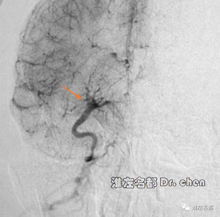
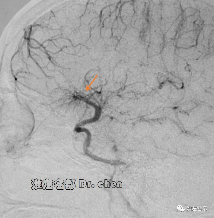
△DSA:右侧额叶异常引流静脉,呈水母头样,提示静脉发育异常
△DSA showed an abnormal vein with a typical medusa head sign in the right frontal lobe, which prompted a diagnosis of the developmental venous anomaly.
静脉发育异常亦称为静脉畸形,是正常脑部解剖的一种变异,发生率达5%。这些畸形静脉具有引流脑组织血流的功能。静脉发育异常见于大脑半球和小脑,可合并海绵状血管畸形。静脉发育异常通常是一良性发现,极少数情况下可致癫痫或出血。
Developmental venous anomaly (DVA), also known as venous angiomas, is a variation of normal brain anatomy and are found in up to 5% of people. They are blood vessels that provide a channel for blood to leave the brain. DVAs are seen in the cerebral hemispheres, cerebellum and can also be associated with cavernous malformations. They are generally benign findings and only rarely spansent with seizures or hemorrhage.


