2021年04月26日发布 | 826阅读

[Long term follow up/SAC]Pcom Aneurysm with a daughter sac

张晓龙

复旦大学附属华山医院

达人收藏

Our case


History

• 62 y/o male
• Sudden onset of right limb weakness on April 9th, followed by right ptosis six days later.
• Muscle strength returned to normal by admission, though cognitive dysfunction remained.
• Medical history: HTN for 11 years. No diabetes. Patient suffered from right central facial paralysis three times (in 2009, 2011 and 2013). Smoking for over 45 years. Stomach bleeding twice.
• 62岁男性
• 患者4月9日突发右侧肢体无力,6天后出现右侧上睑下垂。
• 入院时肌力已恢复正常,但仍有认知功能障碍。

• 既往史:高血压病史11年;否认糖尿病史;吸烟史45年余。患者既往右侧中枢性面瘫三次(2009年、2011年、2013年),胃出血两次。


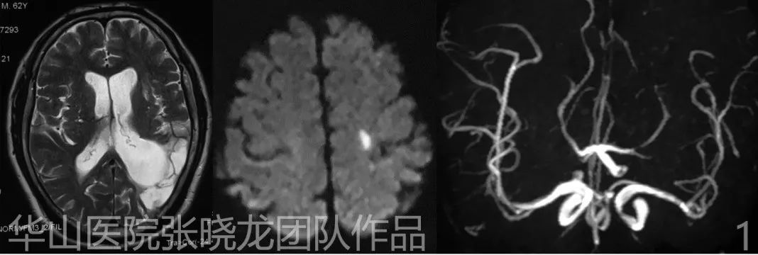
Figure 1. Acute infarction on left spancentral gyrus. Chronic infarction on left temporal lobe. Right PcomA AN. 左侧中央前回急性梗塞灶;左侧颞叶慢性梗塞灶;右侧后交通动脉瘤。


Video 1. Severe stenosis on the  right pericallosal artery and occlusion of the callosomarginal artery. Wide-necked posterior communicating artery aneurysm with an elongated daughter sac. 右侧胼周动脉重度狭窄、胼缘动脉闭塞。宽颈后交通动脉瘤伴狭长的子瘤。


Video 2. Contrast media stagnated in the daughter sac. To reduce comspanssion on the oculomotor nerve, coils were not densely packed in the daughter sac. Therefore a coil was chosen (3mm in diameter) according to the neck width of the main sac. 子瘤内造影剂瘀滞。子囊中弹簧圈未致密填塞,从而减少了对动眼神经的压迫。因此,我们根据动脉瘤主囊的瘤颈宽度选择了一枚直径3mm的弹簧圈。

1

Strategy

The reasons for dual microcatheter technique:
1. The pseudo-aneurysm is irregular and therefore easy to rupture. 
2. The main sac is small and wide-necked.
3. LVIS stents currently unavailable.
4. If the AN ruptures during coiling, the other microcatheter can be used to pack the sac rapidly.

策略:
采用双微导管技术栓塞的原因:
1. 假性动脉瘤形态不规则,易破裂。
2. 动脉瘤主囊小且瘤颈宽。
3. 当时无法获取LVIS支架。
4. 若在栓塞过程中动脉瘤破裂,另一根微导管可用于快速栓塞瘤腔。

2

Operation


Video 3. Dual microcatheter plus stent assisted technique was chosen to densely pack the main sac while only loosely packing the daughter sac. Therefore the microcatheter should not be inserted deeply into the daughter sac. The initial coil (MicroPlex 3mm*8cm) entered the daughter sac spansenting a rupture risk. 采用双微导管加支架辅助技术致密栓塞动脉瘤主囊,而子囊仅疏松栓塞。因此,微导管不应深入子瘤内。首枚填入子囊内的MicroPlex 3mm*8cm弹簧圈存在破裂风险。


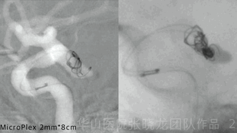
Figure 2 GIF. Insert the second coil (Microplex 2mm*8cm). The microcatheter was readjusted to retrieve the coil and placed it in the main sac, although the microcatheter was quite unstable. 填入第二枚Microplex 2mm*8cm弹簧圈。微导管较为不稳定,重新调整微导管以回收弹簧圈并重新填入动脉瘤主囊内。


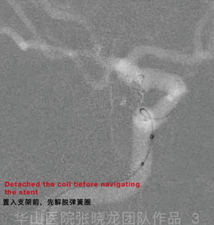
Figure 3 GIF. For a better vision of the neck of aneurysm and deployment of the stent,we changed working projection. Then one Neuroform 4mm*20cm was advanced across the protruded loop. 我们更换支架释放工作角度。然后可见Neuroform 4mm*20cm支架通过之前脱出的襻环


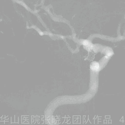
Figure 4 GIF. As coils protruded into the parent artery, one microcatheter was retrieved to free space in the sac. 当弹簧圈突入载瘤动脉时,回撤一根微导管以空出瘤腔内的空间。


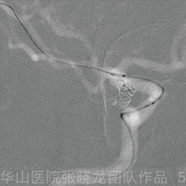
Figure 5 GIF. Neuroform is easy to rebound. So the stent was deployed from the origin of MCA. General heparinization. Neuroform支架容易弹回,所以从大脑中动脉起始部开始释放支架。行全身肝素化。


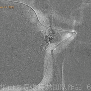
Figure 6 GIF. MicroPlex 2mm*6cm. 


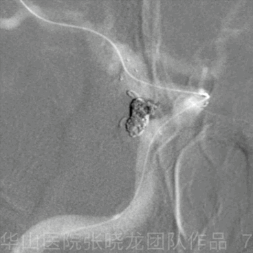
Figure 7 GIF. Microplex 2mm*3cm,four coils in all. Microplex 2mm*3cm,共四枚弹簧圈。


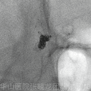
Figure 8 GIF. The way of retrieving the microcatheter after coiling. 弹簧圈栓塞后微导管的回收方法。


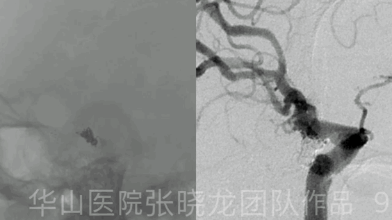
Figure 9 GIF. Post-operative angiography shows densely packing of the aneurysm and the contrast media stagnation in the daughter sac. 术后血管造影显示动脉瘤致密栓塞,子瘤内有造影剂滞留。


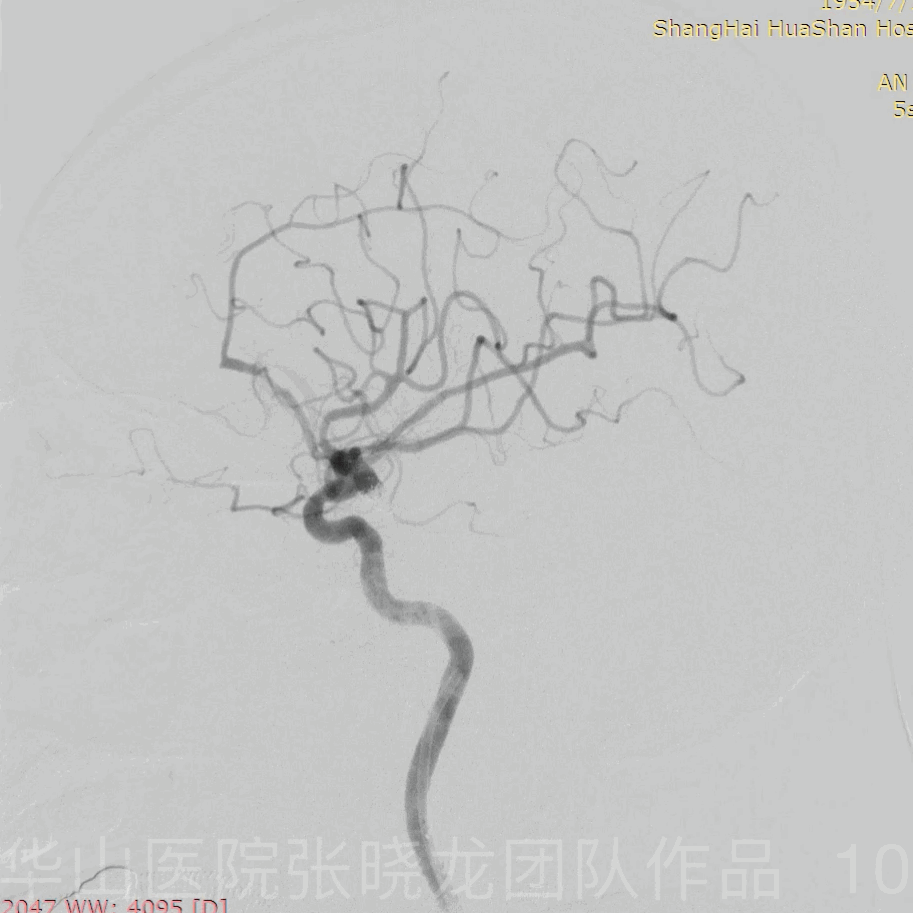
Figure 10 GIF. Intracranial vessels are intact and the parent artery is patent. Tirofiban (Xinweining) 20ml was given via the guiding catheter. 颅内血管显示完整,载瘤动脉通畅。经导引导管给予替罗非班(欣维宁)20ml。


3

Post operation


• No more neurologic deficit.

• PE: GCS 15, alert, right ptosis.
• Medication: Aspirin 100mg qd, Clopidogrel 75mg qd, Atorvastatin 20mg qN. Nifedipine 30mg qd, Irbesartan 150mg qd.

• 无新发现神经功能缺损症状。
• 体格检查:GCS 评分:15,神清,右侧上睑下垂。
• 用药:阿司匹林100mg qd,氯吡格雷75mg qd,阿托伐他汀20mg qn,硝苯地平30mg qd,厄贝沙坦150mg qd。

7 month/4 year follow up
• Still suffered from left side diplopia. The right ptosis significantly improved.
• PE: occasional right ptosis, left side diplopia
• Medication: Aspirin 100mg qd, Atorvastatin 20mg qn, Nifedipine 30mg qd, Irbesartan 150mg qd

术后7个月/4年随访
• 仍有左视复视,右侧上睑下垂显著改善。
• 体格检查:偶有右侧上睑下垂,左侧复视。
• 药物治疗:阿司匹林100mg qd,阿托伐他汀20mg qn,硝苯地平30mg qd,厄贝沙坦150mg qd。


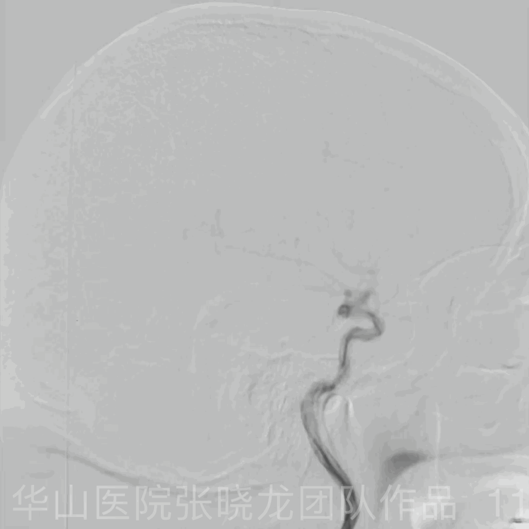
Figure 11 GIF. 4 year follow up angiography shows no relapse of the aneurysm. 4年随访血管造影未见动脉瘤复发。


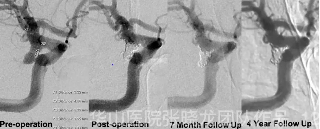
Figure 12. The aneurysm was densely packed without relapse in 4 year follow up angiography. The shape of coils kept stable. 4年随访,血管造影显示:动脉瘤致密栓塞,未见复发。弹簧圈形态保持稳定。


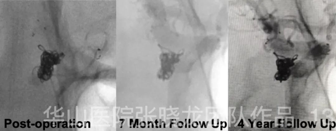
Figure 13. Comparison of coils and stent between post-operation, 7 month follow up and 4 year follow up. 术后即刻、7个月随访和4年随访,弹簧圈和支架的比较。


4

Summary

1. Dual microcatheter plus stent assisted technique was chosen to densely pack the main sac while only loosely packing the daughter sac to reduce the comspanssion by the daughter sac.
2. As the daughter sac is easy to rupture, general heparinization was not performed until the main sac was almost densely packed.
3. Long coil technique and rivet coiling of the aneurysm neck can spanvent recurrence.
4. Next follow up: in 5-8 years.

1. 采用双微导管加支架辅助技术致密栓塞动脉瘤主囊,并疏送栓塞子囊以减少对其的压迫。
2. 由于子瘤易破裂,待动脉瘤主囊致密栓塞后才行全身肝素化。
3. 采用长圈和铆钉技术栓塞动脉瘤颈,预防动脉瘤的复发。
4. 5-8年后进行下一次随访。

声明:脑医汇旗下神外资讯、神介资讯、脑医咨询所发表内容之知识产权为脑医汇及主办方、原作者等相关权利人所有。未经许可,禁止进行转载、摘编、复制、裁切、录制等。经许可授权使用,亦须注明来源。欢迎转发、分享。

最新评论
发表你的评论
发表你的评论