MRI Findings of a Severe Subarachnoid Hemorrhage during the Ultra-early Stage
基本信息 Basic Information
男,54岁
54-year-old male
全麻下行腹主动脉和下肢动脉闭塞血管内介入治疗,术后患者不能苏醒。
After the endovascular treatment for occlusion of abdominal aorta and lower extremity arteries under general anesthesia, this patient could not wake up from the anesthesia.
影像资料
Imaging Data
于手术室的MRI单元紧急行脑MRI评估
Urgent brain MRI in the intraoperative MRI Suite
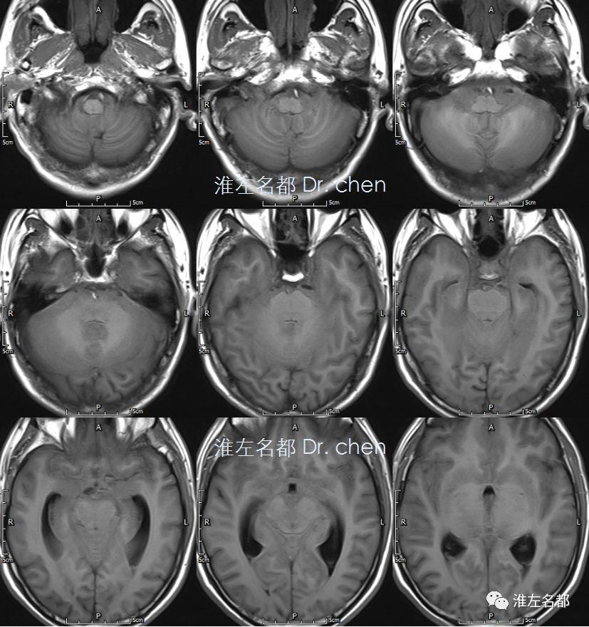
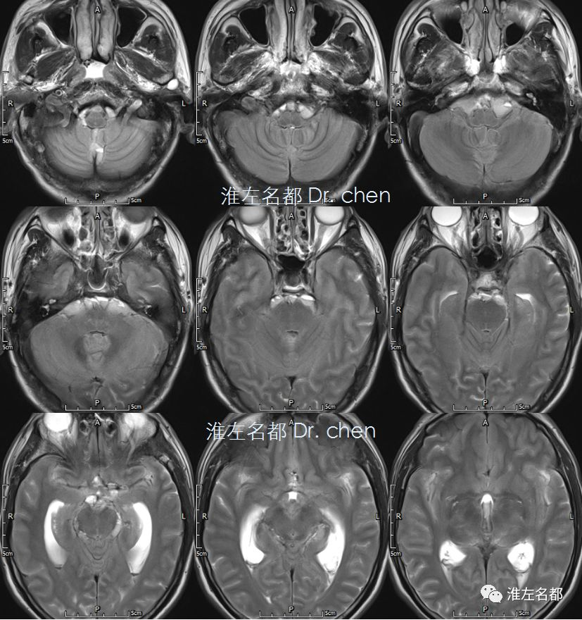
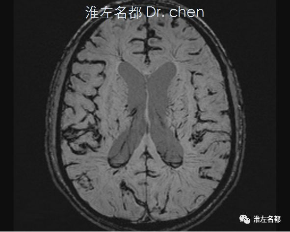
△T1WI和T2WI:幕上脑积水,脑沟、脑池蛛网膜下腔呈等T1和等-高T2信号
△T1WI and T2WI showed a supratentorial hydrocephalus, and that the subarachnoid space of cerebral sulci and cistern spansented with an iso-intense sigal on T1WI and an iso-intense or slightly hyper-intense signal on T2WI
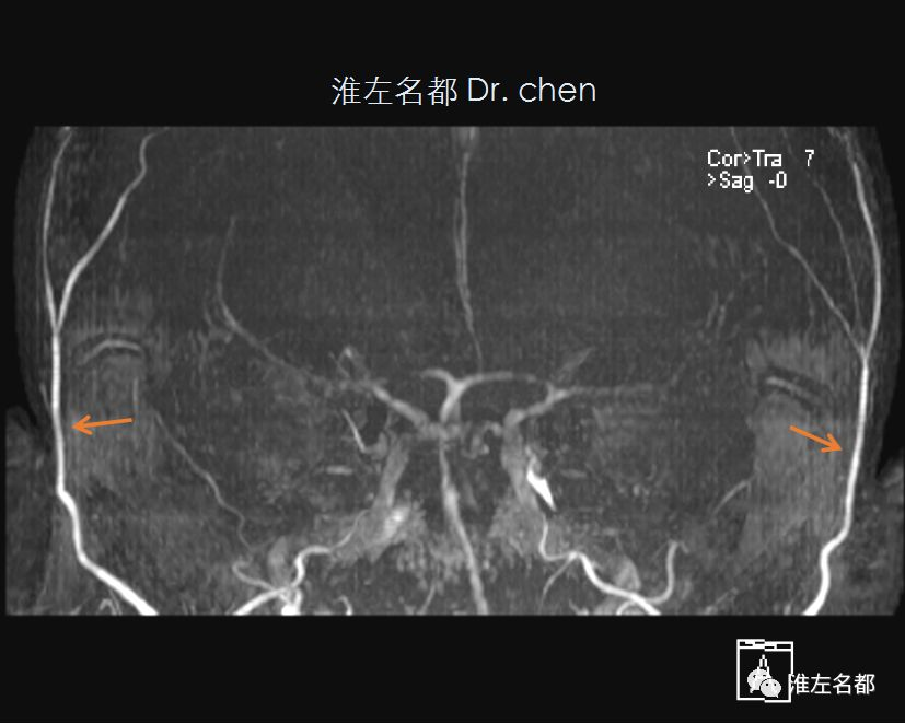
△头颅3D-TOF-MRA:颅内动脉显影极差(提示血流速度缓慢),双侧颞浅动脉显影正常(橙箭)
△Cranial 3D-TOF-MRA: the blood signal of all intracranial arteries was very poor (the blood flow rate was very slow) ; but the blood signal of bilateral superficial temporal arteries was normal (orange arrow)
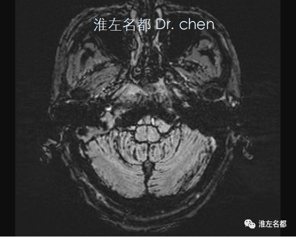
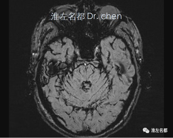
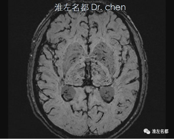
△SWI:广泛的脑沟和脑表面显著低信号(提示出血)
△SWI: significant low signal in the brain sulci and surface of bilateral cerebellar and hemispheres prompting subarachnoid hemorrhage
脑CT评估
Brain CT assessment
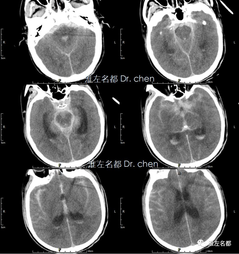
△头颅CT:幕上脑积水,严重蛛网膜下腔出血
△Cranial CT revealed a supratentorial hydrocephalus and a large amount of subarachnoid hemorrhage

扫码进入个人主页