
![]()
引
言
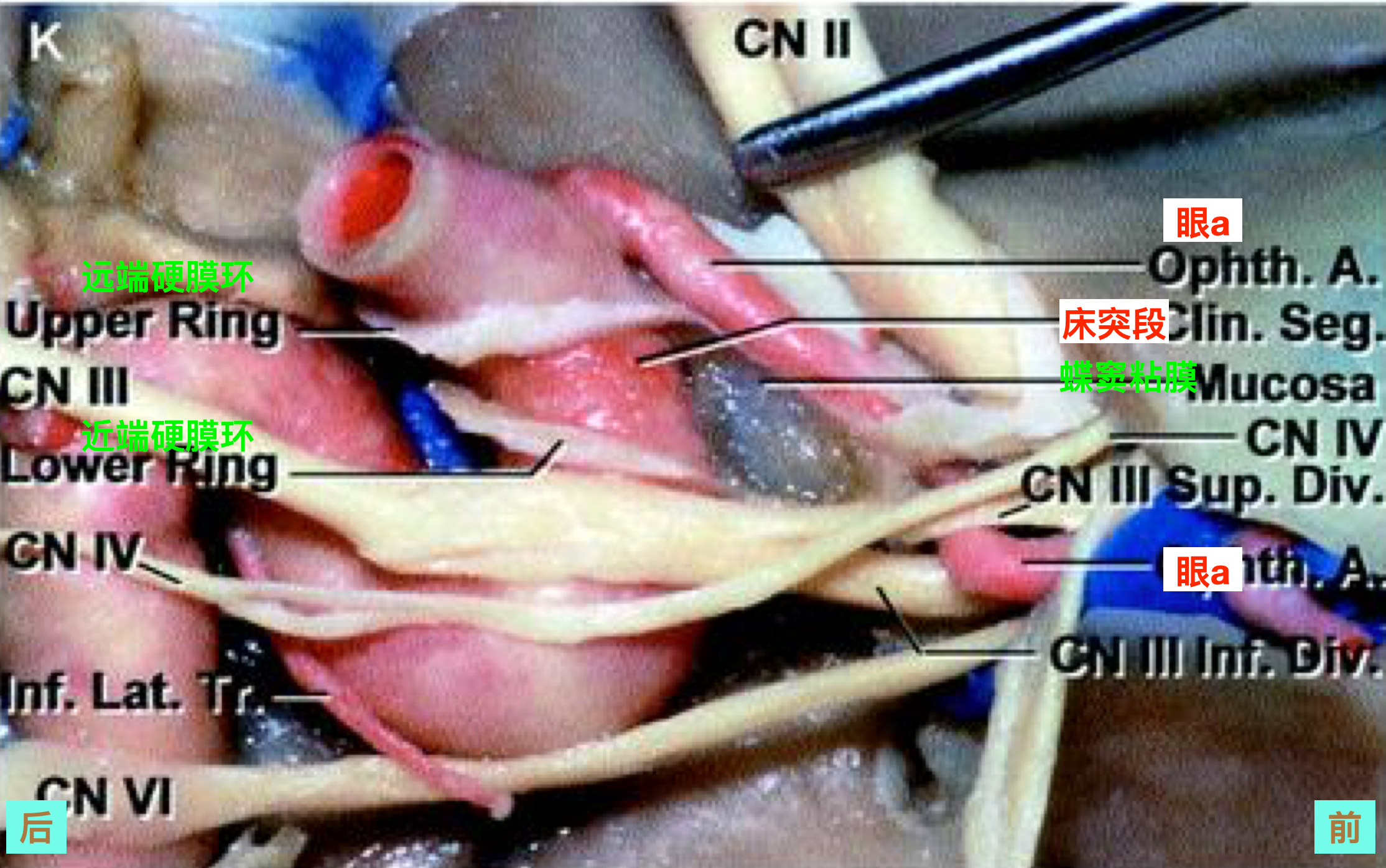
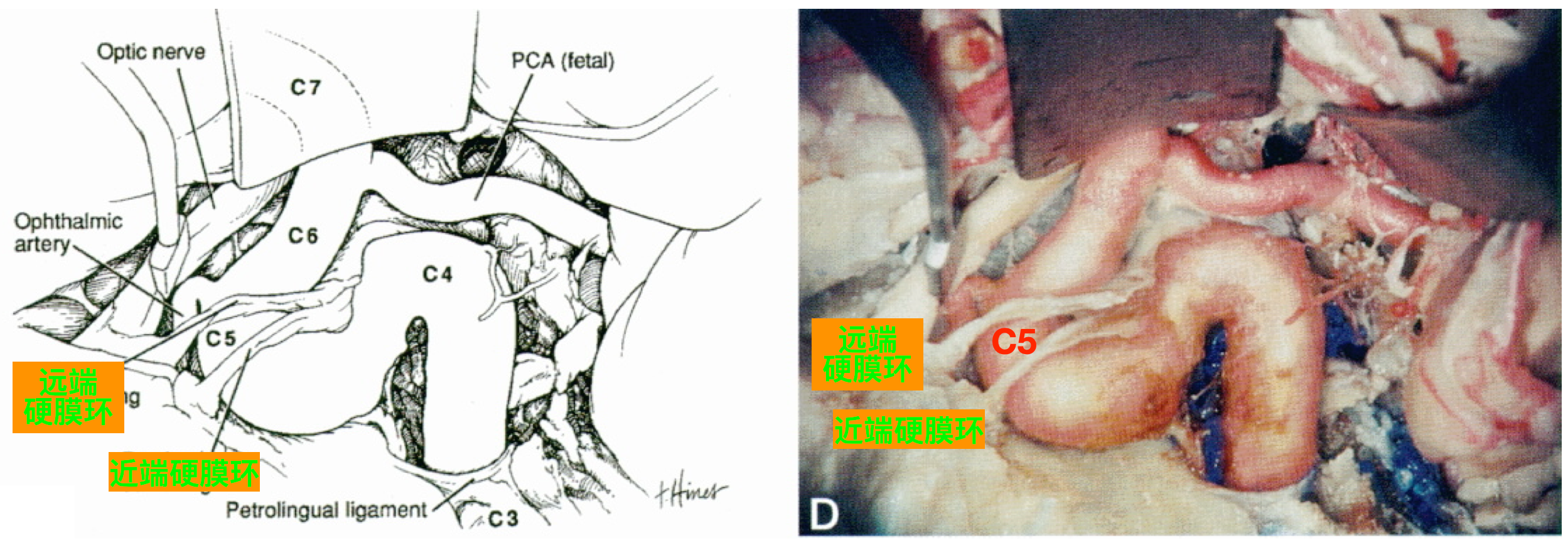
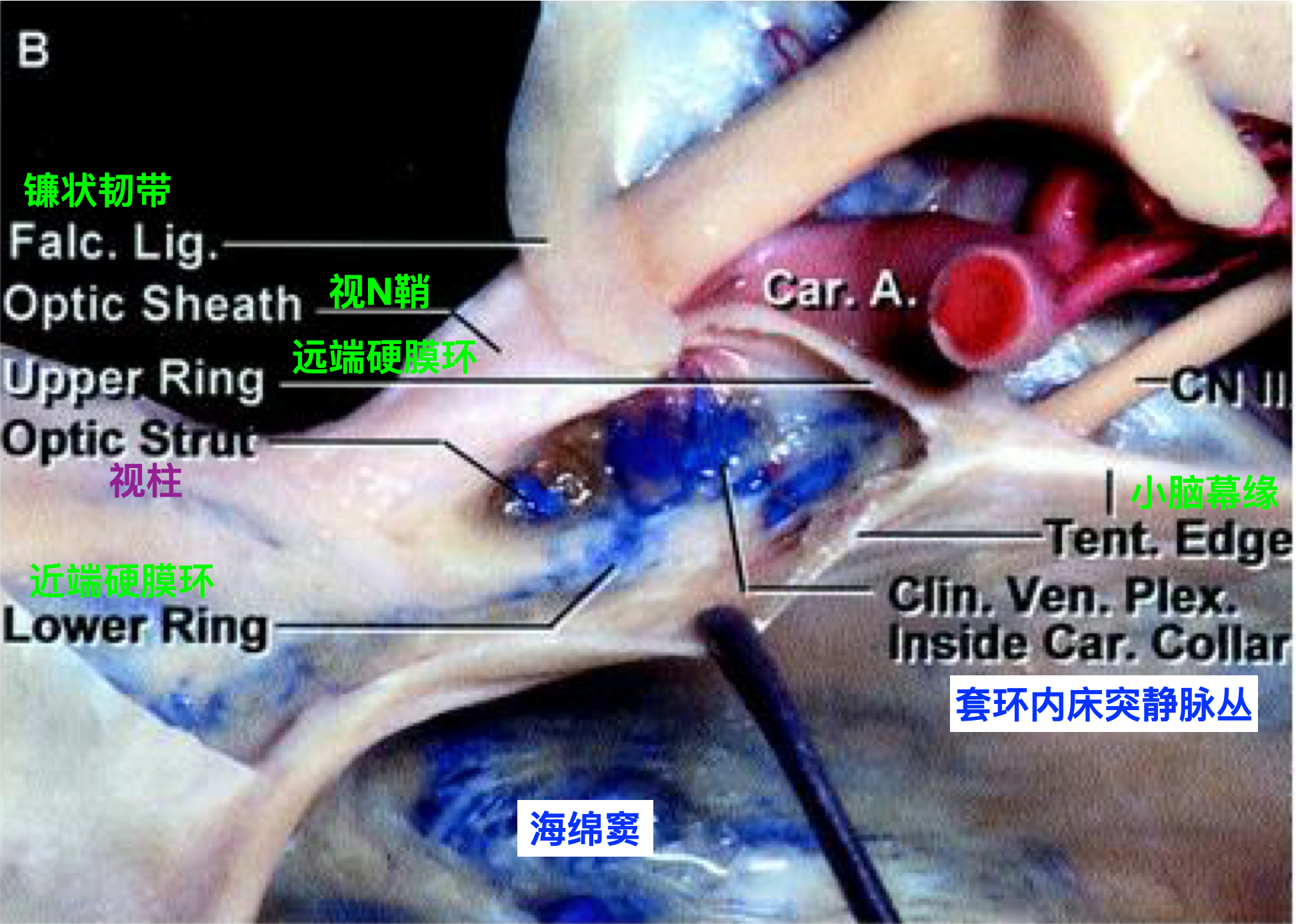
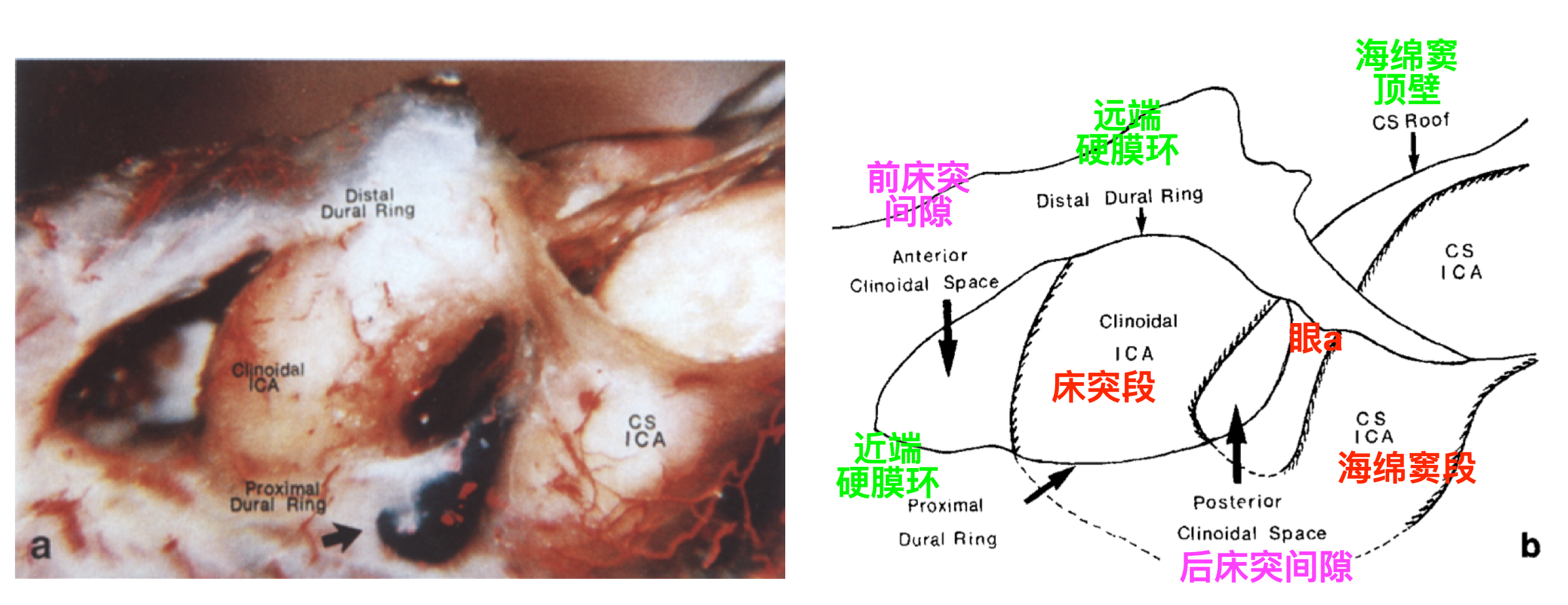
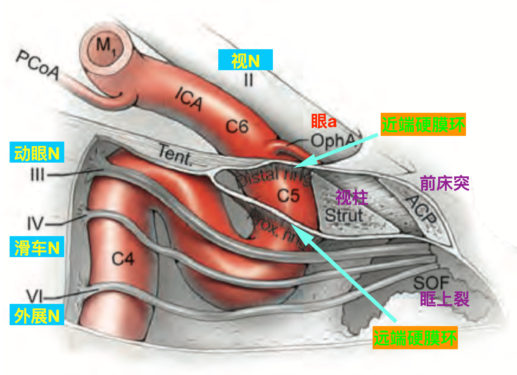
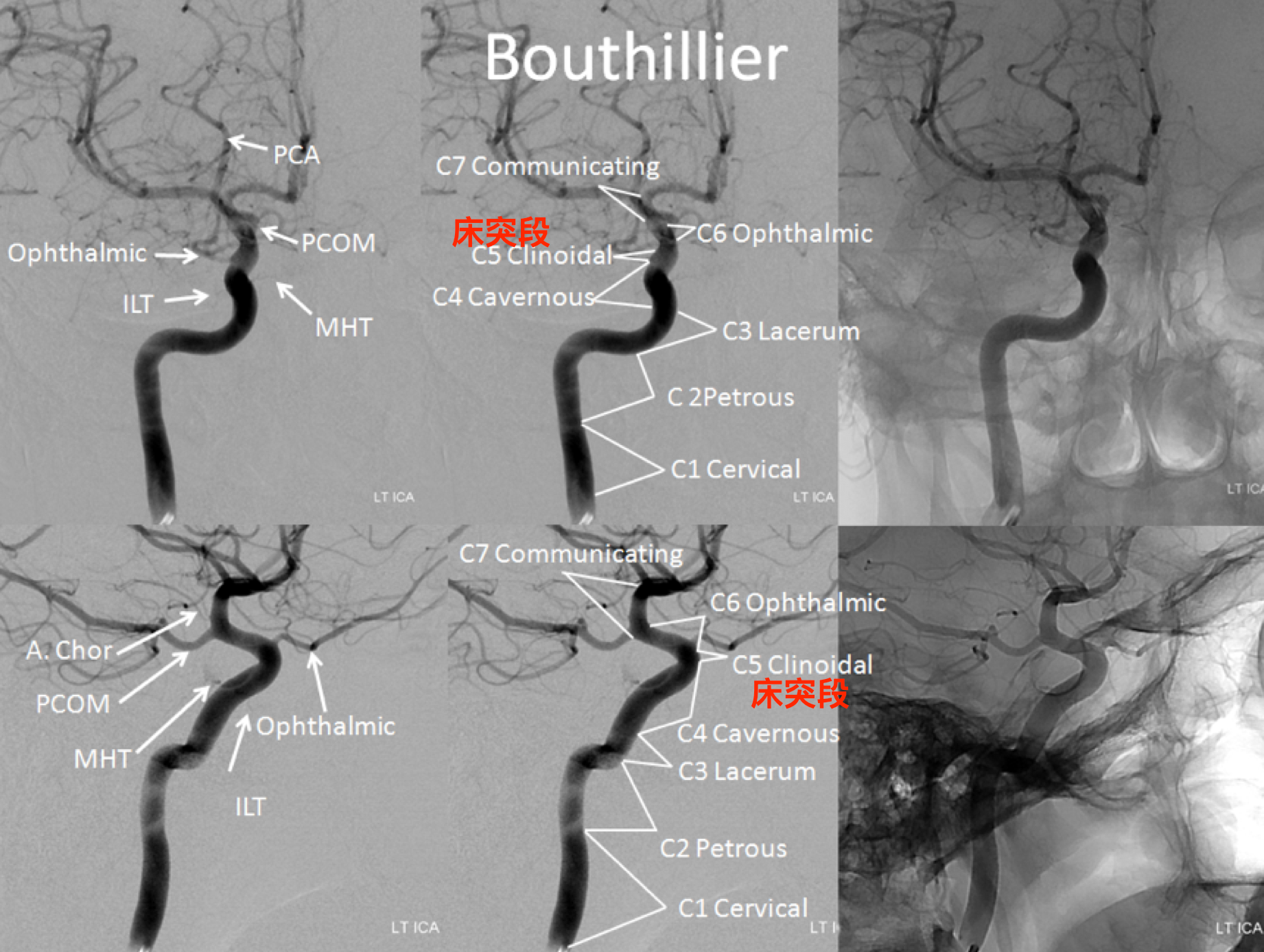
颈内动脉床突段是指位于近端硬膜环和远端硬膜环之间的一段颈内动脉主干。此段颈内动脉与前床突、近端硬膜环和远端硬膜环等关系密切。熟悉其解剖,是处理颈内动脉床突旁动脉瘤的重要基础。
一、相关骨结构
(一)蝶骨
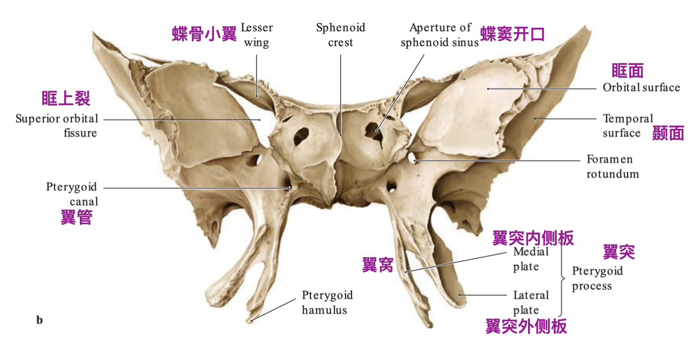
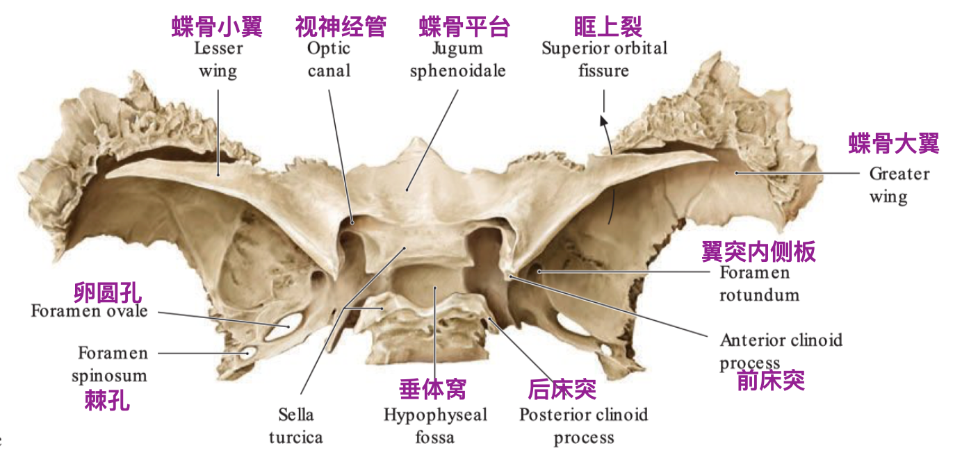
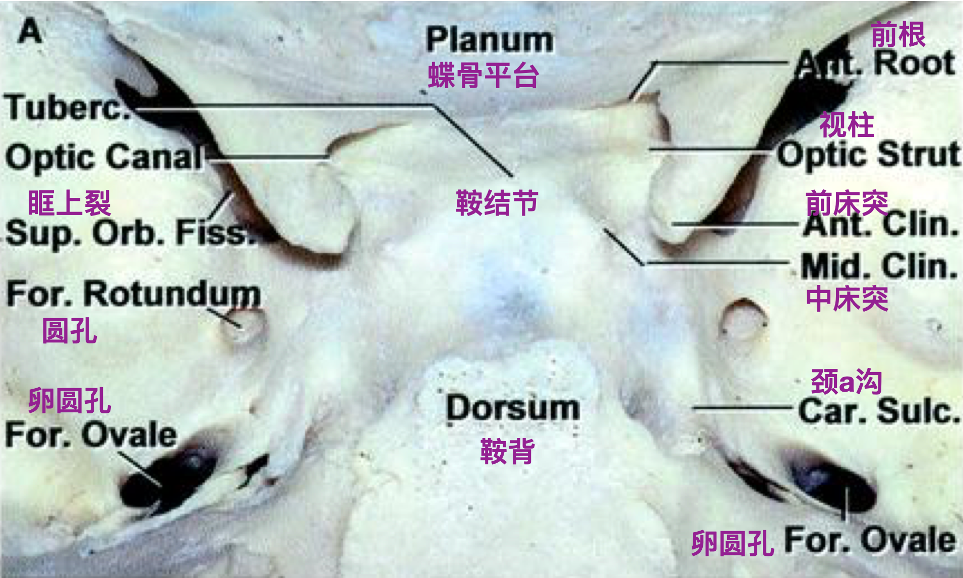
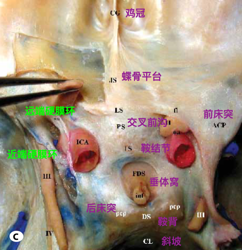
(二)颈动脉沟
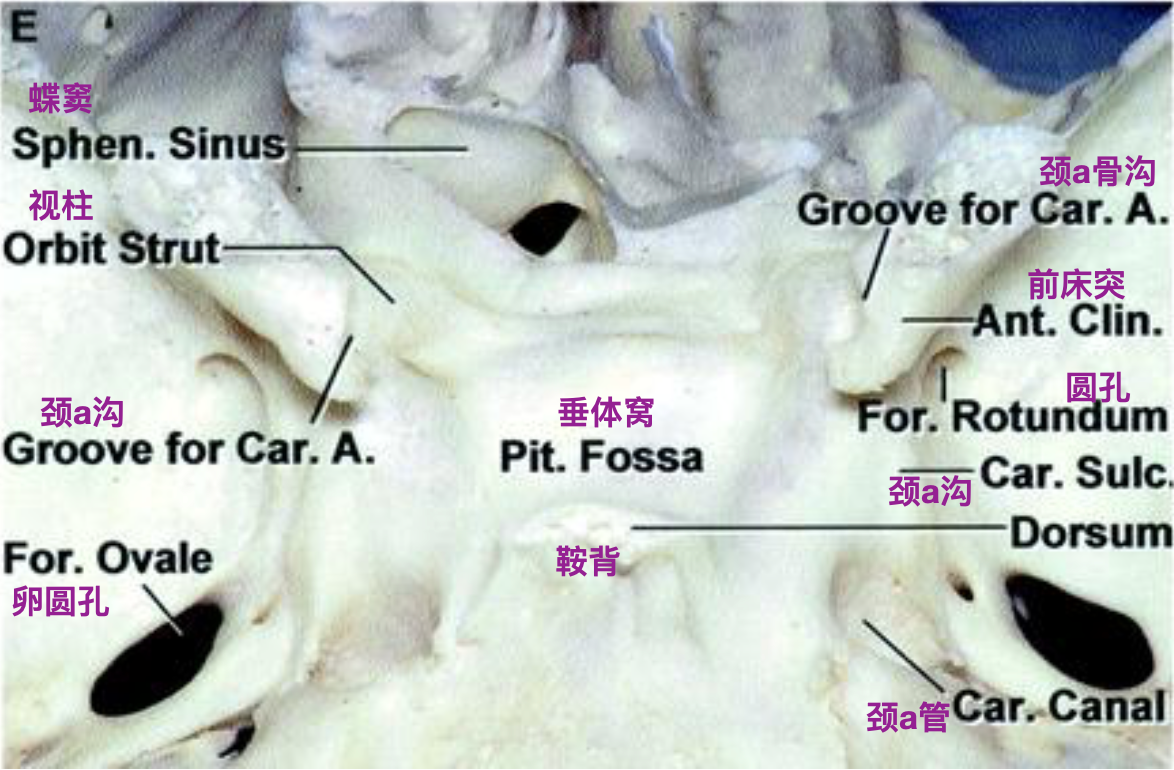
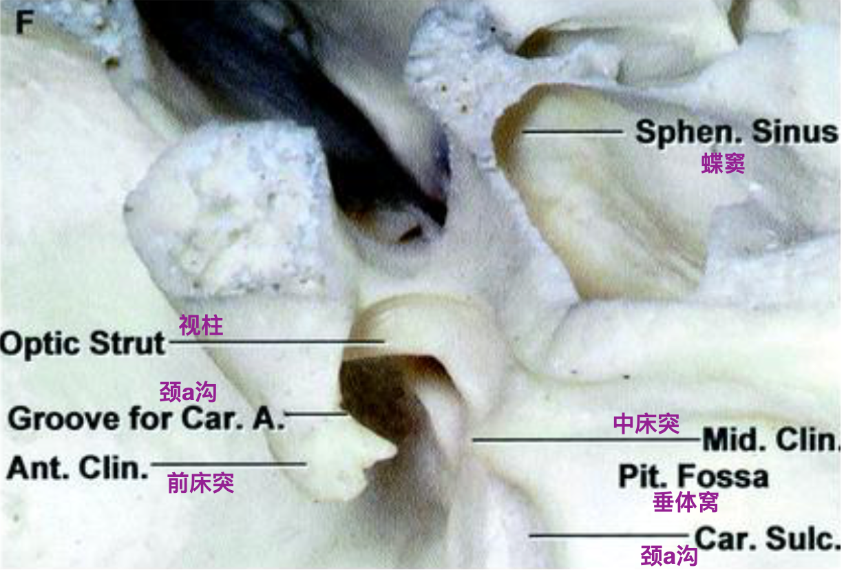
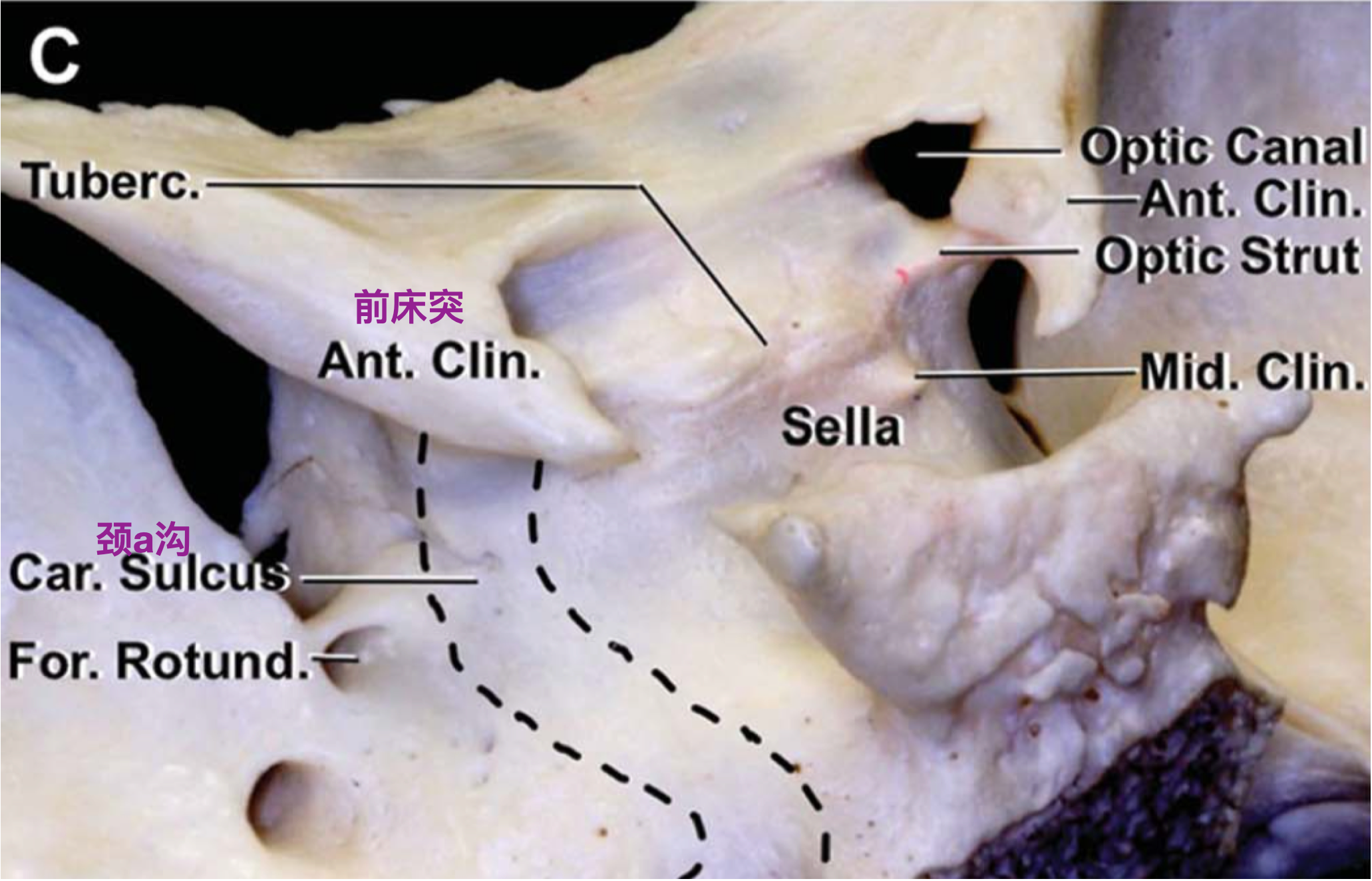
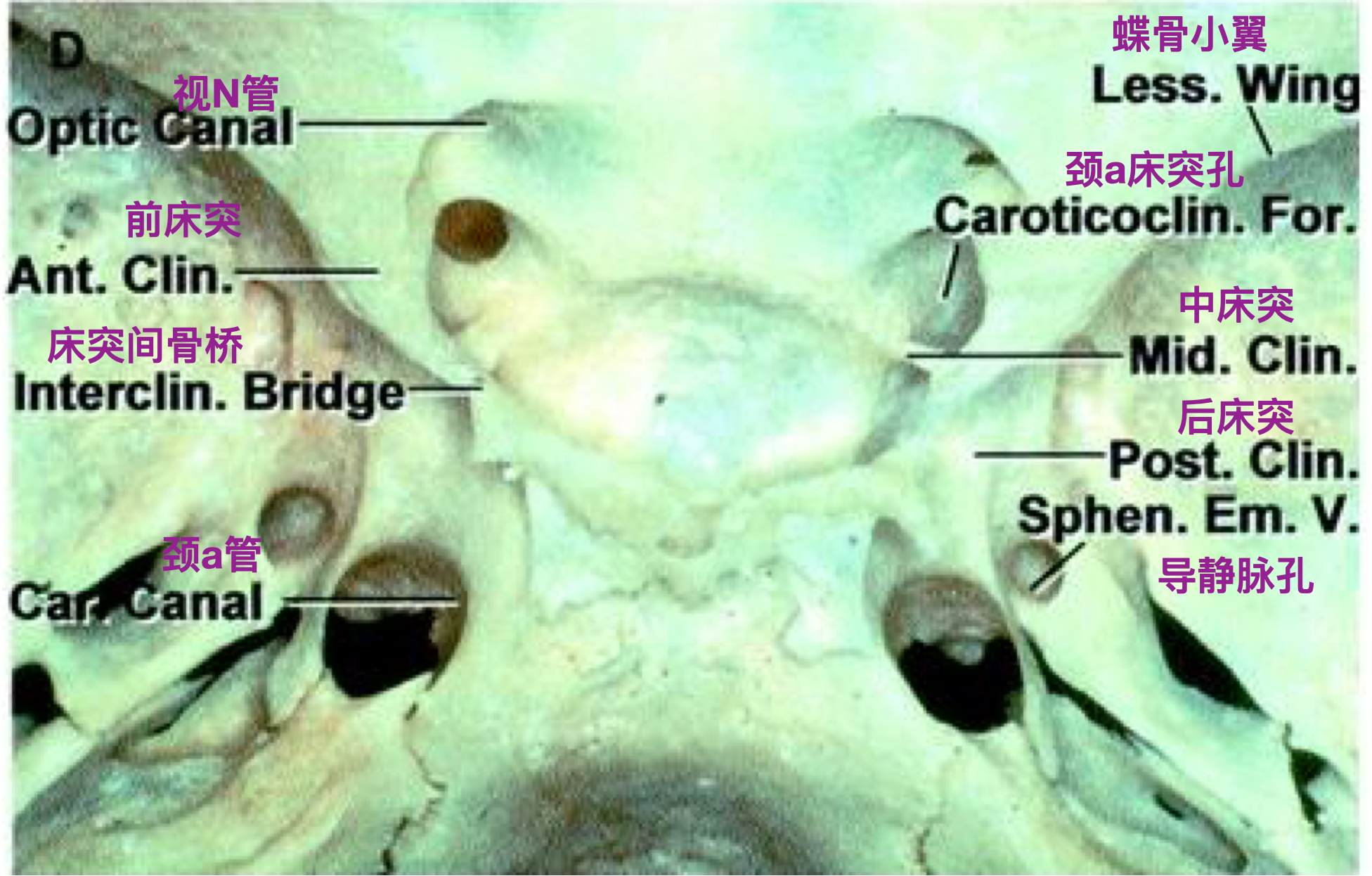
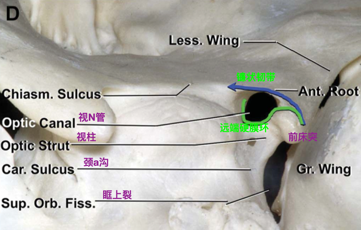
颅骨标本上面观(Rhoton,2002)。蝶骨小翼、前床突基底和视神经管顶壁已磨除。前床突内侧有沟样结构,走行颈内动脉。
(三)前床突
(四)中床突
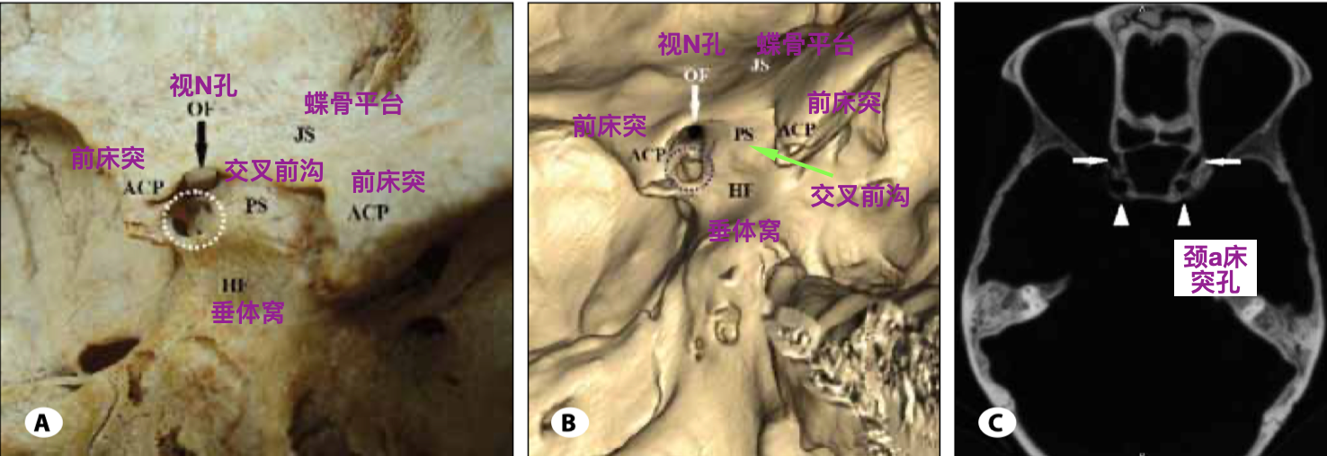

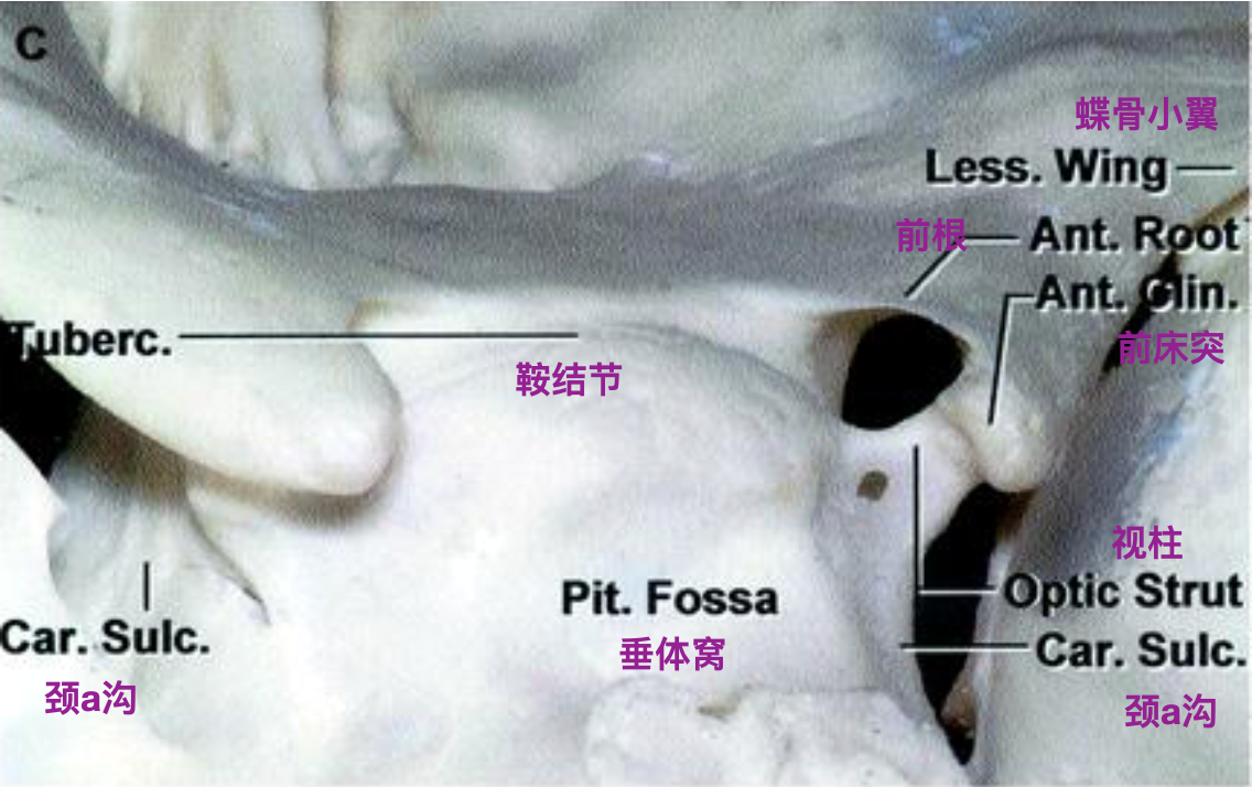
(五)视柱

二、硬脑膜结构
(一)镰状韧带
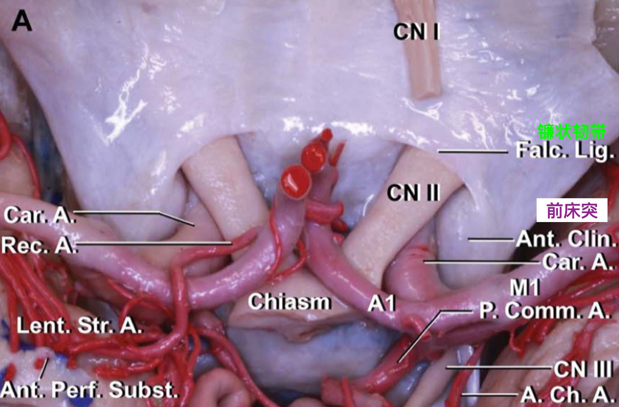
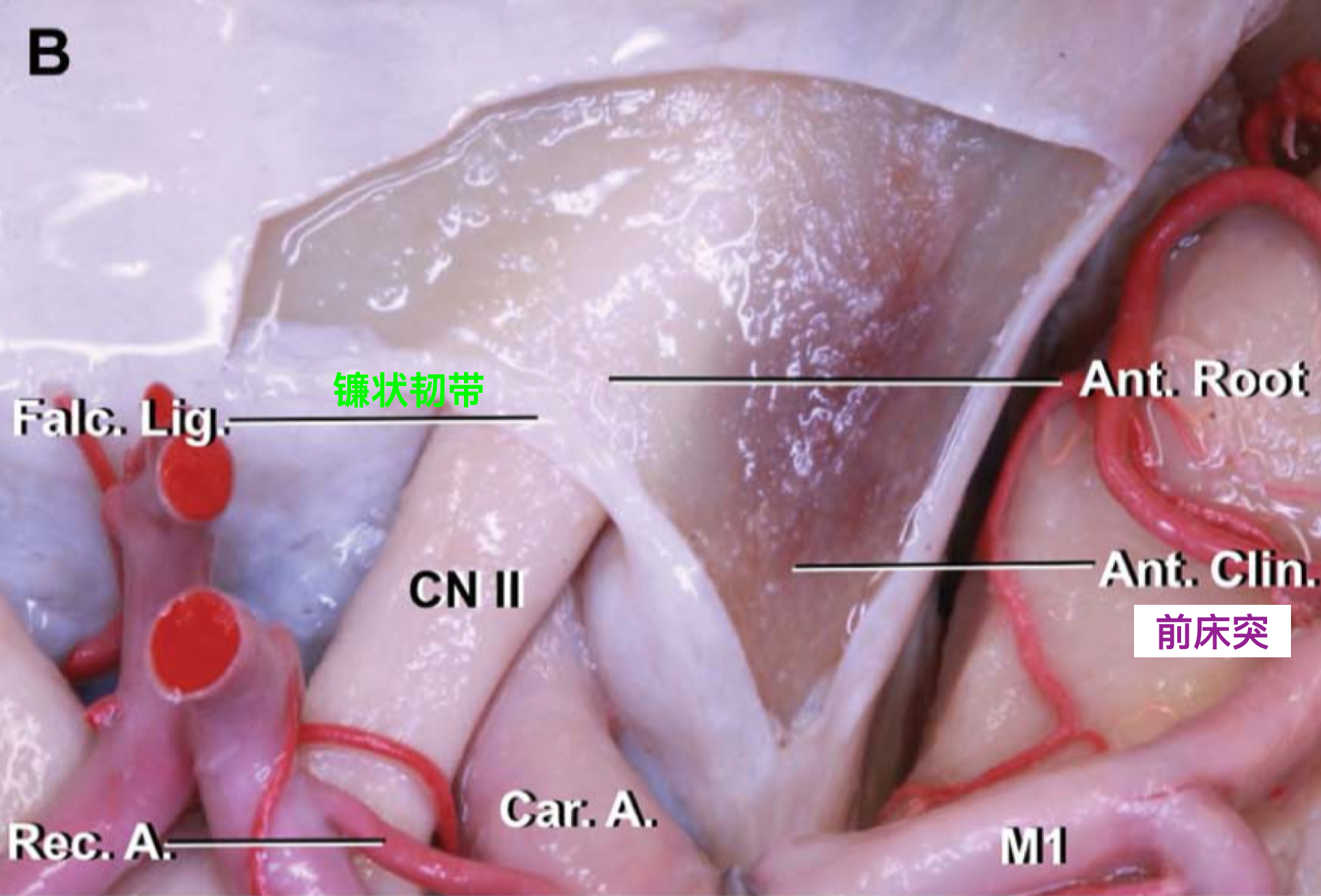
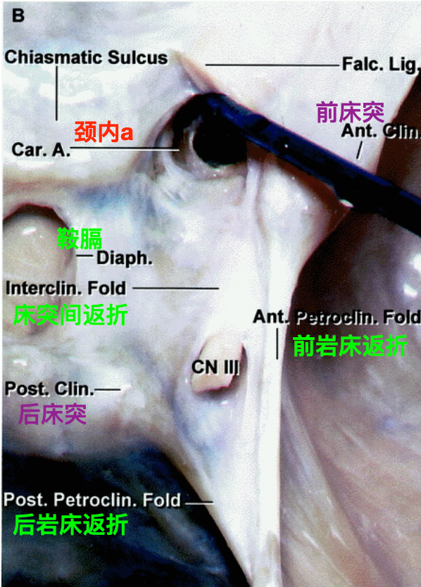
颅底上面观(Joo,2012)。覆盖前床突上表面的硬脑膜在视神经上方延伸,形成镰状韧带(蓝色线条),覆盖前床突上表面的硬脑膜至视柱上缘,形成远端硬膜环的前部(绿色线条)。

(二)远端硬膜环和近端硬膜环
三、颈内动脉床突段
四、小结
五、作者简介
六、参考文献
Bouthillier, A., van Loveren, H. R., & Keller, J. T. (1996). Segments of the internal carotid artery: a new classification. Neurosurgery, 38(3), 425-432; discussion 432-423.
Dagtekin, A., Avci, E., Uzmansel, D., Kurtoglu, Z., Kara, E., Uluc, K., Akture, E., & Baskaya, M. K. (2014). Microsurgical anatomy and variations of the anterior clinoid process. Turk Neurosurg, 24(4), 484-493.
De Jesús, O. (1997). The clinoidal space: anatomical review and surgical implications. Acta Neurochir (Wien), 139(4), 361-365.
Joo, W., Funaki, T., Yoshioka, F., & Rhoton, A. L., Jr. (2012). Microsurgical anatomy of the carotid cave. Neurosurgery, 70(2 Suppl Operative), 300-311; discussion 311-302.
Lawton, M. T. (2011). Seven Aneurysms Tenets and Techniques for Clipping. New York: Thieme.
Rhoton, A. L., Jr. (2002). The cavernous sinus, the cavernous venous plexus, and the carotid collar. Neurosurgery, 51(4 Suppl), S375-410.
Schuenke, M., & Schulte, E. (2016). Thieme Atlas of Anatomy Volume 3 Head, Neck, and Neuroanatomy (2nd ed.). New York: Thieme.


