瘤方百式 指尖沙龙 第六期 艺在南北 圆满收官后,精彩仍在继续,解析南北方专家观点,拓展文献研究证据。

本期特邀北部战区总医院神经外科脑血管病治疗中心高旭教授为您衍生解读“子囊是否需要栓塞”。
自1996年ISUIA研究组首次对伴子囊动脉瘤进行定义以来[1],已发现伴子囊动脉瘤相较囊壁光滑的囊状动脉瘤更易破裂(UCAS HR 1.63, 95% CI:1.08-2.48)[2],这可能是由于子囊处或附近存在较高的剪切应力所致[2,3]。一般认为,动脉瘤的形状取决于囊内血流引起的壁损伤,和瘤壁生物力学反应间的相互作用,还与动脉瘤周围的血管外结构相关[4]。因此,伴子囊动脉瘤也需根据患者年龄、瘤体形态、解剖结构等因素行个体化治疗,而不能将其笼统地归为一类。
病例展示
病例1
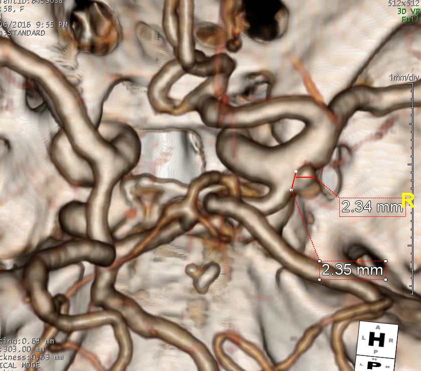
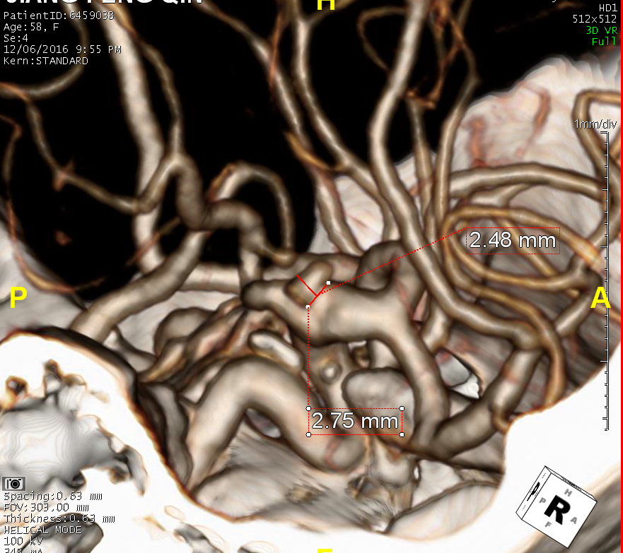
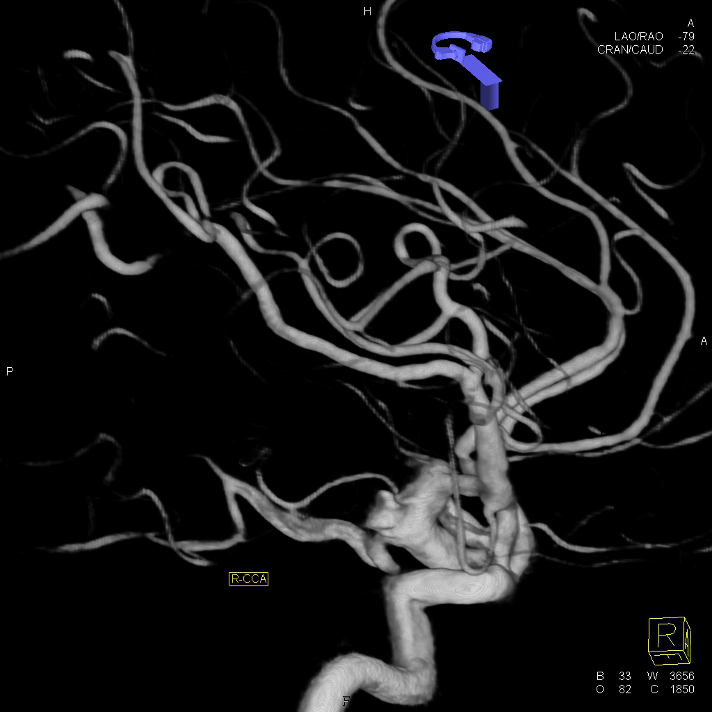
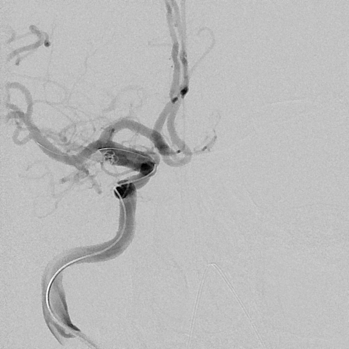
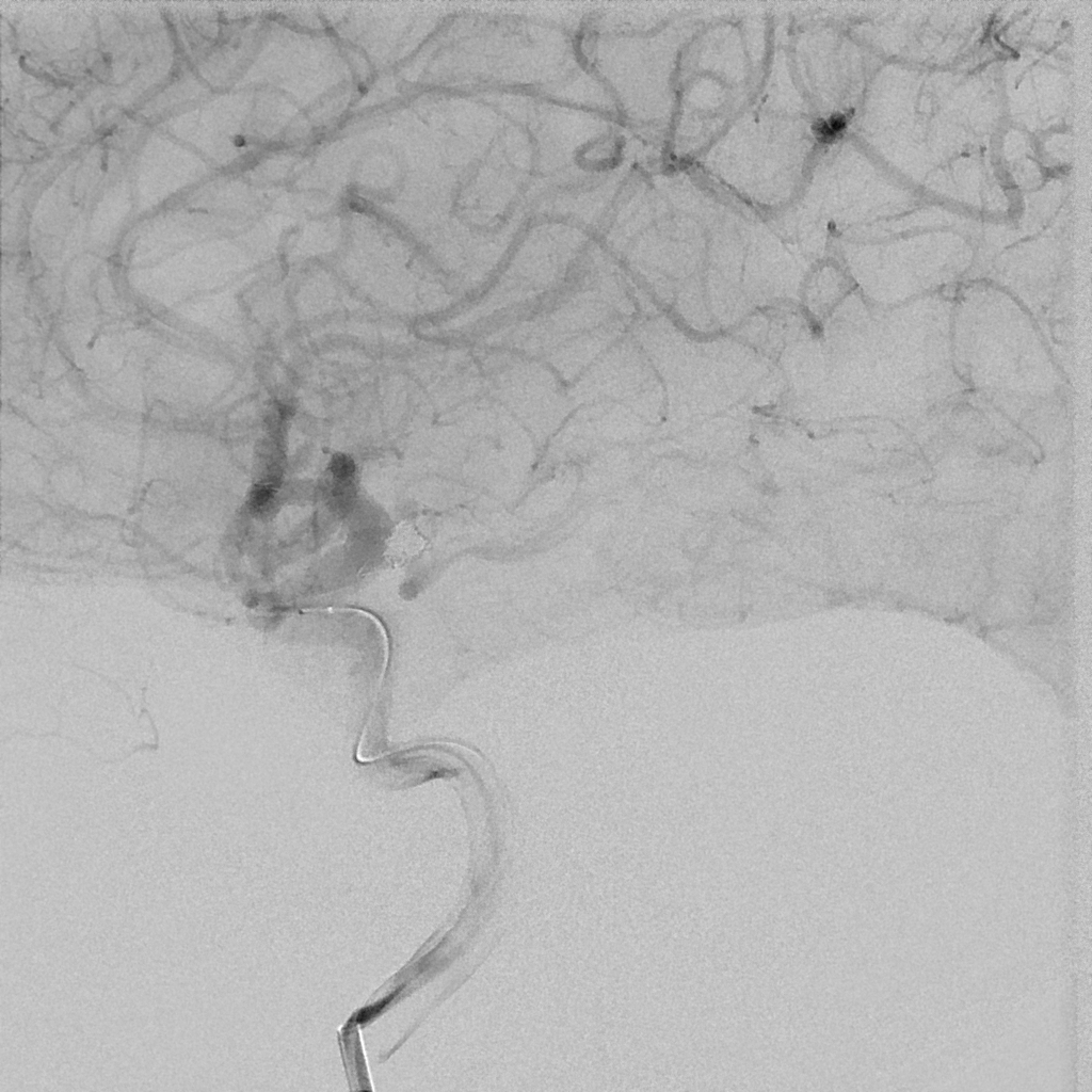
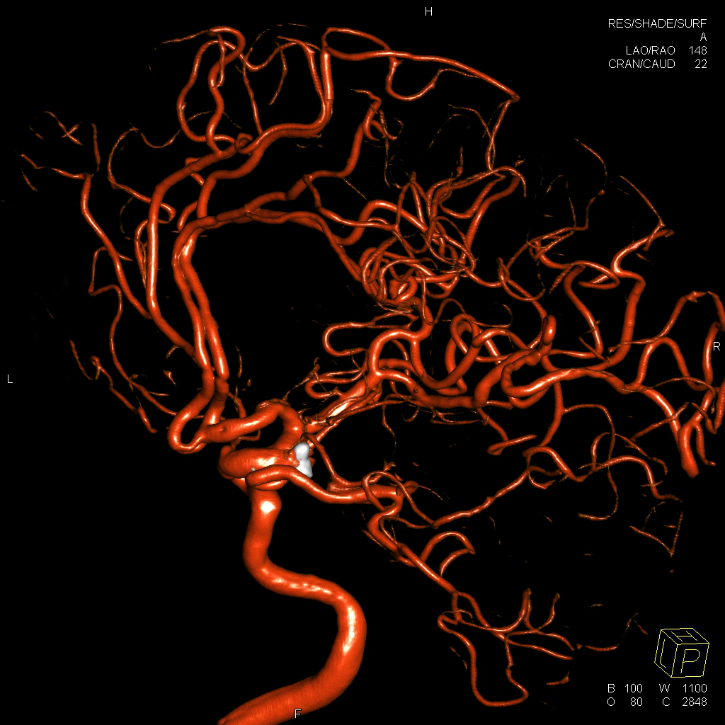
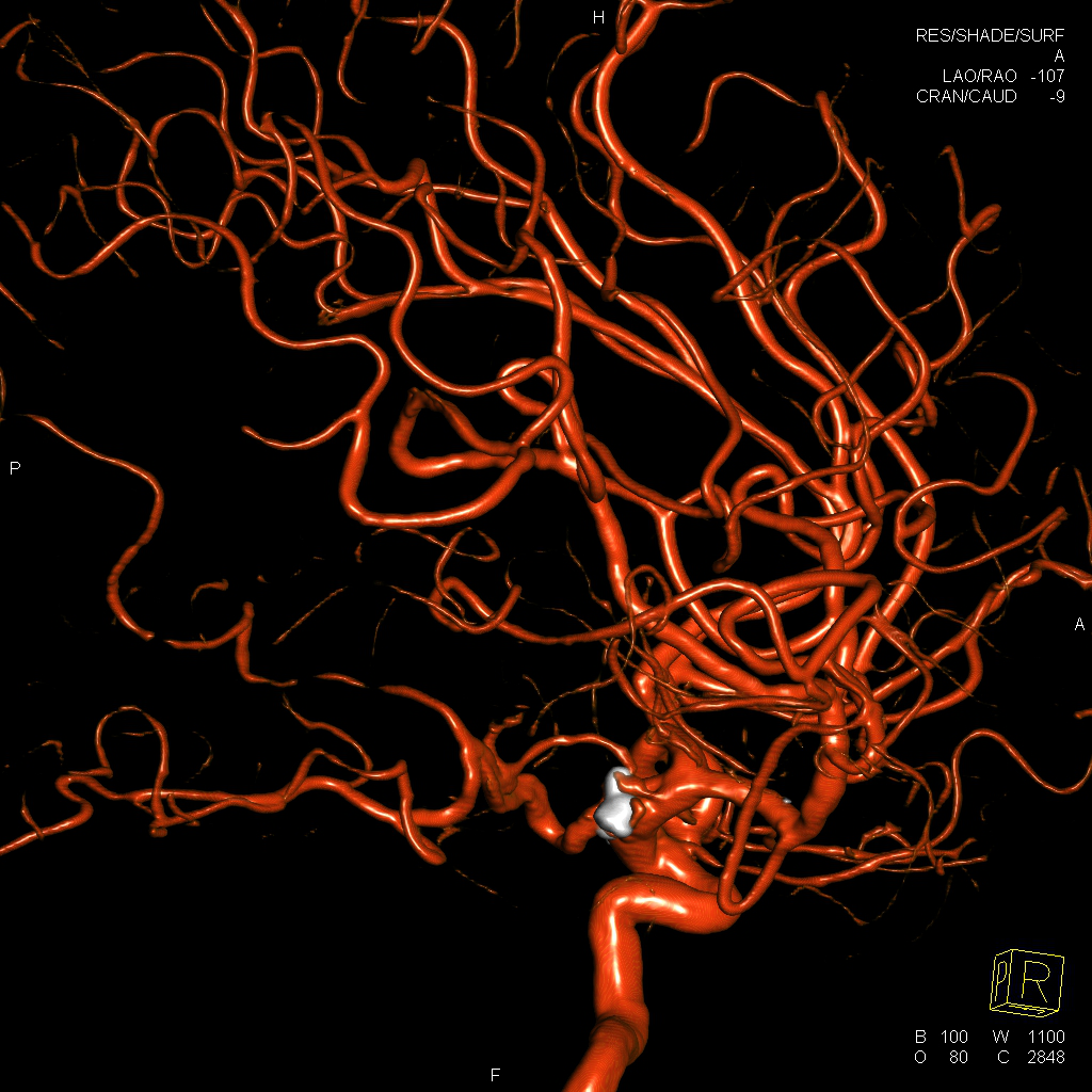
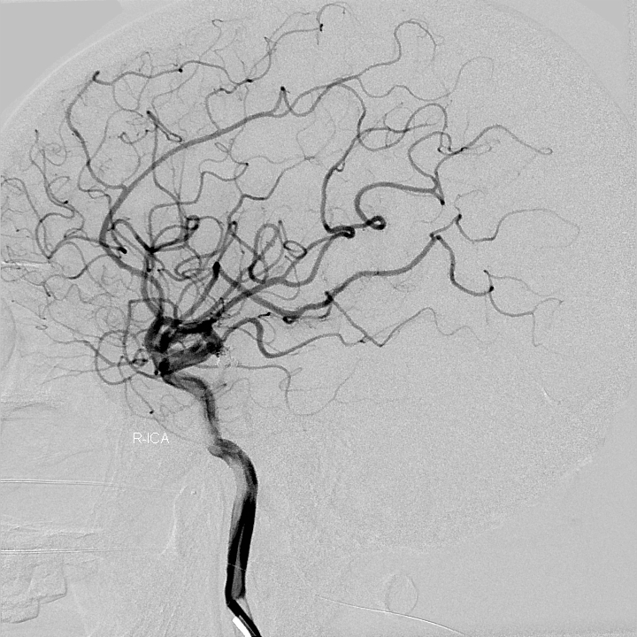
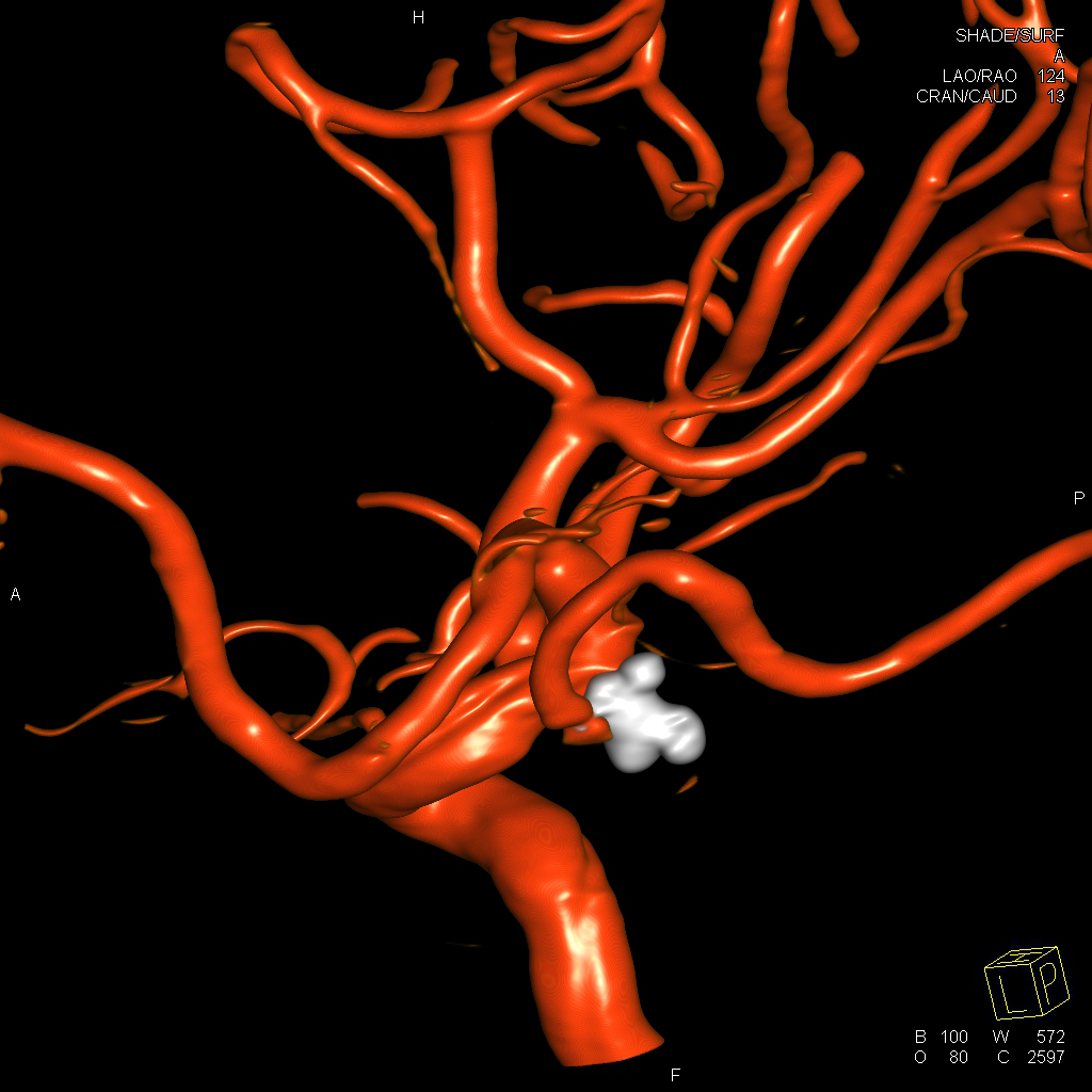
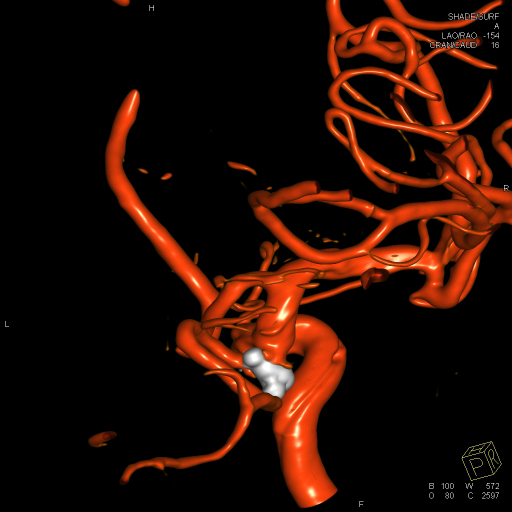
女性,58岁,蛛网膜下腔出血。
右侧后交通伴子囊动脉瘤。
术前影像

手术过程



术后影像

病例2
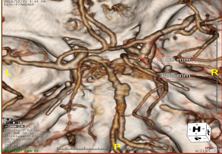
女性,50岁,蛛网膜下腔出血。
右侧后交通伴子囊动脉瘤,基底动脉瘤。
术前影像
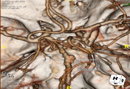


手术过程
右侧后交通动脉瘤栓塞术
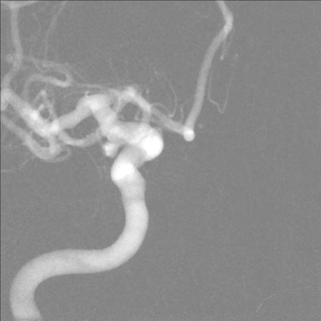
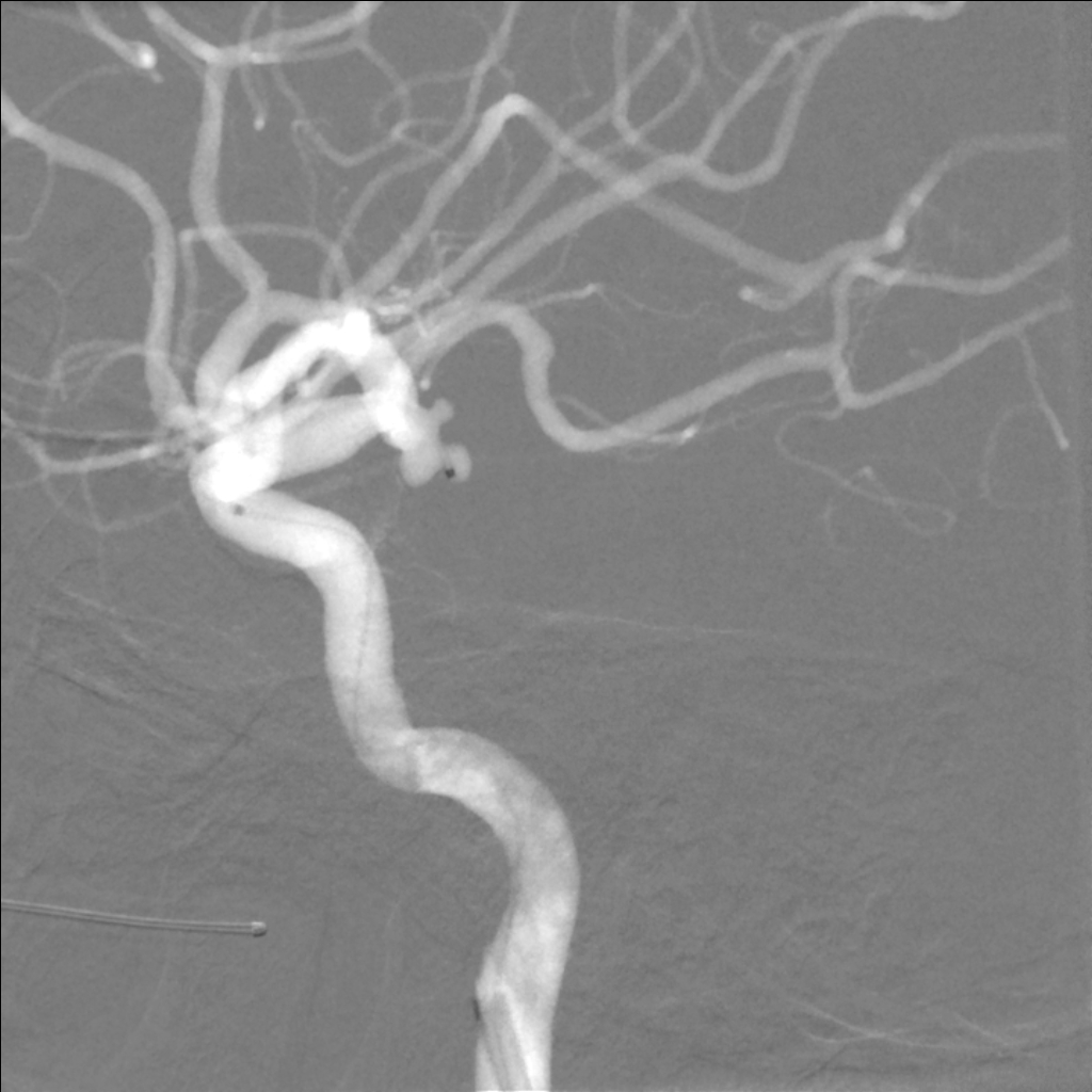
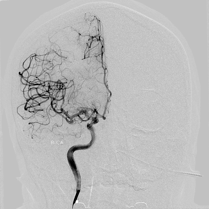
术后影像
专家介绍
引用文献:
[1]. Glenn Forbes, Allan J. Fox, John Huston III, et al. Interobserver variability in angiographic measurement and morphologic characterization of intracranial aneurysms: a report from the International Study of Unruptured Intracranial Aneurysms. AJNR 17:1407-1415, Sep 1996.
[2]. The UCAS Japan Investigators. The natural course of unruptured cerebral aneurysms in a Japanese cohort. N Eng J Med 2012;366:2474-2482.
[3]. Hidetoshi Matsukawa, Akihiro Uemura, Motoharu Fujii, et al. Morphological and clinical risk factors for the rupture of anterior communicating artery aneurysms. J Neurosurg. 2013 May; 118(5):978-83.
[4]. Toru Satoh, Megumi Omi, Chika Ohsako, et al. Influence of perianeurysmal environment on the deformation and bleb formation of the unruptured cerebral aneurysm: assessment with fusion imaging of 3D MRcisternography and 3D MR angiography. AJNR. 2005 Sep;26(8):2010-8.
[5]. Chang-Woo Kang, Hyon-Jo Kwon, Se-Jin Jeong, et al. Intentional sparing of daughter sac from coil packing in the embolization of aneurysms causing the third cranial nerve palsy: initial clinical and radiological results. J Korean Neurosurg Soc 48: 115-118, 2010.
[6]. Jin-Lu Yu, Hong-lei Wang, Kann Xu, et al. Endovascular treatment of an intracranial aneurysm with a ruptured bleb. Neurosciences (Riyadh). 2012 Apr;17(2):127-32.
[7]. Marta Aguilar Perez, Pervinder Bhogal, Rosa Martinez Moreno, et al. The Medina Embolic Device: early clinical experience from a single center. J NeuroIntervent Surg 2016;0:1-12.
[8]. Xintong Zhao, Jiaqiang Liu, Huifang Wang, et al. Endovascular treatment of ruptured aneurysms in anterior cerebral circulation with double microcatheter technique.
[9]. Sin-ichiro Sugiyama, Hidenori Endo, Shunsuke Omodaka, et al. Daughter sac formation related to blood inflow jet in an intracranial aneurysm. World Neurosurg. (2016) 96:396-402.


