最近微信朋友圈看到国内好多老师晒出漂亮的颈静脉球瘤高难度手术照片,作为神外小学生,佩服手术技艺的同时,还是先把一些最基本的问题搞清楚。其中一个最最基本的问题:颈静脉球瘤(glomus jugulare tumor)中的“球(glomus)”和解剖结构颈静脉球(jugular bulb)的“球(bulb)”到底有何联系和区别?而作为副神经节瘤(paraganglioma)的一种,这里的“副(para-)”又是什么含义?略微查了一番wiki和相关文献,总算有了点头绪,赶紧来这咬文嚼字以做小记。
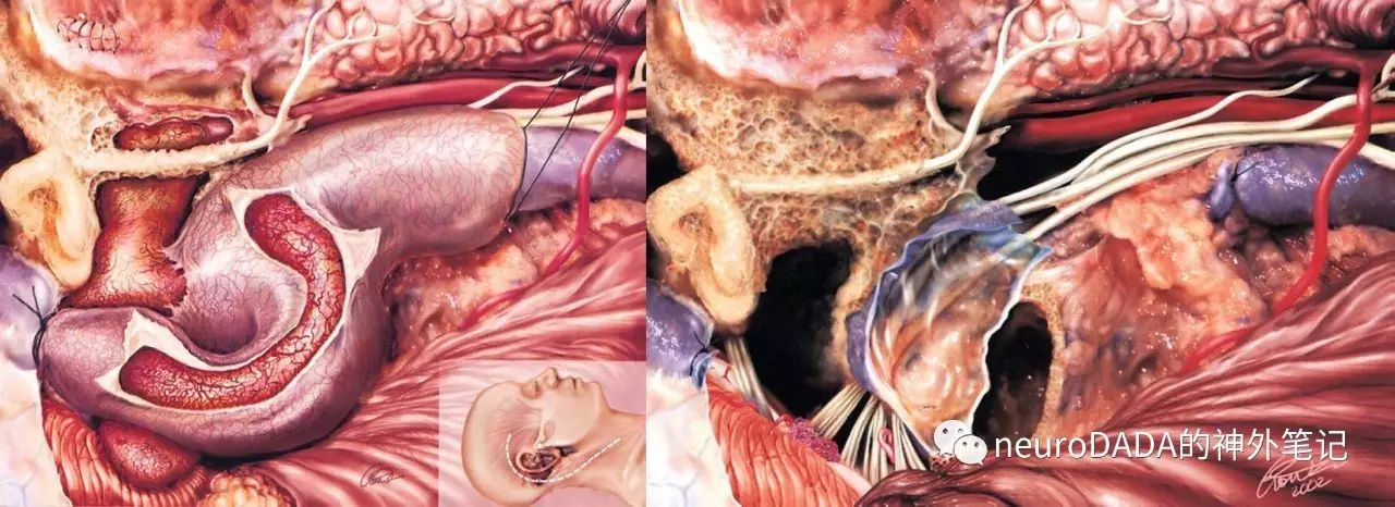
摘自Al-Mefty文献
Paragangliomas are tumors that arise from the paraganglionic system—aggregations of cells found throughout the body associated with vascular and neuronal adventitia. They originate from the neural crest and are related to the autonomic nervous system. The tumors of the head and neck are associated with the parasympathetic division, as opposed to the sympathetic division for the other parts of the body.
Accordingly, the term glomus should be replaced with paraganglioma denoting its location, as outlined in the WHO classification.
这段定义来自意大利ENT界的侧颅底大师Sanna教授2013年的著作《Microsurgery of Skull Base Paragangliomas》一书,从中可以发现,平时常说的“球瘤(glomus tumor)”已经被WHO剔除,而这一类肿瘤的官方名称就是“副神经节瘤(paraganglioma)”。
剔除的原因,查阅wiki可知,真正的“glomus tumor”(血管球瘤)另有所指,其起源于皮肤真皮层(dermis layer)的“glomus body (或glomus apparatus)”(血管球体),后者是结缔组织包绕的动静脉分流器(arteriovenous shunt),大多分布于肢端,作用是通过动静脉分流来调节体温(保暖和散热)。
那为什么平时误称“paraganglioma”(副神经节瘤)为“glomus tumor”(球瘤)呢?因为paraganglioma恰恰起源于“glomus cell”(球细胞)。
已经有点晕了吧……不急,接着刨根究底,看看“paraganglion”和“glomus cell”到底是什么。
A paraganglion (pl. paraganglia) is a group of non-neuronal cells derived of the neural crest. They are named for being generally in close proximity to sympathetic ganglia. They are essentially of two types: chromaffin or sympathetic paraganglia made of chromaffin cells and nonchromaffin or parasympathetic ganglia made of glomus cells. They are neuroendocrine cells, the former with primary endocrine functions and the latter with primary chemoreceptor functions.
这段引自wiki的定义十分清晰地告诉我们,“paraganglion”是起源于神经嵴(neural crest)的一类非神经元性细胞,它的命名是因为其主要(注意是“generally”而非“totally”)位于交感神经节(sympathetic ganglia)的附近。由此可以回答标题里的问题了——这里的“para”究竟该不该翻译为“副”?
众所周知,英语里的“para-”通常有两种含义:“旁”,例如“parahippocampal gyrus”(海马旁回),表示方位;“副”,例如“parasympathetic”(副交感),表示对立。根据上面的定义,“paraganglion”的“para-”显然是“旁”的意思。神经节(ganglion)作为神经系统的一个结构,本来不存在什么对立面啊!另外,“paraganglion”又是以一群细胞而非单个零散细胞为存在形式。因此,个人认为,我们常年习惯用语“副神经节”应改为“神经节旁体”,“副神经节瘤”应改为“神经节旁体瘤”更为恰当。
接着看定义,这群“paraganglion”细胞本身属于神经内分泌细胞(neuroendocrine cell),因此,WHO将其起源的肿瘤归类于神经内分泌肿瘤。而根据功能和嗜铬性(chromaffinity)的不同,分为两类:与交感系统关系密切、具有交感特性、嗜铬染色阳性(分泌儿茶酚胺catecholamines)者;与副交感系统关系密切、具有副交感特性,嗜铬染色阴性者。
前者主要聚集于肾上腺附近,具有肾上腺相似的内分泌功能,起源于此的肿瘤,由于嗜铬染色阳性,即所谓的“嗜铬细胞瘤(pheochromocytomas或chromaffin tumor)”,当然包括其他肾上腺以外部位(腹、胸)的相同来源肿瘤,其中主要为“主动脉旁体瘤”(起源于para-aortic body或organ of Zuckerkandl)。
后者虽然也是神经内分泌细胞,但是其分泌功能(儿茶酚胺)很少表现,因此嗜铬染色常阴性(起源于此的1-3%肿瘤也具有分泌功能而嗜铬染色阳性),取而代之的则是具有化学感受器(chemoreceptor)功能,因此缘于此的肿瘤又曾被称为“化学感受器瘤”(chemodectomas)。由于这种化学感受器最主要的分布为颈动脉小球(carotid glomus,又称颈动脉体 carotid body),因此,这种细胞也被称为球细胞(glomus cell)。这类细胞同样还分布于颅底其他部位,包括鼓室、颈静脉孔舌咽神经和迷走神经走行附近(具体见后)。
这就回答了刚才的问题——平时误称“paraganglioma”(副神经节瘤)为“glomus tumor”(球瘤)的原因。另外也回答了开篇的第一个问题——就算是平时所谓的“球瘤”,此“球”(glomus)也并非指病变多累及颈静脉球(jugular bulb)中的“球”(bulb),只是解剖部位(见下文)恰恰多重合而已。
再补充一下,颈动脉小球作为重要的化学感受器,其组成细胞即球细胞(glomus cell),其中I型才是这里说的神经内分泌细胞,由于需要充分感受血液化学改变,因此这些细胞群高代谢、高灌注(我猜这也是这类肿瘤血供极度丰富的原因);II型为胶质细胞,起到支持作用。颈动脉分叉处另一重要结构,颈动脉窦(carotid sinus),为压力感受器。感受器的传入神经为主要为窦神经,汇入舌咽神经,且与迷走神经、颈交感干有广泛联系。

从左到右:颈动脉体、颈动脉窦、窦神经、舌咽神经、迷走神经、颈交感干
To simplify the terminology, the term pheochromocytoma should be used for those tumors arising in the adrenal, extraadrenal abdominal, and thoracic locations to emphasize their endocrinologically active nature, while paraganglioma refers to those tumors that arise in the head and neck and are nonsecreting in the vast majority of cases.
再回到Sanna教授的引文,我们可以得出结论:“paraganglioma”本应是一大类总称,根据细胞的嗜铬性,分为“pheochromocytomas或chromaffin tumor”(嗜铬细胞瘤)和“nonchromaffin tumors”(非嗜铬细胞瘤),后者因具源自化学感受器细胞而又称为“chemodectomas”(化学感受器瘤)。但是,目前为了方便,在大多数情况下,“paraganglioma”等同于后者,成为逻辑上与嗜铬细胞瘤相对的一个概念。至于“paraganglioma”的中文翻译,是“副神经节瘤”还是“神经节旁体瘤”,相信大家自有定夺。下文都暂用英文来命名。
再来说点相关的解剖。
了解了头颈部paraganglioma的起源,就可以根据它具体起源的解剖部位进行大体的划分了。
tympanojugular paraganglioma:起源于沿Jacobson神经和Arnold神经走行分布的paraganglion
vagal paraganglioma:起源于迷走神经的下神经节(nodose ganglion)区域的paraganglion
carotid body tumor:起源于颈动脉体区域的paraganglion
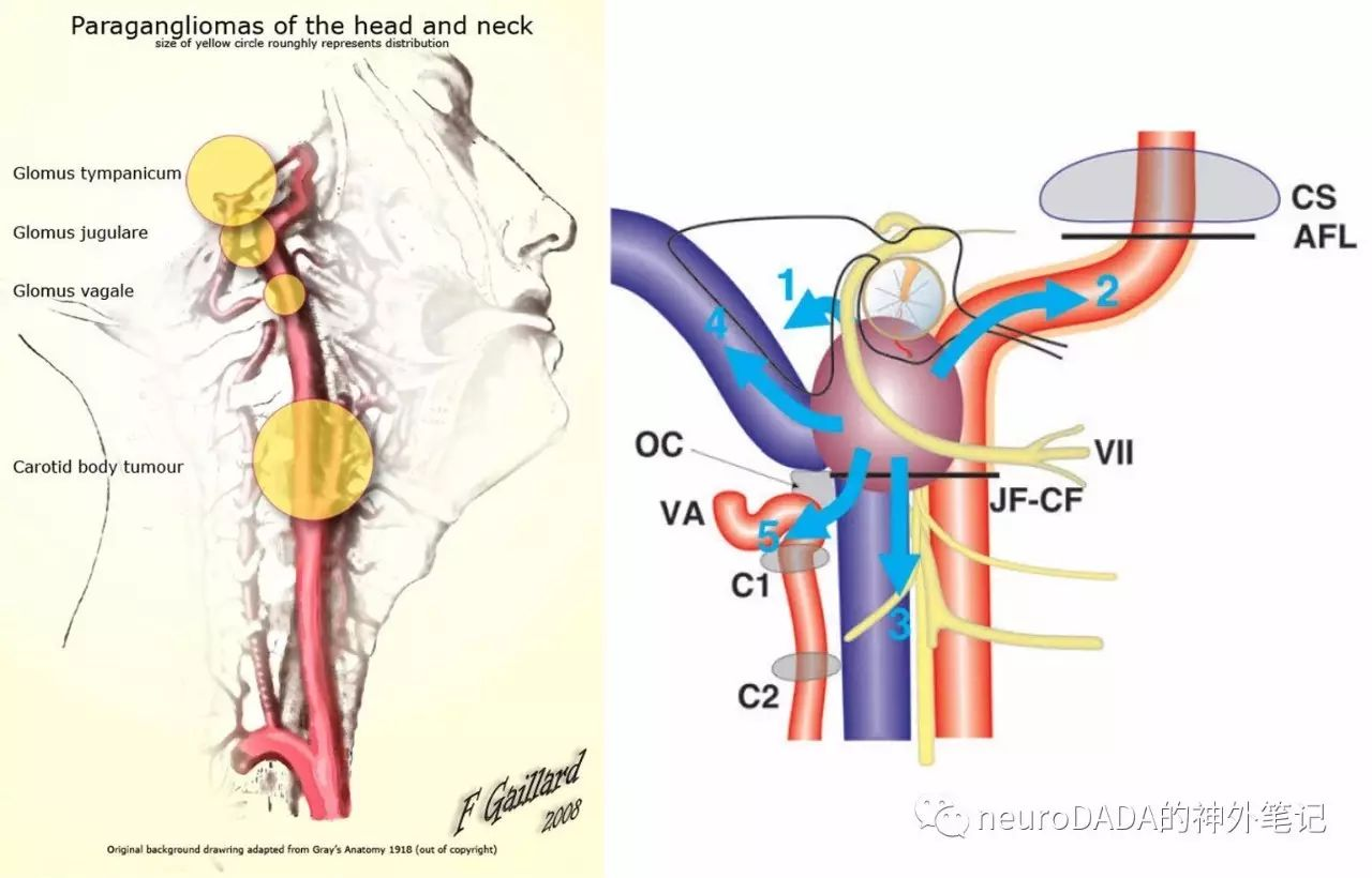
头颈部paraganglioma的解剖学生长特点
tympanojugular paraganglioma,根据modified Fisch分级,根据累及范围越来越广泛,又分为tympanic、tympanomastoid、tympanojugular三大类。这首先需要理解Jacobson神经和Arnold神经的解剖。
Jacobson神经,即舌咽神经的鼓室支(tympanic branch),起自舌咽神经下神经节(petrosal ganglion),经颈静脉孔与颈内动脉管外口之间的骨嵴,颈内嵴(intrajugular ridge)上的鼓室小管(tympanic canaliculus)穿破鼓室底壁,与起自颈内动脉交感丛、穿破鼓室前壁的颈鼓神经(caroticotympanic nerve)在鼓室内形成鼓室丛(tympanic plexus),其中一支穿入中颅窝底,与来自中间神经(nervus intermedius)和Arnold神经(见下文)的交通支汇合,形成岩小神经(lesser petrosal nerve),向前经无名小管(canaliculus innominatus)或棘孔(foramen spinosum)或蝶岩缝(sphenopetrosal suture)进入颞下窝(infratemporal fossa),在耳神经节(otic ganglion)内换元,继续经三叉神经下颌支的耳颞神经(auriculotemporal nerve)支配腮腺。行经耳神经节但不换元的还有来自脑膜中动脉交感丛的交感节后纤维、来自耳颞神经的一般躯体感觉交通支、发自下颌支支配腭帆张肌和鼓膜张肌的运动支。
Arnold神经,及迷走神经的耳支(auricular branch),起自迷走神经上神经节(jugular ganglion),连同起自舌咽神经下神经节(petrosal ganglion),贴行于颈静脉球前壁至颈静脉窝外侧壁的乳突小管(mastoid canaliculus),经此管道入颞骨,横跨面神经乳突段。此处,一部分纤维继续沿面神经乳突段上行,并穿经面神经鼓室段,至膝状神经节(geniculate ganglion)附近,与来自中间神经(nervus intermedius)的交通支一起再与Jacobson神经来源的神经汇合形成岩小神经(lesser petrosal nerve)(见上文);另一部分则在颞骨内下行,经鼓乳缝(tympanomastoid fissure)出颅,一部分汇入面神经出茎乳孔后的耳后神经(posterior auricular nerve),另一部分则分布于耳后及外耳道皮肤。
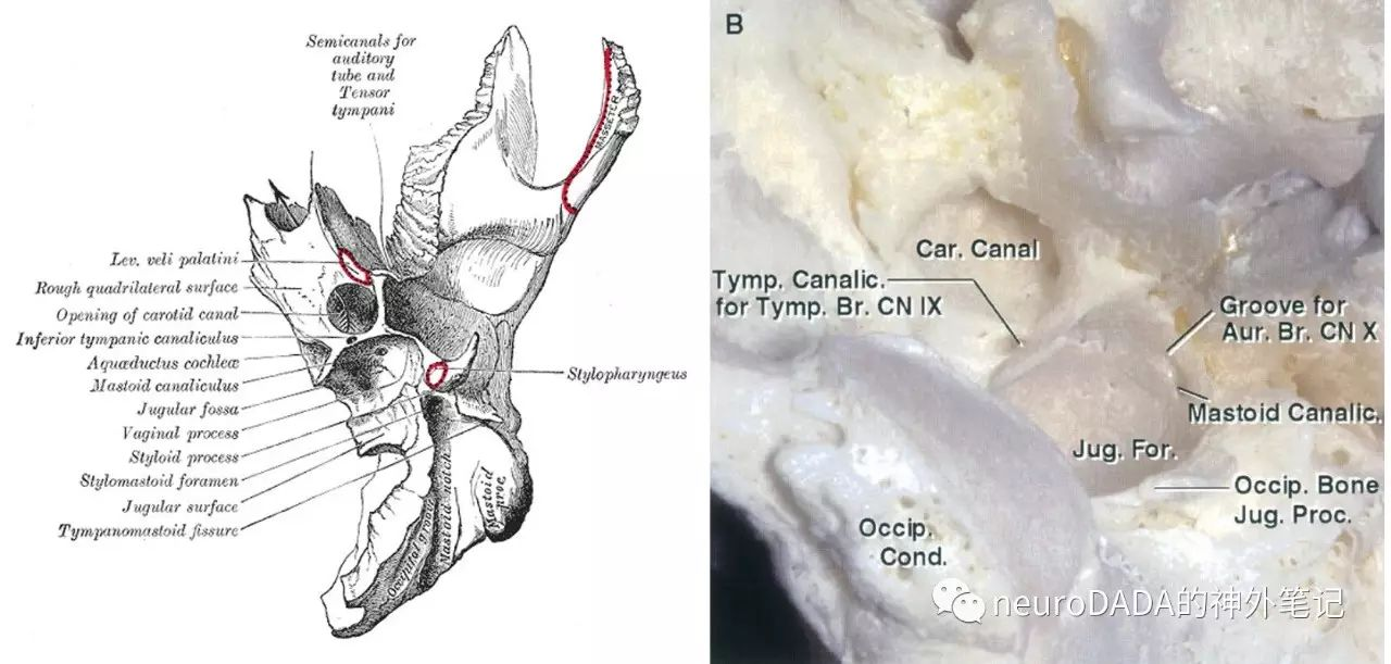
颈静脉孔下面观,可见鼓室小管(tympanic canaliculus)和乳突小管(mastoid canaliculus)
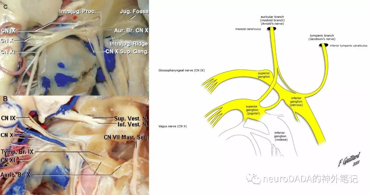
颈静脉孔后面观,可见Jacobson神经和Arnold神经的起源和入颞骨方式
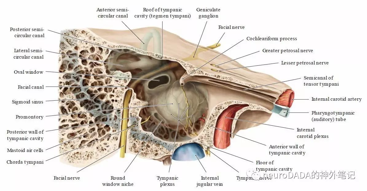
Jacobson神经、鼓室丛、岩小神经的关系
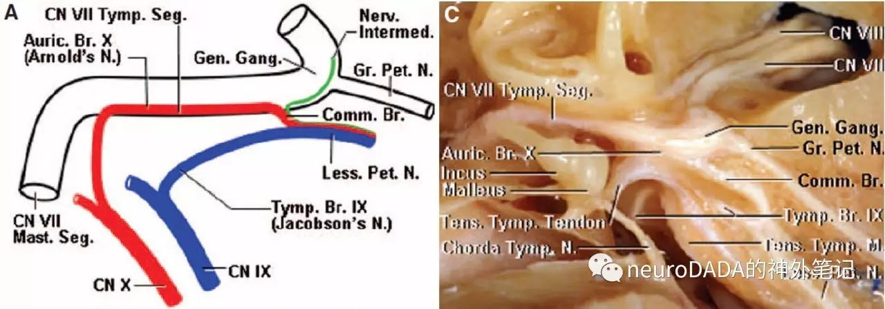
Jacobson神经、Arnold神经、中间神经汇合为岩小神经的关系

岩小神经汇入耳神经节及终末支配
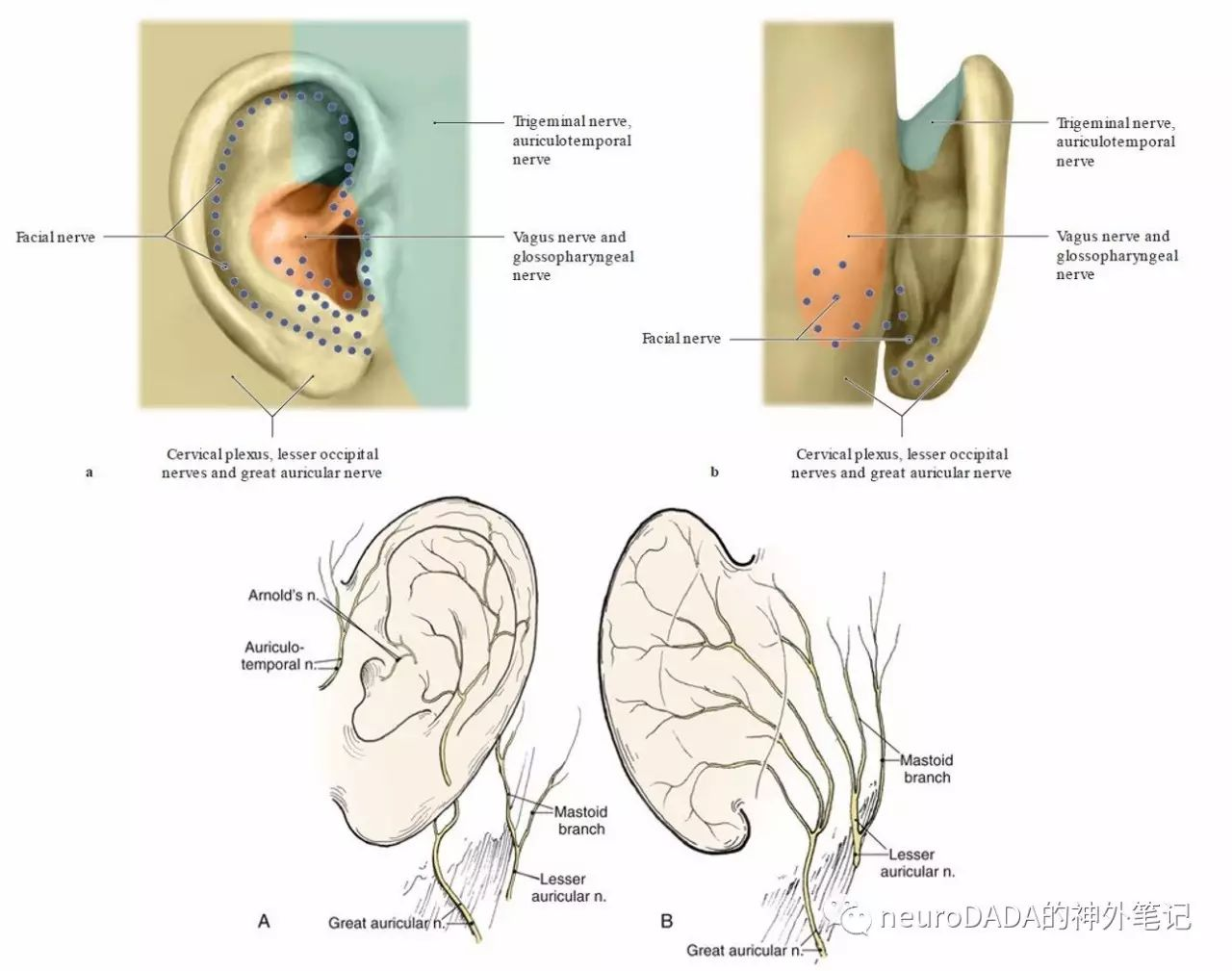
Arnold神经最终支配
了解了这两根神经的走行,才可理解tympanojugular paraganglioma的解剖分级。另外,神经走行过程中,各神经纤维的性质和功能,所查阅文献有限,暂不完全明了,待日后继续学习。
参考文献
1.Al-Mefty O, Teixeira A. Complex tumors of the glomus jugulare: criteria, treatment, and outcome. Journal of neurosurgery. 2002;97(6):1356-66.
2.Kakizawa Y, Abe H, Fukushima Y, Hongo K, El-Khouly H, Rhoton AL, Jr. The course of the lesser petrosal nerve on the middle cranial fossa. Neurosurgery. 2007;61(3 Suppl):15-23; discussion
3.《Rhoton s Cranial Anatomy and Surgical Approaches》2007
4.《Microsurgery of Skull Base Paragangliomas》2013
5.《The Rhoton Collection——Head and Neck Anatomy for Neurosurgeons》2016
6.《THIEME Atlas of Anatomy:Head, Neck, and Neuroanatomy.2ed》2016
7.https://en.wikipedia.org


