

最近《Rhoton Collection》发布了雪藏多年的眶颧入路解剖视频,而JNS也在本月在线发表了Rhoton教授生前署名的一篇关于眶颧的文章,借着这股“眶颧热”,今天就汇总一下大师历来有关额颞眶颧入路(frontotemporal orbitozygomatic approach)的10篇经典文献和美图,并引申其他相关经典文献,加上一些个人读书体会,以作记录和分享。
文献一:廿年前的“眶上入路”
Delashaw JB, Jr., Tedeschi H, Rhoton AL. Modified supraorbital craniotomy: technical note. Neurosurgery, 1992, 30(6):954-6.
这应该是Rhoton教授署名的与额颞眶颧入路相关的第一篇文献,所谓的“modified”是针对1982年Jane首次提出的“supraorbital approach”而言的,本文一是加入了对颧弓的切除,二是提出了对额窦前后壁分层切开的方法,目的是避免中线处钻孔带来的容貌缺陷问题。
这一时期,文献中所谓的“supraorbital”与2005年Hernesniemi Juha教授提出的“lateral supraorbital”(见后文)并非同一个概念,而是可以看做眶颧入路发展至后期mini-OZ的雏形;只不过,在当时颅底外科兴起之时,入路的“扩大化”是认识颅底解剖、开拓颅底外科技术的主要方式,而现今,在对颅底已有较为全面认识的基础上,则是回归“mini化”而实现微创之目的。因此,我想这正是颅底外科这一学科发展的自然过程,也是我们年轻医生学习颅底外科的正常曲线——先了解如何做大,再追求如何做小。

相关其他文献
1.Jane JA, Park TS, Pobereskin LH, Winn HR, Butler AB. The supraorbital approach: technical note. Neurosurgery. 1982;11(4):537-42.
2.Al-Mefty O. Supraorbital-pterional approach to skull base lesions. Neurosurgery, 1987, 21(4):474-7.
3.Smith RR, Al-Mefty O, Middleton TH. An orbitocranial approach to complex aneurysms of the anterior circulation. Neurosurgery, 1989, 24(3):385-91.
4.Al-Mefty O, Smith RR. Tailoring the cranio-orbital approach. Keio J Med, 1990, 39(4):217-24.
文献二:标准翼点入路的经典教材
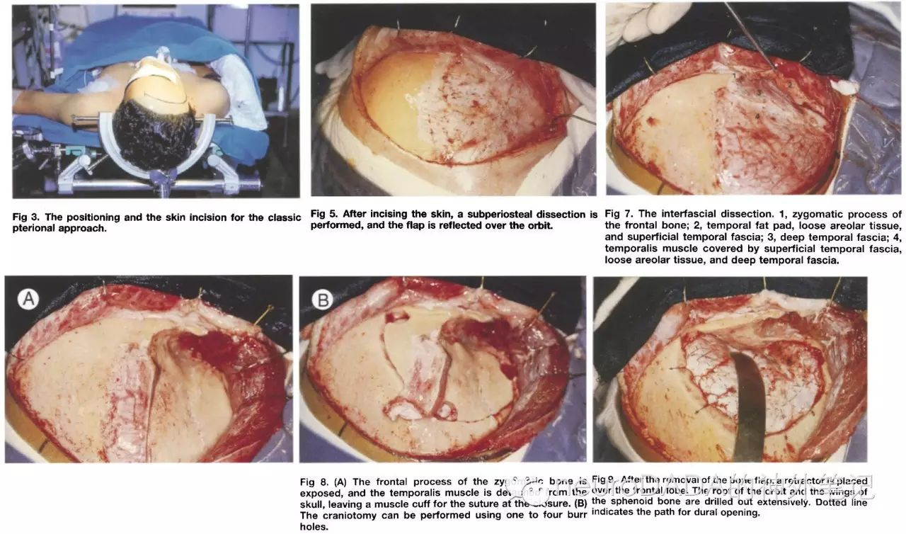
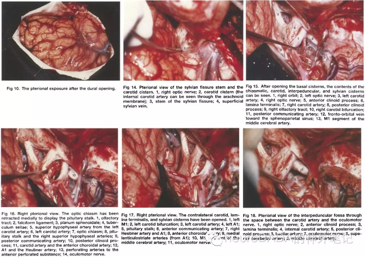
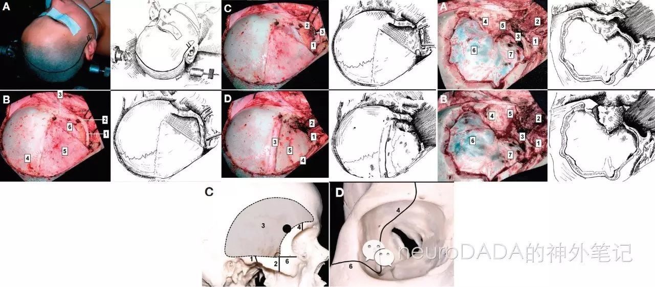
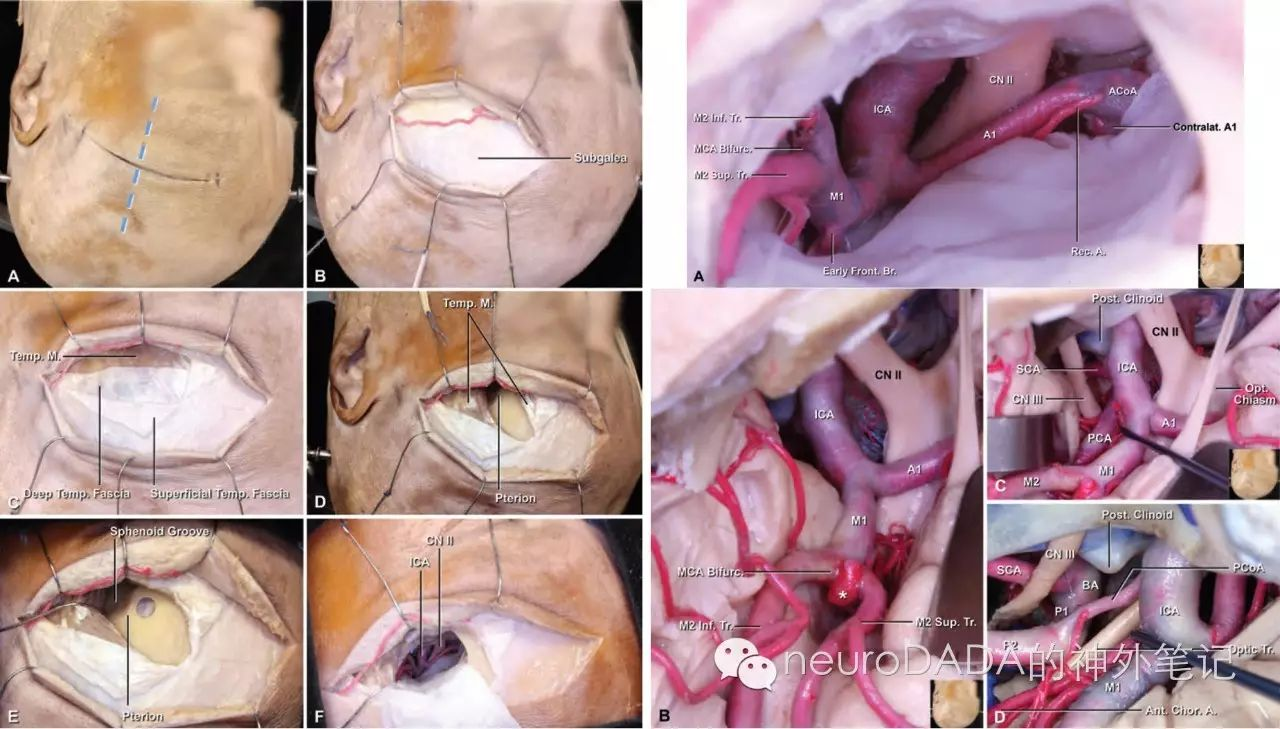
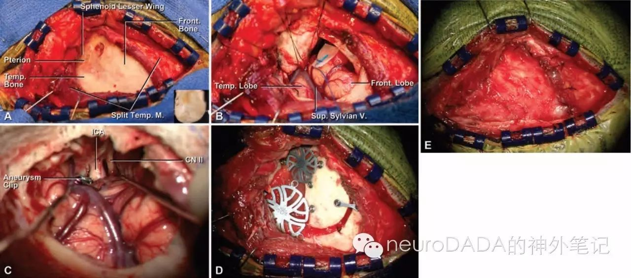
Wen HT, de Oliveira E, Tedeschi H, Andrade FC, Jr., Rhoton AL, Jr. The Pterional Approach: Surgical Anatomy, Operative Technique, and Rationale. Operative Techniques in Neurosurgery. 2001;4(2):60-72.
这篇文献可以看作是Yasargil经典翼点入路的Rhoton版,从解剖到入路,从体位到分离侧裂,都进行了极为详尽的描述。当然,关于侧裂的解剖,Rhoton后期还有较多更为精彩的文献;但对于标准翼点开颅,这绝对是神经外科史上最佳的教科书式文献之一。
其他相关文献:
1.Yasargil MG. Microneurosurgery, Volume IVB.1996
2.Vishteh AG, Marciano FE, David CA, Baskin JJ, Spetzler RF. The Pterional Approach. Operative Techniques in Neurosurge. 1998;1(1):39-49.
文献三、文献四:单骨瓣法眶颧入路重要解剖基础
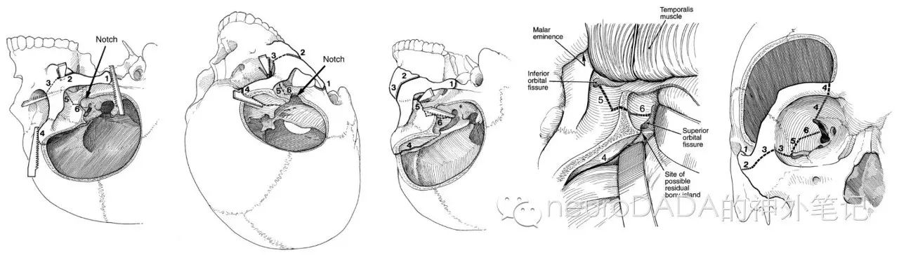
3.Martins C, Li X, Rhoton AL, Jr. Role of the zygomaticofacial foramen in the orbitozygomatic craniotomy: anatomic report. Neurosurgery. 2003;53(1):168-72; discussion 72-3.
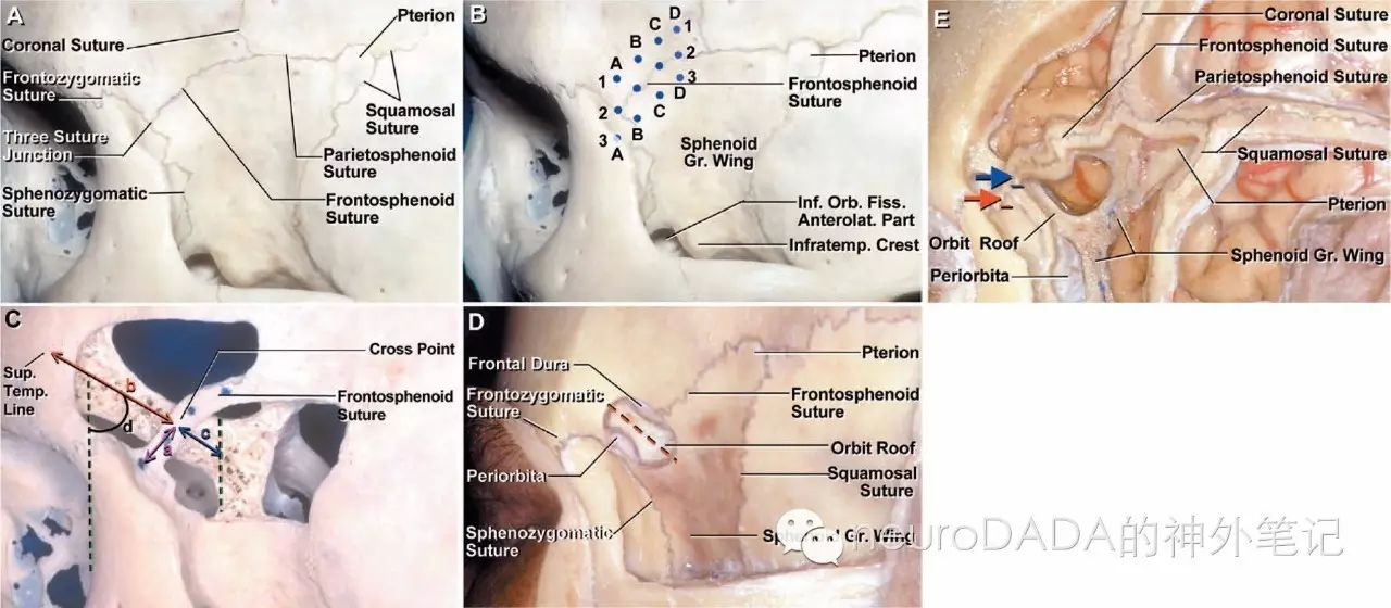
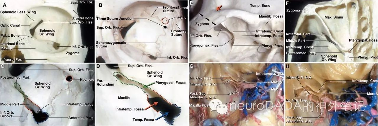
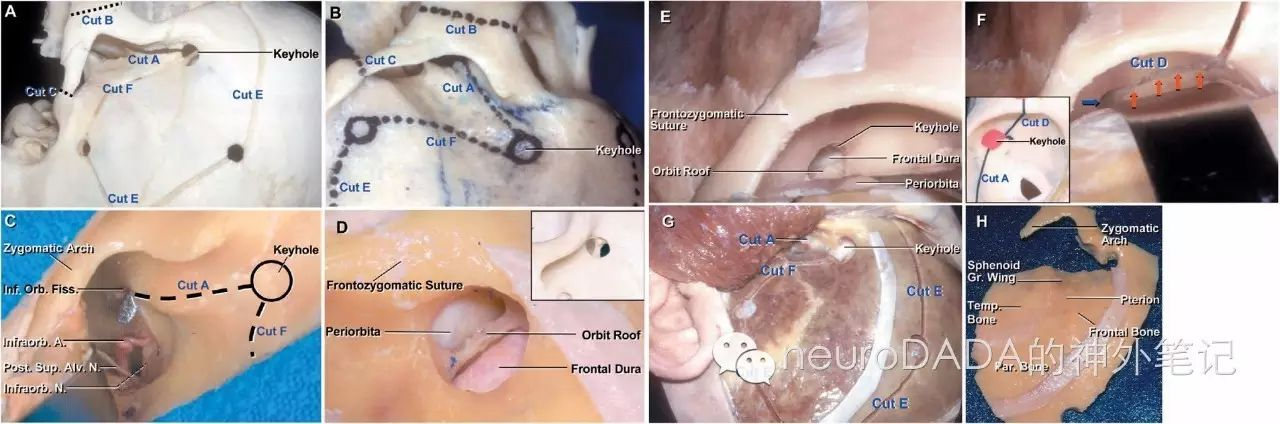
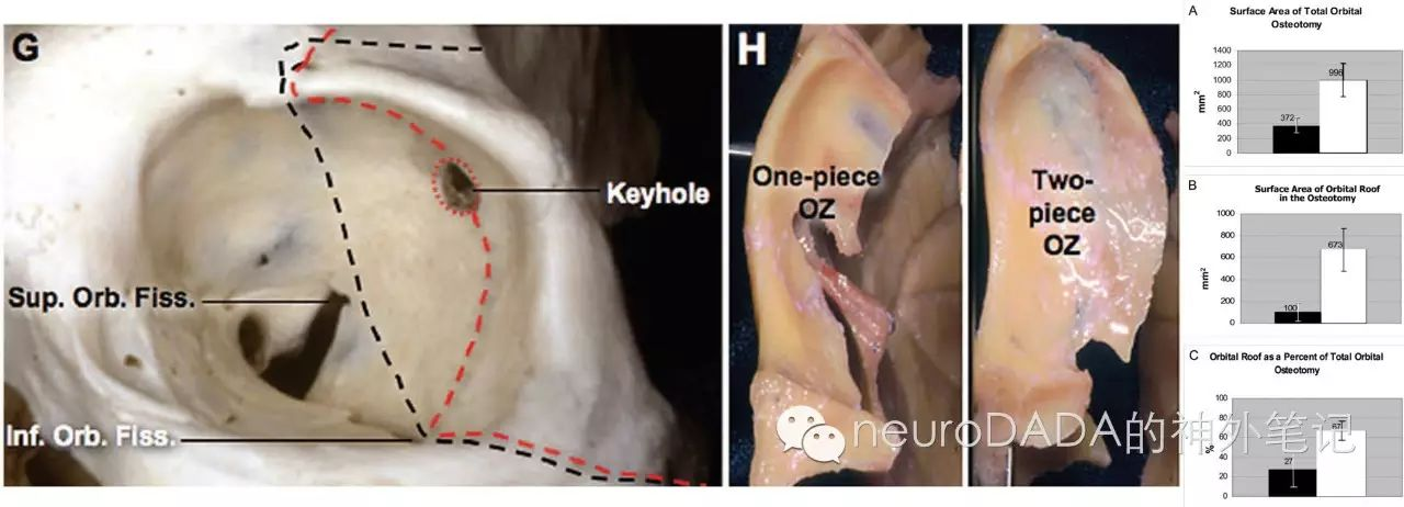
4.Shimizu S, Tanriover N, Rhoton AL, Jr., Yoshioka N, Fujii K. MacCarty keyhole and inferior orbital fissure in orbitozygomatic craniotomy. Neurosurgery. 2005;57(1 Suppl):152-9; discussion -9.
这两篇探讨的是one-piece法(单骨瓣法)眶颧入路的两个重要手术细节。文献3聚焦于经典one-piece法眶颧入路cut 3的定位标志,颧面孔(zygomaticofacial foramen),然而研究得出的结论是,由于解剖变异的存在,这一标志物并不常常可靠。然而,这篇文章也让人重新复习了上颌神经的颧神经的两大分支,即颧面神经(zygomaticofacial nerve)和颧颞神经(zygomaticotemporal nerve)的走行及意义。

文献4则是关于MacCarty孔最为精彩详实的诠释之一,而这正是one-piece法眶颧入路的最关键的“关键孔”;其次是眶下裂的阐述。了解两者解剖、定位、意义,是理解眶颧入路的重要基础。
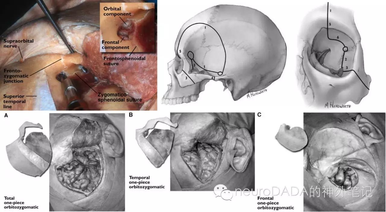
关于one-piece法眶颧入路,不得不提的还有Loveren大神的经典文献(见下),不仅对上述关键环节进行了同样深刻的阐述,同时也将one-piece法眶颧入路根据所需暴露范围的大小不同进行了分类(此后还有新增,并在two-piece法中也有相对应的分类,见下段),这也是前期扩大化之后向mini化改良的基础和开始。
其他相关文献:
1.Tubbs RS, Loukas M, Shoja MM, Cohen-Gadol AA. Refined and simplified surgical landmarks for the MacCarty keyhole and orbitozygomatic craniotomy. Neurosurgery. 2010;66(6 Suppl Operative):230-3.
2.Aziz KM, Froelich SC, Cohen PL, Sanan A, Keller JT, van Loveren HR. The one-piece orbitozygomatic approach: the MacCarty burr hole and the inferior orbital fissure as keys to technique and application. Acta neurochirurgica. 2002;144(1):15-24.
3.Youssef AS, Willard L, Downes A, Olivera R, Hall K, Agazzi S, et al. The frontotemporal-orbitozygomatic approach: reconstructive technique and outcome. Acta neurochirurgica. 2012;154(7):1275-83.
4.Tubbs RS, Mortazavi MM, Shoja MM, Loukas M, Cohen-Gadol AA. The zygomaticotemporal nerve and its relevance to neurosurgery. World neurosurgery. 2012;78(5):515-8.
文献五、文献六:两骨瓣法、三骨瓣法的优势
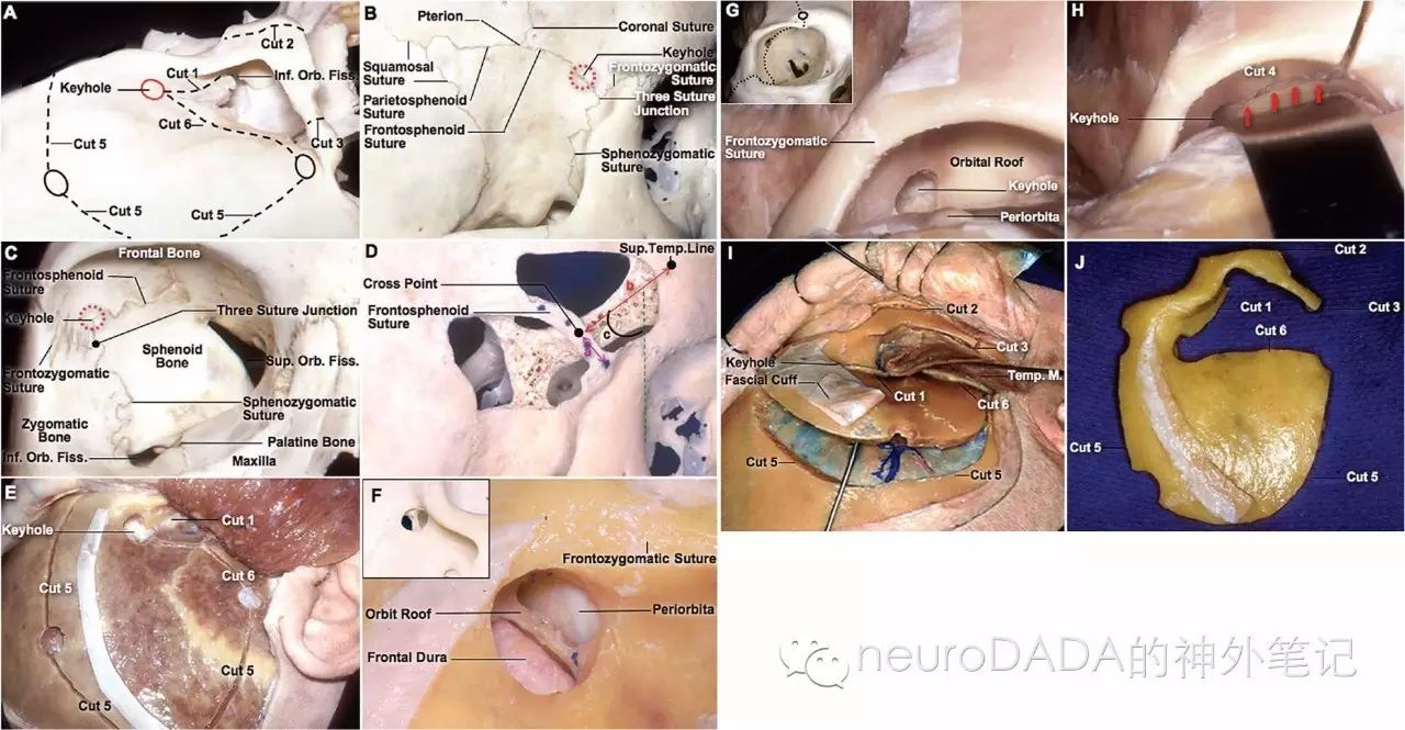
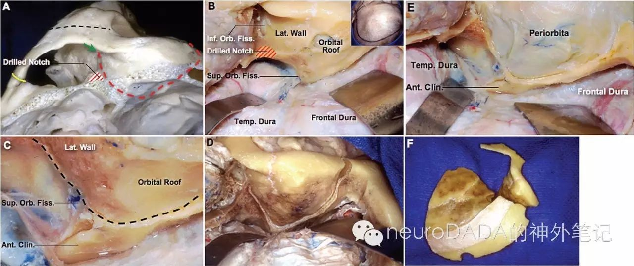
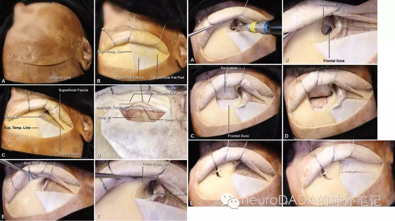
5.Tanriover N, Ulm AJ, Rhoton AL, Jr., Kawashima M, Yoshioka N, Lewis SB. One-piece versus two-piece orbitozygomatic craniotomy: quantitative and qualitative considerations. Neurosurgery. 2006;58(4 Suppl 2):ONS-229-37; discussion ONS-37.
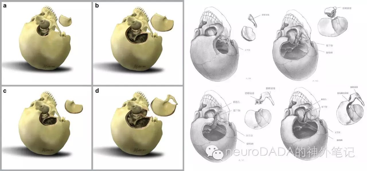
6.Campero A, Martins C, Socolovsky M, Torino R, Yasuda A, Domitrovic L, et al. Three-piece orbitozygomatic approach. Neurosurgery. 2010;66(3 Suppl Operative):E119-20; discussion E20.
这两篇文献关注的问题是:眶颧入路到底用几片法好?文献5首先可作为标准one-piece法和two-piece法的经典教科书文献,同时可以明确地看到两者的差异——开颅后眶壁骨质残余量的不同,导致术后眶壁骨质缺损量的不同,而影响眼球塌陷这一容貌问题。
文献6则是对three-piece法的step by step展示。虽然看着骨瓣数量增多,但实际上,three-piece法从某种意义上来说是“去伪存真”,去除了经典眶颧开颅中不必要的部分,并且由于将颧弓单独离断而可减少对咬肌的损伤,同时也可让人思考,眶颧入路中,到底何时需要去眶,何时需要去颧。
当然,说到two-piece法,不得不提的还有Barrow大神Zabramski的经典文献。另外,Loveren的经典著作中,也将two-piece法根据不同暴露目的进行了分类(与one-piece法相对应)。
其他相关文献
1.Zabramski JM, Kiris T, Sankhla SK, Cabiol J, Spetzler RF. Orbitozygomatic craniotomy. Technical note. Journal of neurosurgery. 1998;89(2):336-41.
2.Tew JM, Jr., Keller JT, van Loveren HR. Atlas of Operative Microneurosurgery - Vol. 2 Brain tumors. 2007
3.Lemole GM, Jr., Henn JS, Zabramski JM, Spetzler RF. Modifications to the orbitozygomatic approach. Technical note. Journal of neurosurgery. 2003;99(5):924-30.
4.Youssef AS, Willard L, Downes A, Olivera R, Hall K, Agazzi S, et al. The frontotemporal-orbitozygomatic approach: reconstructive technique and outcome. Acta neurochirurgica. 2012;154(7):1275-83.
文献七:去除颧弓,海阔天空
7.Campero A, Campero AA, Socolovsky M, Martins C, Yasuda A, Basso A, et al. The transzygomatic approach. Journal of clinical neuroscience : official journal of the Neurosurgical Society of Australasia. 2010;17(11):1428-33.
这篇文献的术式其实可看做two-piece眶颧中的翼点+去颧弓。1984年Pellerin首次进行颧弓切除,去颧弓在眶颧入路中的作用主要是增加颞肌的下翻而使开颅更接近中颅底,从而获得颞下更大的垂直角度。通过这一术式,可联合各种经中颅窝入路进行相关扩展和联合,例如处理蝶岩斜区脑膜瘤、中后颅窝哑铃型三叉神经鞘瘤等。
将这篇文献单独列出的意义还在于,去颧弓的另一作用似乎是独立于眶颧入路范畴以外的,即进行颞下窝的暴露。其中,颞下颌关节如何处理也是一大焦点。Fukushima教授利用这一技术,发展了前、中-耳前经颧弓颞下窝入路(preauricular transzygomatic infratemporal fossa approach),与耳科界侧颅底大师Fisch教授的B、C型颞下窝入路(infratemporal approach B、C)相辅相成,成为处理侧颅底复杂广泛病灶的重要入路。
其他相关文献:
1.Pellerin P, Lesoin F, Dhellemmes P, Donazzan M, Jomin M. Usefulness of the orbitofrontomalar approach associated with bone reconstruction for frontotemporosphenoid meningiomas. Neurosurgery. 1984;15(5):715-8.
2.Nonaka Y, Fukushima T, Watanabe K, Sakai J, Friedman AH, Zomorodi AR. Middle infratemporal fossa less invasive approach for radical resection of parapharyngeal tumors: surgical microanatomy and clinical application. Neurosurgical review. 2016;39(1):87-96; discussion -7.
3.Ohue S, Fukushima T, Kumon Y, Ohnishi T, Friedman AH. Preauricular transzygomatic anterior infratemporal fossa approach for tumors in or around infratemporal fossa lesions. Neurosurgical review. 2012;35(4):583-92; discussion 92.
4.Froelich S, Aziz KA, Levine NB, Tew JM, Jr., Keller JT, Theodosopoulos PV. Extension of the one-piece orbitozygomatic frontotemporal approach to the glenoid fossa: cadaveric study. Neurosurgery. 2008;62(5 Suppl 2):ONS312-6; discussion ONS6-7.
文献八、文献九:筋膜间or筋膜下,一切为了面神经
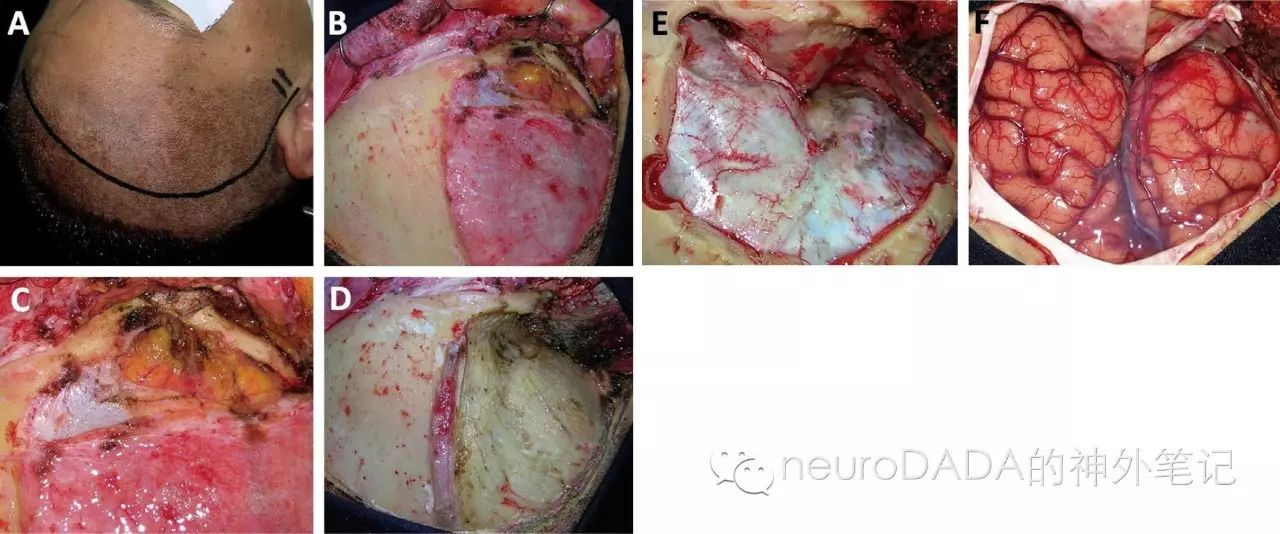
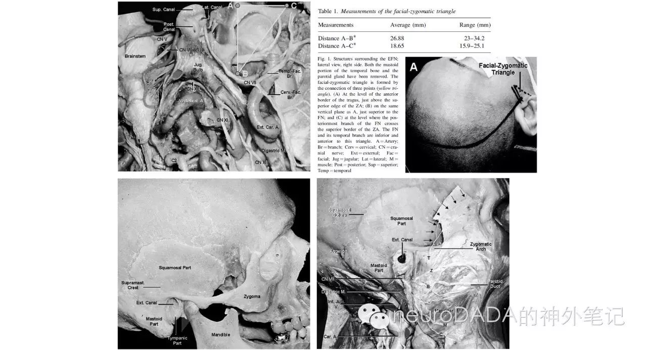
8.Campero A, Socolovsky M, Martins C, Yasuda A, Torino R, Rhoton AL. Facial-zygomatic triangle: a relationship between the extracranial portion of facial nerve and the zygomatic arch. Acta neurochirurgica. 2008;150(3):273-8; discussion 8.
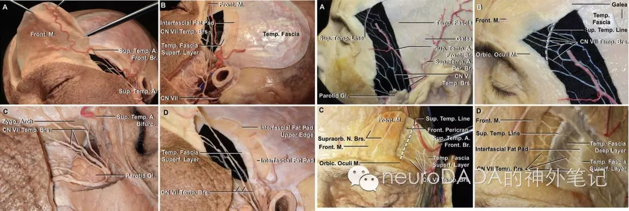
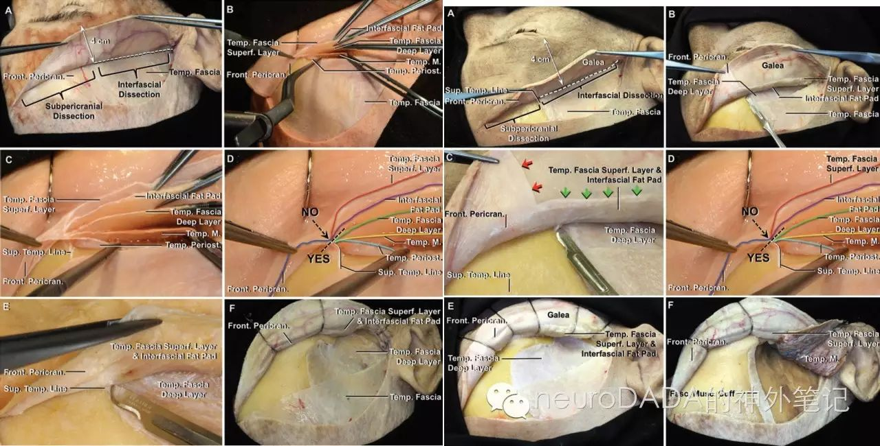
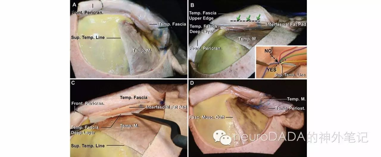
9.Poblete T, Jiang X, Komune N, Matsushima K, Rhoton AL, Jr. Preservation of the nerves to the frontalis muscle during pterional craniotomy. Journal of neurosurgery. 2015;122(6):1274-82.
这两篇文献关注的是额颞开颅中另一焦点问题——面神经的保护。文献8关注的是颧弓下方的面神经总干,文中提出的一个概念“面-颧三角”很少听闻,却提供了颧弓下方皮肤切口长度的依据。
文献9关注的是颧弓上方的面神经颞支。这部分内容历来有大量文献述及,然而无论是命名和结论本身都存在许多争议。这篇文献应该是目前最为细致和权威的参考,不仅明确了各层次命名、解剖、各标志点的距离数值,还非常详细地step by step展示了筋膜间(interfascial)和筋膜下(subfascial)分离颞肌筋膜的技术。相关内容也有《Rhoton collection》相关视频发布。
另外,关于颞肌的剥离,以及如何在关颅时复位缝合,以防止术后颞肌萎缩,也有众多经典文献为据(见下)。
其他相关文献:
1.Yasargil MG, Reichman MV, Kubik S. Preservation of the frontotemporal branch of the facial nerve using the interfascial temporalis flap for pterional craniotomy. Technical article. Journal of neurosurgery. 1987;67(3):463-6.
2.Spetzler RF, Lee KS. Reconstruction of the temporalis muscle for the pterional craniotomy. Technical note. Journal of neurosurgery. 1990;73(4):636-7.
3.Oikawa S, Mizuno M, Muraoka S, Kobayashi S. Retrograde dissection of the temporalis muscle preventing muscle atrophy for pterional craniotomy. Technical note. J Neurosurg. 1996;84(2):297-9.
4.Kadri PA, Al-Mefty O. The anatomical basis for surgical preservation of temporal muscle. Journal of neurosurgery. 2004;100(3):517-22.
5.Krayenbuhl N, Isolan GR, Hafez A, Yasargil MG. The relationship of the fronto-temporal branches of the facial nerve to the fascias of the temporal region: a literature review applied to practical anatomical dissection. Neurosurgical review. 2007;30(1):8-15; discussion
6.Youssef AS, Ahmadian A, Ramos E, Vale F, van Loveren HR. Combined subgaleal/myocutaneous technique for temporalis muscle dissection. Journal of neurological surgery Part B, Skull base. 2012;73(6):387-93.
文献十:以小见大,殊途同归
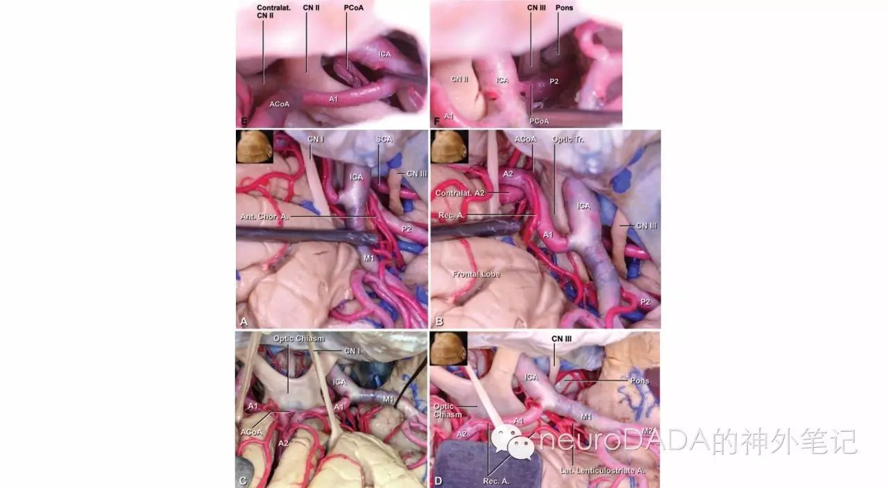
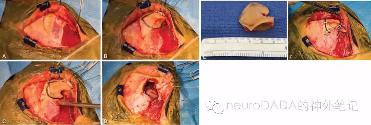
10.Yagmurlu K, Safavi-Abbasi S, Belykh E, Kalani MY, Nakaji P, Rhoton AL, Jr., et al. Quantitative anatomical analysis and clinical experience with mini-pterional and mini-orbitozygomatic approaches for intracranial aneurysm surgery. Journal of neurosurgery. 2016:1-14.
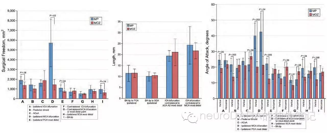
这是本月刚刚在线发表于JNS的最新文献,与篇首24年前的“古文”相比,视角已截然不同——从“大”到“小”,以“小”见“大”。文中对mini翼点入路(mini-pterional approach)和mini眶颧入路(mini-OZ approach)进行step by step的描述以及多参数对比,以明确各自更为擅长的适应症。结果表明,对于动脉瘤夹闭,mini翼点更适合处理同侧病变,mini眶颧更适合控制对侧血管;mini翼点更适合处理MCA分叉部及后交通动脉瘤,mini眶颧更适合处理前交通和ICA分叉部动脉瘤。
随着锁孔技术的出现,各种mini化的额颞眶颧入路也随之涌现,例如近年来大热的由Hernesniemi Juha教授倡导的外侧眶上入路(lateral supraorbital approach,LSO)、Reisch和Perneczky大神倡导的眉弓入路(eyebrow approach),其他诸如蝶骨嵴锁孔入路(sphenoid ridge keyhole approach),以及本文提到的mini翼点入路和mini眶颧入路(mini-OZ approach)等等。这也促使近年来“定量解剖分析”(quantitative anatomical analysis)类解剖学文章大量发表。我想,定量比较的目的,并不是要比出个孰优孰劣,更不是对经典翼点入路和眶颧入路的摒弃,而是要明确每一部分颅骨去除后,对目标结构暴露的差异,最终目的是对不同的病例tailor出不同的、最高效、最微创、最个性化的手术入路,此乃“殊途同归”。
其他相关文献:
1.Nathal E, Gomez-Amador JL. Anatomic and surgical basis of the sphenoid ridge keyhole approach for cerebral aneurysms. Neurosurgery. 2005;56(1 Suppl):178-85; discussion -85.
2.Reisch R, Perneczky A. Ten-year experience with the supraorbital subfrontal approach through an eyebrow skin incision. Neurosurgery. 2005;57(4 Suppl):242-55; discussion -55.
3.Hernesniemi J, Ishii K, Niemela M, Smrcka M, Kivipelto L, Fujiki M, et al. Lateral supraorbital approach as an alternative to the classical pterional approach. Acta Neurochir Suppl. 2005;94:17-21.
4.Balasingam V, Noguchi A, McMenomey SO, Delashaw JB, Jr. Modified osteoplastic orbitozygomatic craniotomy. Technical note. J Neurosurg. 2005;102(5):940-4.
5.Figueiredo EG, Deshmukh V, Nakaji P, Deshmukh P, Crusius MU, Crawford N, et al. An anatomical evaluation of the mini-supraorbital approach and comparison with standard craniotomies. Neurosurgery. 2006;59(4 Suppl 2):ONS212-20; discussion ONS20.
6.Figueiredo EG, Deshmukh P, Nakaji P, Shu EB, Crawford N, Spetzler RF, et al. An anatomical analysis of the mini-modified orbitozygomatic and supra-orbital approaches. Journal of clinical neuroscience : official journal of the Neurosurgical Society of Australasia. 2012;19(11):1545-50.
7.Figueiredo EG, Bor-Seng-Shu E, Spetzler RF, Preul MC. Analytical anatomy: quantifying surgical access with and without orbital bar removal--cadaver and surgical phantom studies. Acta neurochirurgica. 2014;156(8):1633-4.
8.Figueiredo EG, Teixeira MJ, Spetzler RF, Preul MC. Clinical and surgical experience with the minipterional craniotomy. Neurosurgery. 2014;75(3):E324-5.
最后还是以Rhoton教授那句名言结尾:

